

 PDF
PDF ePub
ePub Citation
 Print
Print


Abstract
Purpose
Nowadays, checkbite methods and a digital sensor are used to analyze the movement of mandible. However, there are no study comparing two methods. Therefore, this study has compared measuring the condylar inclination methods by using the new ARCUSdigma 2 system and the checkbite method.
Materials and methods
Young 20 adults without any orthodontic treatment experiences, missing teeth, and restorations with the change of occlusal plane were tested. Angles of condylar path were measured 3 times each, based on Camper's line, by using two methods. KaVo PROTAR Evo 7 semi-adjustable articulator was used and the data were statistically analyzed.
Results
1. The anterior sagittal condylar inclination by ARCUSdigma 2 system were measured as 26.97° (±7.38°) on the left side and 29.80° (±8.19°) on the right side. The lateral condylar inclination were measured as 5.75° (±3.47°) on the left side and 8.10° (±4.98°) on the right side. 2. The anterior sagittal condylar inclination by checkbite method were measured as 25.20° (±6.53°) on the left side and 28.18° (±7.38°) on the right side. The lateral condylar inclination were measured as 10.97° (±5.63°) on the left side and 12.03° (±5.22°) on the right side. There was no statistically significant difference between male and female (P>.05). 3. The lateral condylar inclinations of ARCUSdigma 2 were statistically significantly smaller than that of checkbite method (P<.05).
Go to : 
REFERENCES
1.Dentists' Supply Co. of New York. The Gysi face bow; a description of the instrument and accessories and directions for their use. New York,. 1935.
2.Pelletier LB., Campbell SD. Comparison of condylar control settings using three methods: a bench study. J Prosthet Dent. 1991. 66:193–200.

3.Pelletier LB., Campbell SD. Evaluation of the relationship between anterior and posterior functionally disclusive angles. Part II: Study of a population. J Prosthet Dent. 1990. 63:536–40.
4.Mullick SC., Stackhouse JA Jr., Vincent GR. A study of interocclusal record materials. J Prosthet Dent. 1981. 46:304–7.
5.Olsson A., Posselt U. Relationship of various skull reference lines. J Prosthet Dent. 1961. 11:1045–9.
6.el-Gheriani AS., Winstanley RB. Graphic tracings of condylar paths and measurements of condylar angles. J Prosthet Dent. 1989. 61:77–87.
7.Zamacona JM., Otaduy E., Aranda E. Study of the sagittal condylar path in edentulous patients. J Prosthet Dent. 1992. 68:314–7.
8.Johnson A., Winstanley RB. Recording sagittal condylar angles using a mandibular facebow. J Oral Rehabil. 1997. 24:904–8.
9.Beard CC., Donaldson K., Clayton JA. A comparison of articulator settings to age and sex. J Prosthet Dent. 1986. 56:551–4.
10.Corbett NE., DeVincenzo JP., Huffer RA., Shryock EF. The relation of the condylar path to the articular eminence in mandibular protrusion. Angle Orthod. 1971. 41:286–92.
11.Aull AE. Condylar determinants of occlusal patterns. J Prosthet Dent. 1965. 15:826–49.
12.Donegan SJ., Christensen LV. Sagittal condylar guidance as determined by protrusion records and wear facets of teeth. Int J Prosthodont. 1991. 4:469–72.
13.dos Santos J Jr., Nelson S., Nowlin T. Comparison of condylar guidance setting obtained from a wax record versus an extraoral tracing: a pilot study. J Prosthet Dent. 2003. 89:54–9.
Go to :
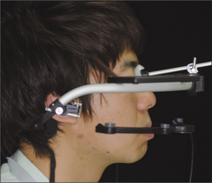
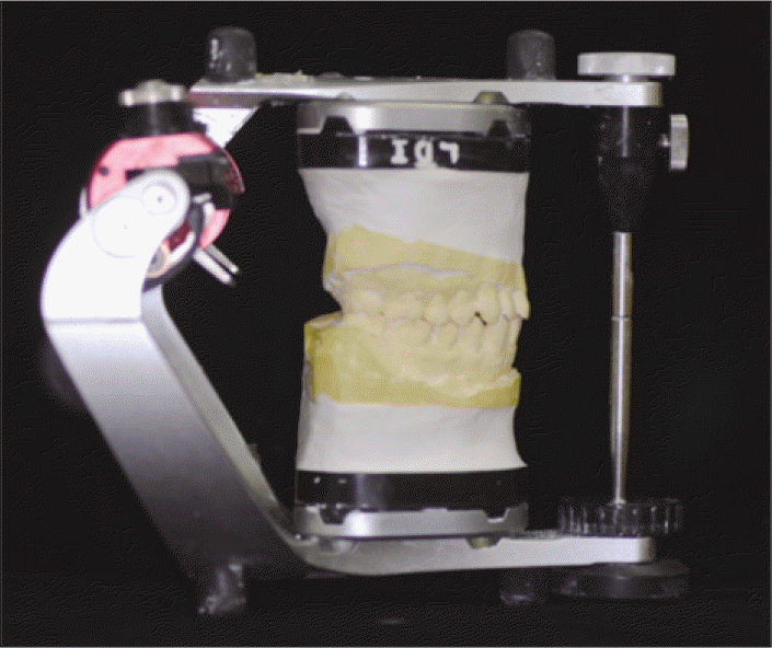
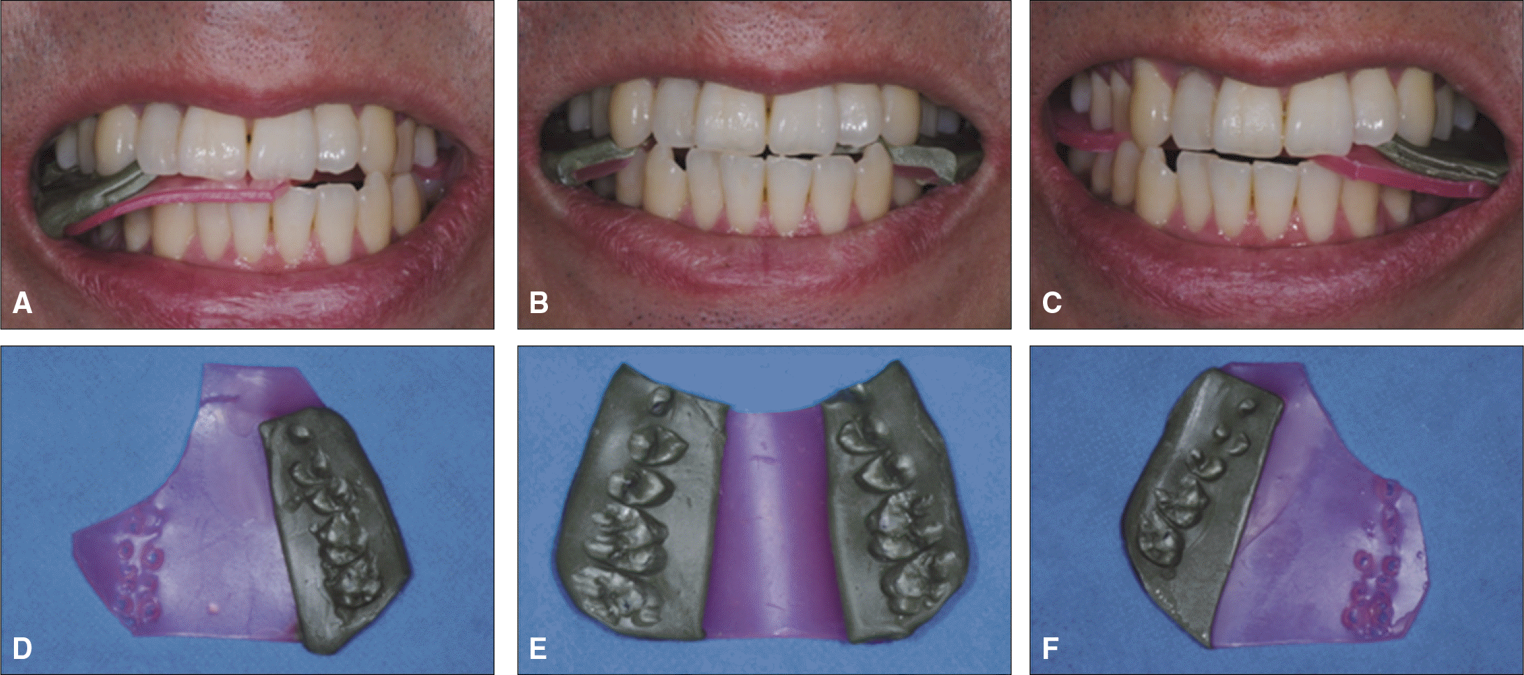
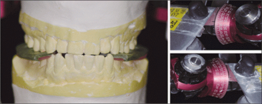
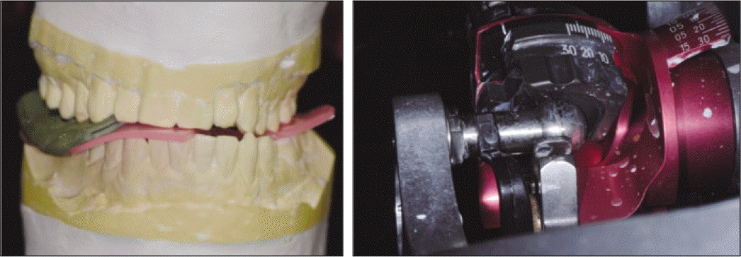
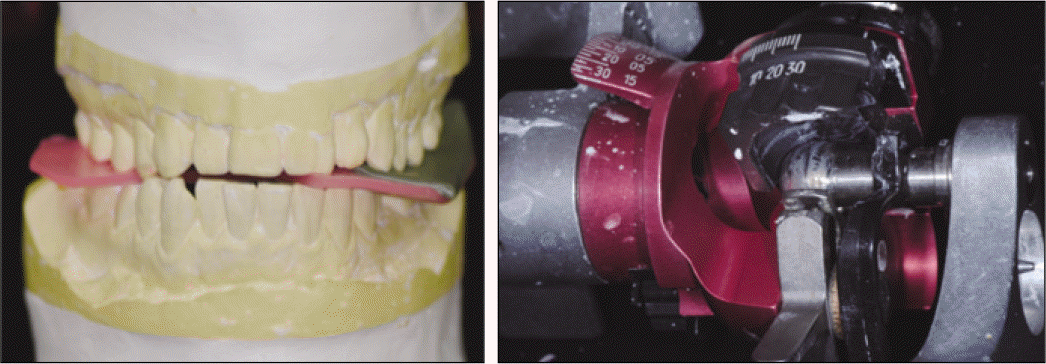
Table 1.
Participants in the study
| Number | Mean age (years) | |
|---|---|---|
| Male | 10 | 28.8 |
| Female | 10 | 30.4 |
| Total | 20 | 29.6 |
Table 2.
Using ARCUSdigma2, Mean sagittal condylar inclination, lateral condylar inclination and standard deviations
Table 3.
Using Checkbite methods, Mean sagittal condylar inclination, lateral condylar inclination and standard deviations
 XML Download