

 PDF
PDF ePub
ePub Citation
 Print
Print


Abstract
Purpose
The aim of this study was to propose the position of maxillary anterior teeth and intercanine width measurements based on the incisive papilla in accordance with the cephalic type and gender of dentate Korean adult with normal teeth alignment.
Materials and methods
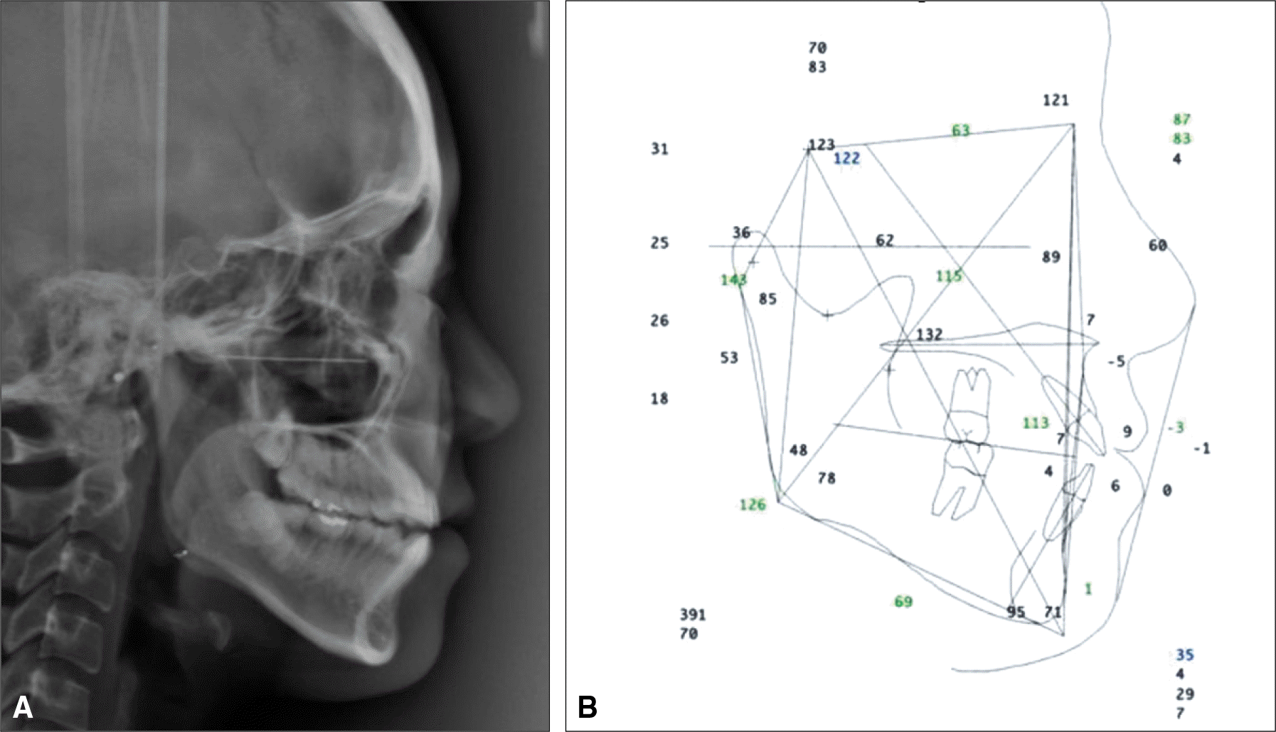
42 students with Class I normal occlusion, without crowding or spacing, were selected from the Chonnam National University School of Dentistry. The lateral skull radiographs of the subjects were taken and were classified as different cephalic types, based on their PFH / AFH ratios. 42 casts of their maxilla were prepared and both the distance between the upper central incisors and the middle of the incisor papilla was measured with a vernier caliper (A) and the distance between the maxillary canine cusp tips (B) were measured. Statistical analysis was performed using SPSS version 15 and their significance was investigated.
Results
For dolichocephalic group, the mean values for A and B were 8.43 (SD: 0.61) and 36.73 (SD: 2.17), respectively. The mean value for A was 8.51 (SD: 1.27) for the mesocephalic group and 8.76 (SD 1.03) for the brachycephalic group. The mean value for B was 35.91 (SD: 1.86) for the mesocephalicgroup and 37.34 (SD: 2.23) for the brachycephalic group. For the male group, the mean A value was 8.86 (SD: 1.04) and the mean B value was 37.60 (SD: 0.24). For the female group, the mean A value was 8.41 (SD: 0.93) and the mean B value was 36.18 (SD: 2.01). The difference between male and female group in A values were not statistically significant (P>.05). The B values of the male subjects were greater than those of the female subjects and was statistically significant (P<.05).
Conclusion
42 students with normal dentition and occlusion in korea, the distance from the incisive papilla and the incisal edge of maxillary central incisors had no difference in cephalic type or gender. However, the distance between the cusp tip of both canines had significant difference in gender where the male showed higher values than the female, while having no difference in cephalic types. (J Korean Acad Prosthodont 2013;51:147-52)
Go to : 
REFERENCES
1.Harper RN. The incisive papilla. J Dent Res. 1948. 27:661–8.

2.Hickey JC., Boucher CO., Woelfel JB. Responsibility of the dentist in complete dentures. J Prosthet Dent. 1962. 12:637–53.
3.Martone AL., Black JW. An approach to prosthodontics through speech science: Part V. Speech science research of prosthodontic significance. J Prosthet Dent. 1962. 12:629–36.
4.Murray CG. Anterior tooth positions in prosthodontics. Aust Dent J. 1977. 22:113–9.
5.Mavroskoufis F., Ritchie GM. Nasal width and incisive papilla as guides for the selection and arrangement of maxillary anterior teeth. J Prosthet Dent. 1981. 45:592–7.
6.Schiffman P. Relation of the maxillary canines to the incisive papilla. J Prosthet Dent. 1964. 14:469–72.
7.Johnson K. A three-year study of the dimensional changes occurring in the maxilla following immediate denture treatment. Aust Dent J. 1967. 12:152–9.
8.Watt DM., Likeman PR. Morphological changes in the denture bearing area following the extraction of maxillary teeth. Br Dent J. 1974. 136:225–35.
9.Suwannachat J. The relationship of interalar width, width of mouth and maxillary intercanine width in Kalasin adult population. Khon Kaen Hosp Med J. 2008. 32:145–52.
10.Jarabak JR., Fizzell JA. Technique and treatment with lightwire edgewise appliance. St Louis: CV Mosby;1972.
11.Sawiris MM. The role of anthropometric measurements in the design of complete dentures. J Dent. 1977. 5:141–8.
12.Lau GC., Clark RF. The relationship of the incisive papilla to the maxillary central incisors and canine teeth in southern Chinese. J Prosthet Dent. 1993. 70:86–93.
13.Heo YS., Shin SW. A study on the positioning of the maxillary central incisor in Koreans. J Korean Acad Prosthodont. 1995. 33:85–97.
14.Ahn HJ., Yang HS., Park HO. A study on the selection of the maxillary anterior artificial teeth in Korean adults. J Korean Acad Prosthodont. 2002. 40:484–92.
Go to :
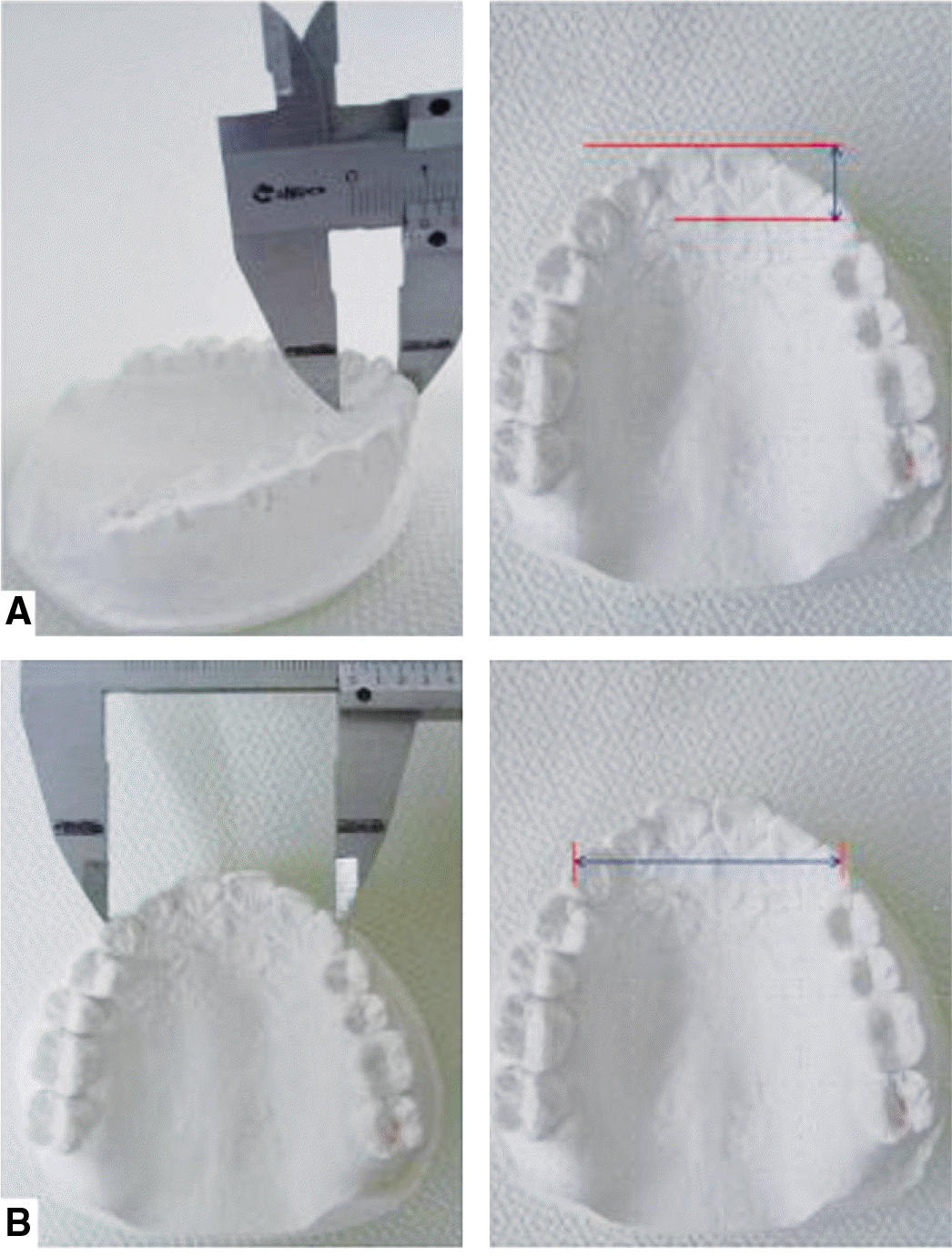 | Fig. 2.A: Measurement of the horizontal distance between the tip of maxillary central incisors and the midpoint of incisive papilla, B: Measurement of the distance between cusp tip of canines. |
Table 1.
Cephalic type
| Male | Female | Total | ||||
|---|---|---|---|---|---|---|
| N | % | N | % | N | % | |
| Dolicocephalic | 0 | 0 | 10 | 23.8 | 10 | 23.8 |
| Mesocephalic | 5 | 11.9 | 5 | 11.9 | 10 | 23.8 |
| Brachycephalic | 15 | 35.7 | 7 | 16.7 | 22 | 52.4 |
Table 2.
Distance between maxillary central incisor and incisive papilla (A) and intercanine width (B) by cephalic types
Table 3.
Distance between maxillary central incisor and incisive papilla (A) and intercanine width (B) by gender and maxillary central incisor (A) and maxillary intercanine width (B)
| Cephalic type | N | A (mm) | B (mm) | ||
|---|---|---|---|---|---|
| Average | Standard deviation | Average | Standard deviation | ||
| Male | 20 | 8.86 | 1.04 | 37.60 | 0.24 |
| Female | 22 | 8.41 | 0.93 | 36.18 | 2.01 |
| Total | 42 | 8.62 | 1.00 | 36.85 | 2.16 |
 XML Download