

 PDF
PDF ePub
ePub Citation
 Print
Print


Abstract
Many of the patients with extensive abrasion need comprehensive restorative treatment. The abrasion is usually caused by attrition, besides of it, there are many reasons for it. The plan of treatment should be started on assessment of the type of attrition and the etiologic analysis. Patient with well-developed masticatory muscle, alveolar process, and high occlusal force and also with little muscle length difference between the stable and the contracted state should be carefully assessed for the vertical dimensional loss and the restoration should be carefully designed. Decrease of tooth length can be compensated by the growth of the alveolar bone height; therefore, consistency of the occlusal vertical dimension is maintained. Accordingly, a careless increase of the vertical dimension can produce muscle fatigue, depressed tooth and pain, and fracture of the restoration. In this case, the patient with multiple tooth abrasion and clenching habit, the edentulous maxillary area is restored with amalgam inserted RPD, and the dentulous area of the maxilla and mandible are treated with fixed restoration accompanying with the increase of vertical dimension. Consequently, we are going to report about the satisfying result in both functional and esthetic aspects. (J Korean Acad Prosthodont 2013;51:119-24)
Go to : 
REFERENCES
1.Turner KA., Missirlian DM. Restoration of the extremely worn dentition. J Prosthet Dent. 1984. 52:467–74.

2.Kiliaridis S., Johansson A., Haraldson T., Omar R., Carlsson GE. Craniofacial morphology, occlusal traits, and bite force in persons with advanced occlusal tooth wear. Am J Orthod Dentofacial Orthop. 1995. 107:286–92.
3.Dawson PE. Functional occlusion: from TMJ to smile design. Mosby;2007. p. 430–52.
4.Vergani CE., Giampaolo ET., Cucci AL. Composite occlusal surfaces for acrylic resin denture teeth. J Prosthet Dent. 1997. 77:328–31.
5.Sowter JB., Bass RE. Increasing the efficiency of resin posterior teeth. J Prosthet Dent. 1968. 19:465–8.
6.Prasad S., Kuracina J., Monaco EA Jr. Altering occlusal vertical dimension provisionally with base metal onlays: a clinical report. J Prosthet Dent. 2008. 100:338–42.
7.Craddock HL., Lynch CD., Franklin P., Youngson CC., Manogue M. A study of the proximity of the Broadrick ideal occlusal curve to the existing occlusal curve in dentate patients. J Oral Rehabil. 2005. 32:895–900.
8.Hoyle DE. Fabrication of a customized anterior guide table. J Prosthet Dent. 1982. 48:490–1.
9.Katsoulis J., Nikitovic SG., Spreng S., Neuhaus K., Mericske-Stern R. Prosthetic rehabilitation and treatment outcome of partially edentulous patients with severe tooth wear: 3-years results. J Dent. 2011. 39:662–71.
10.Johansson A. A cross-cultural study of occlusal tooth wear. Swed Dent J Suppl. 1992. 86:1–59.
11.Mohlin B., Sagne S. The frequency of malocclusion and the craniofacial morphology in a medieval population in Southern Sweden. OSSA (Int J Hum Animal Osteol). 1978. 5:57–84.
12.Gibbs CH., Anusavice KJ., Young HM., Jones JS., Esquivel-Upshaw JF. Maximum clenching force of patients with moderate loss of posterior tooth support: a pilot study. J Prosthet Dent. 2002. 88:498–502.
Go to :
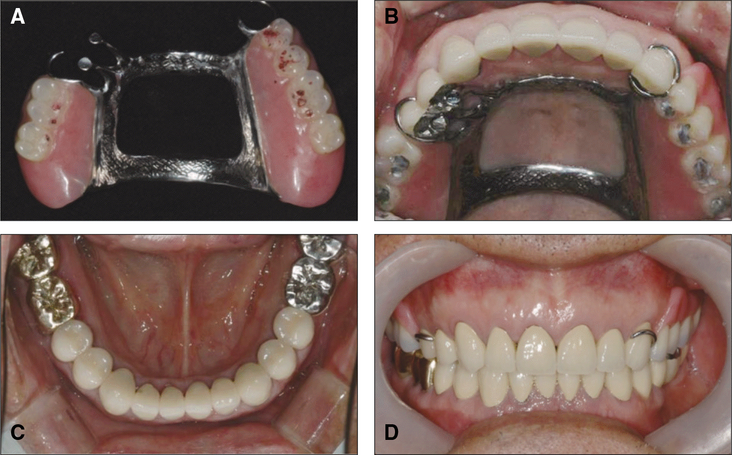
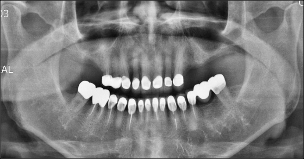
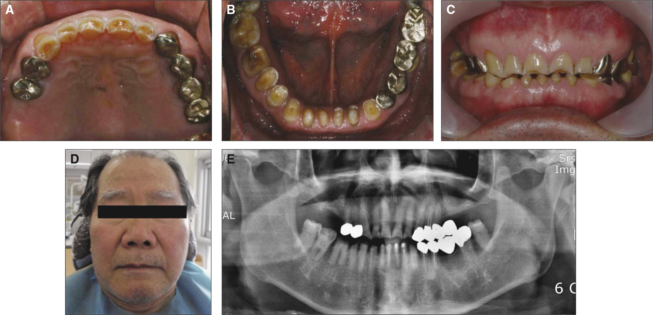 | Fig. 1.Initial photograph. A: Maxillary occlusal view, B: Mandibular occlusal view, C: Frontal view, D: Extraoral view, E: Radiograph panoramic view. |
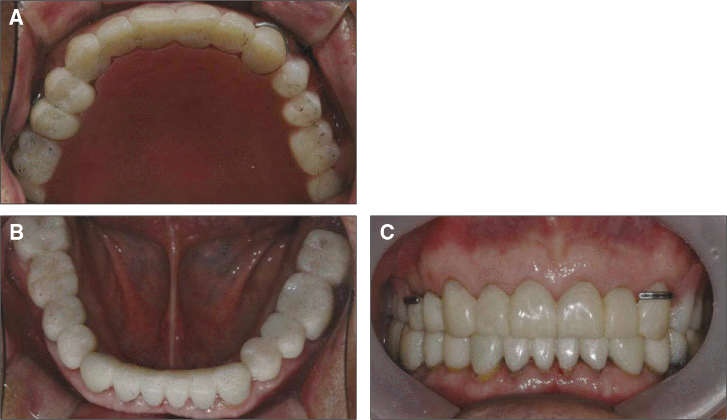 | Fig. 4.Temporary prostheses. A: Maxillary occlusal view, B: Mandibular occlusal view, C: Frontal view. |
 XML Download