

 PDF
PDF ePub
ePub Citation
 Print
Print


Abstract
Fixed dental prostheses such as inlay, onlay, crown, and bridge fabricated by CAD/CAM technique combined with digital impressions is getting popular due to the recent rapid progress of digital impression taking system. For the scope of implant prosthesis, however, digital intra-oral scan hasn't been actively utilized for the fabrication of superstructures. In this case report, 6 cases of titanium-milled custom abutment based on the iTero intra-oral scan data were introduced, five of them were restored with screw-type prosthesis after cementation (SCRP) and the clinical results were satisfactory on restoring the function and esthetics.
Figures and Tables
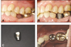
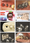 | Fig. 1Clinical pictures of case 1.
A: Peri-implant soft tissue shape after tissue-molding, B: Scan body adaptation for digital impression taking, C: Buccal bite registration by iTero intra-oral scanner, D: Computer-aided design of customized abutment using PowerMILL CAD solution, E: Polyurethane dies and model milled at the milling center, F: Milled titanium customized abutment, G: Customized abutments and SCRP-type porcelain fused-to gold prosthesis, H: Final prosthesis after excess cement removal and polishing.
|
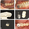
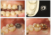 | Fig. 2Intra-oral and model view of case 2.
A: Occluding scan body due to insufficient inter-arch distance, B: Polyurethane die and customized abutment, C: Customized abutment try-in, D: Final implant prosthesis after cementation.
|
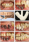 | Fig. 3Clinical pictures of case 3.
A: Intra-oral view after implant surgery, B: Scan body adaptation (note the tissue swelling around implants), C: Computer-aided design of customized abutments, D: Polyurethane model fabricated at milling center, E: Customized abutment tryin, F: Provisional prosthesis built on customized abutments, G: Corrected margin after pick-up impression taking and additional milling, H: Cementation-type implant prosthesis.
|
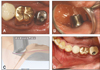 | Fig. 4Clinical pictures on case 4.
A: Customized abutment try-in (occlusal view), B: Supragingival margin (lingual view), C: The amount of marginal discrepancy was around 1.5 mm, D: Completed implant prosthesis.
|
 | Fig. 5Clinical pictures of case 5.
A: Initial intraoral view, B: Occluding scan body due to insufficient inter-arch distance, C: Marginal discrepancy between customized abutment and crown milled from Pt-Ag-In alloy block, D: SCRP-type final implant prosthesis.
|
 | Fig. 6Clinical pictures of case 6.
A: Initial intraoral view, B: Scan body adaptation, C: Polyurethane model milled at milling center, D: Preparation for the cementation between customized abutment and full-zirconia crown Teflon tape is useful for temporary sealing of screw hole, E: Marginal discrepancy shown on screw-type implant prosthesis after cementation, F: Cemented final implant prosthesis.
|
References
1. Mörmann WH, Brandestini M. Die Cerec. Computer reconstruction: Inlays, Onlays and Veneers. 1989. Berlin: Quintessence;75–97.
2. Leinfelder KF, Isenberg BP, Essig ME. A new method for generating ceramic restorations: a CAD-CAM system. J Am Dent Assoc. 1989. 118:703–707.

3. Syrek A, Reich G, Ranftl D, Klein C, Cerny B, Brodesser J. Clinical evaluation of all-ceramic crowns fabricated from intraoral digital impressions based on the principle of active wavefront sampling. J Dent. 2010. 38:553–559.
4. Mörmann WH, Bindl A. The Cerec 3--a quantum leap for computer-aided restorations: initial clinical results. Quintessence Int. 2000. 31:699–712.
5. Herrguth M, Wichmann M, Reich S. The aesthetics of all-ceramic veneered and monolithic CAD/CAM crowns. J Oral Rehabil. 2005. 32:747–752.
6. Heymann HO, Bayne SC, Sturdevant JR, Wilder AD Jr, Roberson TM. The clinical performance of CAD-CAM-generated ceramic inlays: a four-year study. J Am Dent Assoc. 1996. 127:1171–1181.
7. Mörmann WH. The evolution of the CEREC system. J Am Dent Assoc. 2006. 137:7S–13S.
8. Garg AK. Cadent iTero's digital system for dental impressions: the end of trays and putty? Dent Implantol Update. 2008. 19:1–4.
9. Christensen GJ. The challenge to conventional impressions. J Am Dent Assoc. 2008. 139:347–349.
10. Hinds KF. Custom impression coping for an exact registration of the healed tissue in the esthetic implant restoration. Int J Periodontics Restorative Dent. 1997. 17:584–591.
 XML Download