

 PDF
PDF ePub
ePub Citation
 Print
Print


Abstract
Purpose
The purpose of this study is to ascertain the stability of the implant by comparing the effects of the change of implant diameter, length and design on implant stability quotient.
Materials and methods
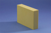
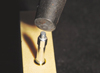
To remove the variable due to the difference of bone quality, the uniform density (0.48 g/cm3) Polyuretane foam blocks (Sawbones®, Pacific Research Laboratories Inc, Vashon, Washington) were used. Implants (Implantium®, Dentium, Seoul, Korea) were placed with varying diameters (ϕ3.8, ϕ4.3 and ϕ4.8) and length (8 mm, 10 mm and 12 mm), to assess the effect on implant stability index (ISQ). Also the influence of the design of the submerged and the non-submerged (SimplelineII®, Dentium, Seoul, Korea) on ISQ was evaluated. To exclude the influence of insertion torque, a total of 60 implants (n = 10) were placed with same torque to 35 N. Using Osstell™ mentor (Integration Diagnostic AB, Sweden) ISQ values were recorded after measuring the resonant frequency, one-way ANOVA and Tukey HSD test results were analyzed. (α=0.05).
Results
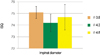
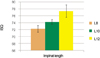
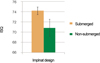
1. The change of the diameter of the implant did not affect the ISQ (P>.05), but the increase of implant length increased the ISQ(P<.001). 2. The change in implant design were correlated with the ISQ, and the ISQ of submerged design was significantly higher than that of the non-submerged design(P<.05).
Figures and Tables
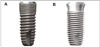 | Fig. 2Implant fixtures used in this experiment(A: Implantium®, B: Simpleline II®, Dentium, Seoul, Korea).
|


References
1. Brånemark PI, Adell R, Breine U, Hansson BO, Lindström J, Ohlsson A. Intra-osseous anchorage of dental prostheses. I. Experimental studies. Scand J Plast Reconstr Surg. 1969. 3:81–100.
2. Adell R, Lekholm U, Rockler B, Brånemark PI. A 15-year study of osseointegrated implants in the treatment of the edentulous jaw. Int J Oral Surg. 1981. 10:387–416.

3. Adell R, Eriksson B, Lekholm U, Brånemark PI, Jemt T. Long-term follow-up study of osseointegrated implants in the treatment of totally edentulous jaws. Int J Oral Maxillofac Implants. 1990. 5:347–359.
4. Meredith N. Assessment of implant stability as a prognostic determinant. Int J Prosthodont. 1998. 11:491–501.
5. Atsumi M, Park SH, Wang HL. Methods used to assess implant stability: current status. Int J Oral Maxillofac Implants. 2007. 22:743–754.
6. Javed F, Almas K, Crespi R, Romanos GE. Implant surface morphology and primary stability: is there a connection? Implant Dent. 2011. 20:40–46.
7. Trisi P, De Benedittis S, Perfetti G, Berardi D. Primary stability, insertion torque and bone density of cylindric implant ad modum Branemark: is there a relationship? An in vitro study. Clin Oral Implants Res. 2011. 22:567–570.
8. Friberg B, Sennerby L, Meredith N, Lekholm U. A comparison between cutting torque and resonance frequency measurements of maxillary implants. A 20-month clinical study. Int J Oral Maxillofac Surg. 1999. 28:297–303.
9. Sennerby L, Meredith N. measuring implant stability and osseointegration. Compend Contin Educ Dent. 1998. 19:493–498. 500502
10. Meredith N, Alleyne D, Cawley P. Quantitative determination of the stability of the implant-tissue interface using resonance frequency analysis. Clin Oral Implants Res. 1996. 7:261–267.
11. Friberg B, Sennerby L, Linden B, Gröndahl K, Lekholm U. Stability measurements of one-stage Brånemark implants during healing in mandibles. A clinical resonance frequency analysis study. Int J Oral Maxillofac Surg. 1999. 28:266–272.
12. Ohta K, Takechi M, Minami M, Shigeishi H, Hiraoka M, Nishimura M, Kamata N. Influence of factors related to implant stability detected by wireless resonance frequency analysis device. J Oral Rehabil. 2010. 37:131–137.
13. Lachmann S, Jäger B, Axmann D, Gomez-Roman G, Groten M, Weber H. Resonance frequency analysis and damping capacity assessment. Part I: an in vitro study on measurement reliability and a method of comparison in the determination of primary dental implant stability. Clin Oral Implants Res. 2006. 17:75–79.
14. Lachmann S, Laval JY, Axmann D, Weber H. Influence of implant geometry on primary insertion stability and simulated periimplant bone loss: an in vitro study using resonance frequency analysis and damping capacity assessment. Int J Oral Maxillofac Implants. 2011. 26:347–355.
15. Sim CP, Lang NP. Factors influencing resonance frequency analysis assessed by Osstell mentor during implant tissue integration: I. Instrument positioning, bone structure, implant length. Clin Oral Implants Res. 2010. 21:598–604.
16. Han J, Lulic M, Lang NP. Factors influencing resonance frequency analysis assessed by Osstell mentor during implant tissue integration: II. Implant surface modifications and implant diameter. Clin Oral Implants Res. 2010. 21:605–611.
17. Saadoun AP, LeGall ML. Clinical results and guidelines on Steri-Oss endosseous implants. Int J Periodontics Restorative Dent. 1992. 12:486–495.
18. Jaffin RA, Berman CL. The excessive loss of Branemark fixtures in type IV bone: a 5-year analysis. J Periodontol. 1991. 62:2–4.
19. Langer B, Langer L, Herrmann I, Jorneus L. The wide fixture: a solution for special bone situations and a rescue for the compromised implant. Part 1. Int J Oral Maxillofac Implants. 1993. 8:400–408.
20. Ivanoff CJ, Sennerby L, Johansson C, Rangert B, Lekholm U. Influence of implant diameters on the integration of screw implants. An experimental study in rabbits. Int J Oral Maxillofac Surg. 1997. 26:141–148.
21. Chan HL, El-Kholy K, Fu JH, Galindo-Moreno P, Wang HL. Implant primary stability determined by resonance frequency analysis in surgically created defects: a pilot cadaver study. Implant Dent. 2010. 19:509–519.
22. Fenner M, Vairaktaris E, Stockmann P, Schlegel KA, Neukam FW, Nkenke E. Influence of residual alveolar bone height on implant stability in the maxilla: an experimental animal study. Clin Oral Implants Res. 2009. 20:751–755.
23. Abrahamsson I, Linder E, Lang NP. Implant stability in relation to osseointegration: an experimental study in the Labrador dog. Clin Oral Implants Res. 2009. 20:313–318.
24. Tabassum A, Meijer GJ, Wolke JG, Jansen JA. Influence of surgical technique and surface roughness on the primary stability of an implant in artificial bone with different cortical thickness: a laboratory study. Clin Oral Implants Res. 2010. 21:213–220.
25. Tabassum A, Meijer GJ, Wolke JG, Jansen JA. Influence of the surgical technique and surface roughness on the primary stability of an implant in artificial bone with a density equivalent to maxillary bone: a laboratory study. Clin Oral Implants Res. 2009. 20:327–332.
26. Al-Nawas B, Wagner W, Grötz KA. Insertion torque and resonance frequency analysis of dental implant systems in an animal model with loaded implants. Int J Oral Maxillofac Implants. 2006. 21:726–732.
27. Turkyilmaz I, Tözüm TF, Tumer C, Ozbek EN. Assessment of correlation between computerized tomography values of the bone, and maximum torque and resonance frequency values at dental implant placement. J Oral Rehabil. 2006. 33:881–888.
28. Devlin H, Horner K, Ledgerton D. A comparison of maxillary and mandibular bone mineral densities. J Prosthet Dent. 1998. 79:323–327.
29. Isoda K, Ayukawa Y, Tsukiyama Y, Sogo M, Matsushita Y, Koyano K. Relationship between the bone density estimated by cone-beam computed tomography and the primary stability of dental implants. Clin Oral Implants Res. 2012. 23:832–836.
30. Bardyn T, Gédet P, Hallermann W, Büchler P. Quantifying the influence of bone density and thickness on resonance frequency analysis: an in vitro study of biomechanical test materials. Int J Oral Maxillofac Implants. 2009. 24:1006–1014.
31. Krennmair G, Waldenberger O. Clinical analysis of wide-diameter frialit-2 implants. Int J Oral Maxillofac Implants. 2004. 19:710–715.
32. Grunder U, Polizzi G, Goené R, Hatano N, Henry P, Jackson WJ, Kawamura K, Köhler S, Renouard F, Rosenberg R, Triplett G, Werbitt M, Lithner B. A 3-year prospective multicenter follow-up report on the immediate and delayed-immediate placement of implants. Int J Oral Maxillofac Implants. 1999. 14:210–216.
33. Bilhan H, Geckili O, Mumcu E, Bozdag E, Sünbüloğlu E, Kutay O. Influence of surgical technique, implant shape and diameter on the primary stability in cancellous bone. J Oral Rehabil. 2010. 37:900–907.
34. Kido H, Schulz EE, Kumar A, Lozada J, Saha S. Implant diameter and bone density: effect on initial stability and pull-out resistance. J Oral Implantol. 1997. 23:163–169.
 XML Download