Abstract
Purpose
This study evaluated the effect of glass fiber pre-impregnated with light-curing resin on the fracture strength and fracture modes of a maxillary complete denture.
Materials and methods
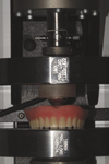
Maxillary acrylic resin complete dentures reinforced with glass fiber pre-impregnated with light-curing resin (SES MESH, INNO Dental Co., Yeoncheon-gun, Korea) and without reinforcement were tested. The reinforcing material was embedded in the denture base resin and placed different regions (Control, without reinforcement; Group A, center of anterior ridge; Group B, rugae area; Group C, center of palate; Group D, full coverage of denture base). The fracture strength and fracture modes of a maxillary complete denture were tested using Instron test machine (Instron Co., Canton, MA, USA) at a 5.0 mm/min crosshead speed. The flexure load was applied to center of denture with a 20 mm diameter ball attachment. When fracture occurred, the fracture mode was classified based on fracture lines. The data were analyzed with one-way ANOVA at the significance level of 0.05.
Figures and Tables
 | Fig. 4Location of SES MESH of the maxillary complete denture: (A) the anterior ridge lap region, (B) the rugae region, (C) the middle region, (D) full coverage, (E) without reinforcement. |
 | Fig. 6Fracture mode of dentures : (A) anteroposterior fracture in group A, (B) anterior fracture in group B, (C) partial fracture on center area in group C, (D) posterior fracture in group D, (E) no significant fracture line in group E. |
References
1. Yoshida K, Takahashi Y, Shimizu H. Effect of embedded metal reinforcements and their location on the fracture resistance of acrylic resin complete dentures. J Prosthodont. 2011. 20:366–371.

2. Smith LT, Powers JM, Ladd D. Mechanical properties of new denture resins polymerized by visible light, heat, and microwave energy. Int J Prosthodont. 1992. 5:315–320.
3. Jeong CM. A comparative study on the several metal reinforcement methods of maxillary complete acrylic resin denture base. J Korean Acad Prosthodont. 1996. 34:363–372.
4. Morris JC, Khan Z, von Fraunhofer JA. Palatal shape and the flexural strength of maxillary denture bases. J Prosthet Dent. 1985. 53:670–673.

5. Lambrecht JR, Kydd WL. A functional stress analysis of the maxillary complete denture base. J Prosthet Dent. 1962. 12:865–872.

6. Vallittu PK, Lassila VP, Lappalainen R. Evaluation of damage to removable dentures in two cities in Finland. Acta Odontol Scand. 1993. 51:363–369.

7. Vallittu PK, Lassila VP. Reinforcement of acrylic resin denture base material with metal or fibre strengtheners. J Oral Rehabil. 1992. 19:225–230.

8. Narva KK, Lassila LV, Vallittu PK. The static strength and modulus of fiber reinforced denture base polymer. Dent Mater. 2005. 21:421–428.

9. Vallittu PK. Glass fiber reinforcement in repaired acrylic resin removable dentures: preliminary results of a clinical study. Quintessence Int. 1997. 28:39–44.
10. Narva KK, Vallittu PK, Helenius H, Yli-Urpo A. Clinical survey of acrylic resin removable denture repairs with glass-fiber reinforcement. Int J Prosthodont. 2001. 14:219–224.
11. Pollet JC, Burns SJ. An analysis of slow crack propagation data in PMMA and brittle materials. Int J Fract. 1977. 13:775–786.

12. Ezrin M. Failure analysis and test procedures. Plastics Failure Guide. 1996. Cincinnati, OH, USA: Hanser Gardner Publ;210–225.

13. Huang GC, Lee CH, Lee JK. Thermal and mechanical properties of short fiber-reinforced epoxy composites. Polym Korea. 2009. 33:530–536.
14. Shin IJ, Lee DJ. Reinforcing characteristics of ductile short-Fiber in brittle matrix composites. Trans KSME A. 2000. 24:250–258.
15. Jang J, Han S. Mechanical properties of glass-bre mat/PMMA functionally gradient composite. Trans Korean Soc Mech Eng. A. 1999. 30:1045–1053.


 PDF
PDF ePub
ePub Citation
 Print
Print


 XML Download