

 PDF
PDF ePub
ePub Citation
 Print
Print


Abstract
Purpose
The purpose of this study was to measure the absorbed dose and to calculate the effective dose for one periapical radiography using the portable and wall type dental X-ray machines.
Materials and methods
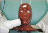
Thermoluminescent chips were placed at 25 sites throughout the layers of the head and neck of a tissue-equivalent human skull phantom. The man phantom was exposed with the portable and wall type dental X-ray machines. For one periapical radiography taken by portable dental X-ray machine, the exposure setting was 60 kVp, 2 mA and 0.2 seconds, while for one periapical radiography taken by wall type dental X-ray machine, exposure setting was 70 kVp, 8 mA and 0.074 seconds. Absorbed dose measurements were performed and equivalent doses to individual organs were summed using ICRP 103 to calculate effective dose.
Results
In the upper anterior periapical radiography using portable dental X-ray machine and in the lower posterior periapical radiography using both machines, the highest absorbed dose was recorded at the mandible body. The effective dose in upper anterior periapical radiography using portable and wall type dental X-ray machines was 4 µSv, 2 µSv, respectively. In the lower posterior periapical radiography, the effective dose for each portable and wall type dental X-ray machines was 6 µSv, 2 µSv.
Figures and Tables
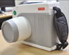
 | Fig. 4Wall-type dental X-ray machine (Kodak2200 intraoral X-ray system, Carestream Health, Inc., Rochester, USA).
|
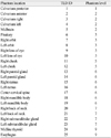
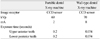
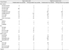
Table 3
Estimated percentage of tissue irradiated and TLDs used to calculated mean dose to a tissue or organ
![]()
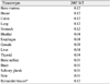
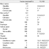
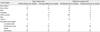
Table 5
Absorbed dose to tissues/organs in head and neck from periapical radiography using the portable and wall-type dental X-ray machine (µGy)
![]()

References
1. Coy J, Vandre RH, Davidson WR. Use of the hand-held dental X-ray machine during joint operation, NATO exercise Display Determination-92. Mil Med. 1997. 162:575–577.

2. Varghese S, Kimmel A, Radmer T, Bradley TG, Bahcall J. In vitro evaluation of the XR-15 portable x-ray unit for forensic odontology. J Forensic Odontostomatol. 2004. 22:5–8.
3. Hermsen KP, Jaeger SS, Jaeger MA. Radiation safety for the NOMAD portable X-ray system in a temporary morgue setting. J Forensic Sci. 2008. 53:917–921.
4. Charlton DG. Portable dental equipment: dental units and x-ray equipment. Gen Dent. 2009. 57:336–341.
5. Kim EK. Effect of the amount of battery charge on tube voltage in different hand-held dental x-ray systems. Imaging Sci Dent. 2012. 42:1–4.
6. Lee W. Current status of medical radiation exposure and regulation efforts. J Korean Med Assoc. 2011. 54:1248–1252.
7. Ludlow JB, Davies-Ludlow LE, Brooks SL, Howerton WB. Dosimetry of 3 CBCT devices for oral and maxillofacial radiology: CB Mercuray, NewTom 3G and i-CAT. Dentomaxillofac Radiol. 2006. 35:219–226.
8. Ludlow JB, Davies-Ludlow LE, Brooks SL. Dosimetry of two extraoral direct digital imaging devices: NewTom cone beam CT and Orthophos Plus DS panoramic unit. Dentomaxillofac Radiol. 2003. 32:229–234.
9. Cho JY, Han WJ, Kim EK. Absorbed and effective dose from periapical radiography by portable intraoral x-ray machine. Korean J Oral Maxillofac Radiol. 2007. 37:149–156.
10. White SC, Pharoah MJ. Oral radiology: principles and interpretation. 2008. 6th ed. Mosby.
11. ICRP. ICRP Publication 103. The 2007 recommendations of the international commission on radiological protection. 2007. Elsevier.
12. Van Dis ML, Miles DA, Parks ET, Razmus TF. Information yield from a hand-held dental x-ray unit. Oral Surg Oral Med Oral Pathol. 1993. 76:381–385.
13. Kim EK. Leakage and scattered radiation from hand-held dental x-ray unit. Korean J Oral Maxillofac Radiol. 2007. 37:65–68.
14. Avendanio B, Frederiksen NL, Benson BW, Sokolowski TW. Effective dose and risk assessment from detailed narrow beam radiography. Oral Surg Oral Med Oral Pathol Oral Radiol Endod. 1996. 82:713–719.
15. Choi SC, Choi HM. Absorbed dose in the full-mouth periapical radiography, panoramic radiography and zonography. J Korean Acad Oral Maxillofac Radiol. 1999. 29:255–260.
16. Underhill TE, Chilvarquer I, Kimura K, Langlais RP, McDavid WD, Preece JW, Barnwell G. Radiobiologic risk estimation from dental radiology. Part I. Absorbed doses to critical organs. Oral Surg Oral Med Oral Pathol. 1988. 66:111–120.
17. Han WJ. Absorbed and effective dose from periapical radiography by portable intraoral x-ray machine and panoramic radiography. J Korean Dent Assoc. 2012. 50:148–158.
 XML Download