

 PDF
PDF ePub
ePub Citation
 Print
Print


Abstract
Purpose
This study was performed to compare the stress distribution pattern of abutment-fixture connection area using 3-dimensional finite element model analysis when 5 different implant systems which have internal connection.
Materials and methods
For the analysis, a finite element model of implant was designed to locate at first molar area. Stress distribution was observed when vertical load of 200 N was applied at several points on the occlusal surfaces of the implants, including center, points 1.5 mm, 3.0 mm away from center and oblique load of 200 N was applied 30° inclined to the implant axis. The finite element model was analyzed by using of 3G. Author (PlassoTech, California, USA).
Results
The DAS tech implant (internal step with no taper) showed more favorable stress distribution than other internally connected implants. AS compare to the situations when the loading was applied within the boundary of implants and an oblique loading was applied, it showed higher equivalent stress and equivalent elastic strain when the loading was applied beyond the boundary of implants. Regardless of loading condition, the abutments showed higher equivalent stress and equivalent elastic strain than the fixtures.
Conclusion
When the occlusal contact is afforded, the distribution of stress varies depending on the design of connection area and the location of loading. More favorable stress distribution is expected when the contact load was applied within the diameter of fixtures and the DAS tech implant (internal step with no tapering) has more benefits than the other design of internally connected implants.
Figures and Tables
 | Fig. 1Abutment and fixture modeling. DSA: JOY (DAS tech, Gwangju, Korea), AST: Osseospeed TX (Astra tech, Mölndal, Sweden), FRI: Ankylos C/X (Friadent, Mannheim, Germany), SST: Standard Implant (Straumann, Waldenburg, Switzerland), SBL: Bone Level Implant (Straumann, Waldenburg, Switzerland).
|

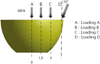 | Fig. 3Loading conditions. A: Loading A condition, B: Loading B condition, C: Loading C condition, D: Loading D condition.
|
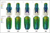

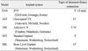
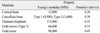
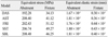
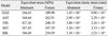
Table 3
Maximum equivalent stress and maximum equivalent strain of various models in loading A condition
![]()
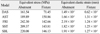
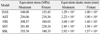
Table 4
Maximum equivalent stress and maximum equivalent strain of various models in loading B condition
![]()
References
1. Brånemark PI, Zarb GA, Albrektsson T, editors. Tissue integrated prostheses. Osseointegration in clinical dentistry. 1985. Chicago: Quintessence.
2. Adell R, Lekholm U, Pockler B, Brånemark PI. A 15 year study of osseointegrated implants in the treatment of the edentulous jaw. Int J Oral Surg. 1981. 10:387–416.

3. Duyck J, Naert IE, Van Oosterwyck H, Van der Sloten J, De Cooman M, Lievens S, Puers B. Biomechanics of oral implants: a review of the literature. Technol Health Care. 1997. 5:253–273.
4. Adell R, Lekholm U, Rockler B, Brånemark P-I, Lidhe J, Eriksson B, Sbordone L. Marginal tissue reactions at osseointegrated titanium fixtures (I) A 3 year longitudinal prospective study. Int J Oral Surg. 1986. 15:39–52.
5. Lindquist LW, Rockler B, Carlsson GE. Bone resorption around fixtures in edentulous patients treated with mandibular fixed tissue-integrated prostheses. J Prosthet Dent. 1988. 59:59–63.
6. Block MS, Gardiner D, Kent JN, Misiek DJ, Finger IM, Guerra L. Hydroxyapatite-coated cylindrical implants in the posterior mandible: 10-year observations. Int J Oral Maxillofac Implants. 1996. 11:626–633.
7. van Steenberghe D, Lekholm U, Bolender C, Folmer T, Henry P, Herrmann I, Higuchi K, Laney W, Linden U, Astrand P. Applicability of osseointegrated oral implants in the rehabilitation of partial edentulism: a prospective multicenter study on 558 fixtures. Int J Oral Maxillofac Implants. 1990. 5:272–281.
8. Isidor F. Loss of osseointegration caused by occlusal load of oral implants. A clinical and radiographic study in monkeys. Clin Oral Implants Res. 1996. 7:143–152.
9. Brunski JB. Biomaterials and biomechanics in dental implant design. Int J Oral Maxillofac Implants. 1988. 3:85–97.
10. Zarb GA, Schmitt A. The longitudinal clinical effectiveness of osseointegrated dental implants: the Toronto study. Part III: Problems and complications encountered. J Prosthet Dent. 1990. 64:185–194.
11. Merz BR, Hunenbart S, Belser UC. Mechanics of the implant-abutment connection: an 8-degree taper compared to a butt joint connection. Int J Oral Maxillofac Implants. 2000. 15:519–526.
12. Becker W, Becker BE. Replacement of maxillary and mandibular molars with single endosseous implant restorations: a retrospective study. J Prosthet Dent. 1995. 74:51–55.
13. Jemt T, Laney WR, Harris D, Henry PJ, Krogh PH Jr, Polizzi G, Zarb GA, Herrmann I. Osseointegrated implants for single tooth replacement: a 1-year report from a multicenter prospective study. Int J Oral Maxillofac Implants. 1991. 6:29–36.
14. Levine RA, Clem DS 3rd, Wilson TG Jr, Higginbottom F, Saunders SL. A multicenter retrospective analysis of the ITI implant system used for single-tooth replacements: preliminary results at 6 or more months of loading. Int J Oral Maxillofac Implants. 1997. 12:237–242.
15. Han SU, Park HO, Yang HS. Stress analysis of supporting tissues and implants according to implant fixture shapes and implant-abutment connections. J Korean Acad Prosthodont. 2004. 42:226–237.
16. Levine RA, Clem D, Beagle J, Ganeles J, Johnson P, Solnit G, Keller GW. Multicenter retrospective analysis of the solid-screw ITI implant for posterior single-tooth replacements. Int J Oral Maxillofac Implants. 2002. 17:550–556.
17. Norton MR. An in vitro evaluation of the strength of an internal conical interface compared to a butt joint interface in implant design. Clin Oral Implants Res. 1997. 8:290–298.
18. Norton MR. Assessment of cold welding properties of the internal conical interface of two commercially available implant systems. J Prosthet Dent. 1999. 81:159–166.
19. Sones AD. Complications with osseointegrated implants. J Prosthet Dent. 1989. 62:581–585.
20. Kallus T, Bessing C. Loose gold screws frequently occur in full-arch fixed prostheses supported by osseointegrated implants after 5 years. Int J Oral Maxillofac Implants. 1994. 9:169–178.
21. Haas R, Mensdorff-Pouilly N, Mailath G, Watzek G. Brånemark single tooth implants: a preliminary report of 76 implants. J Prosthet Dent. 1995. 73:274–279.
22. Kim KT, Jo KH, Lee CH, Yu WJ, Lee KB. Cervical design effect of dental implant on stress distribution in crestal cortical bone studied by finite element analysis. J Korean Acad Prosthodont. 2009. 47:385–393.
23. Boggan RS, Strong JT, Misch CE, Bidez MW. Influence of hex geometry and prosthetic table width on static and fatigue strength of dental implants. J Prosthet Dent. 1999. 82:436–440.
24. Merz BR, Hunenbart S, Belser UC. Mechanics of the implant-abutment connection: an 8-degree taper compared to a butt joint connection. Int J Oral Maxillofac Implants. 2000. 15:519–526.
25. Walton JN, MacEntee MI. A prospective study on the maintenance of implant prostheses in private practice. Int J Prosthodont. 1997. 10:453–458.
26. Kim YL, Cho HW, Lee JB. Three dimensional finite element analysis of internally connected implant systems. J Korean Acad Prosthodont. 2006. 44:85–102.
27. Geng JP, Tan KB, Liu GR. Application of finite element analysis in implant dentistry: a review of the literature. J Prosthet Dent. 2001. 85:585–598.
28. Weinstein AM, Klawitter JJ, Anand SC, Schuessler R. Stress analysis of porous rooted dental implants. J Dent Res. 1976. 55:772–777.
29. Ahn JK, Kay KS, Chung CH. Finite element stress analysis of implant prosthesis with internal connection between the implant and the abutment. J Korean Acad Prosthodont. 2004. 42:356–372.
30. Richter EJ. In vivo vertical forces on implants. Int J Oral Maxillofac Implants. 1995. 10:99–108.
31. Carlsson GE, Haraldson T. Brånemark PI, Zarb GA, Albrektsson T, editors. Functional response. Tissue integrated prostheses. Osseointegration in clinical dentistry. 1986. 4th ed. Chicago: Quintessence;74–78.
32. Craig RG. Restorative Dental Materials. 1980. 6th ed. St. Louis: Mosby;60–61.
33. Lee HS, Kim MR, Park JM, Kim SJ. A 3-dimensional finite element analysis of tapered internal connection implant system (Avana SS III(R)) on different abutment connections. J Korean Acad Prosthodont. 2010. 48:181–188.
34. Ban JH, Shin SW, Kim SJ, Lee JY. Three-dimensional finite element analysis on stress distribution of the mandibular implant-supported cantilever prostheses depending on the designs. J Korean Acad Prosthodont. 2009. 47:70–81.
35. Rangert B, Jemt T, Jörneus L. Forces and moments on Brånemark implants. Int J Oral Maxillofac Implants. 1989. 4:241–247.
36. Rangert B, Krogh PH, Langer B, Van Roekel N. Bending overload and implant fracture: a retrospective clinical analysis. Int J Oral Maxillofac Implants. 1995. 10:326–334.
37. Kwon JH, Choi MH, Kim YL, Cho HW. Three-dimensional finite element stress analysis of single implant restoration using different fixture and abutment screw diameters. J Korean Acad Prosthodont. 2005. 43:105–119.
38. Yu SH, Park WH, Park JJ, Lee YS. A study on the various implant systems using the finite element stress analysis. J Korean Acad Prosthodont. 2006. 44:207–216.
 XML Download