

 PDF
PDF ePub
ePub Citation
 Print
Print


Abstract
Purpose
Dentists suffer back, neck and shoulder pain during their careers due to bad operating posture. If dentists have a good operating posture ergonomically, there would be less pain and discomfort in the shoulder and back. Therefore, dentists should learn the Home position which enables dentists to approach a stable posture ergonomically. This study was to compare tooth preparation in the Home position and the Random position, and evaluate the clinical efficacy of the Home position.
Materials and methods
Tooth preparation for fixed partial denture was performed on the maxillary left 2nd premolar and maxillary left 2nd molar at the two different operating positions were compared. The amount of occlusal reduction, marginal width, subgingival margin depth, and convergence angle were measured. A T-test was performed separately to compare the results of the Random position and the Home position.
Results
1. The amounts of average thickness of occlusal reduction on fossa were deficient to the ordered ones in the Random position and the Home position (P > .05). 2. The average subgingival margin depth of prepared margin on maxillary left 2nd premolar, maxillary left 2nd molar were excessive in the Random position than in the Home position. On the maxillary left 2nd premolar, there was no statistical difference in the Random position and the Home position except Distal midline, DL line angle, Lingual midline, ML line angle (P < .05). On the maxillary left 2nd molar, there was no statistical difference in the Random position and the Home position (P < .05). 3. Average convergence angle in the Random position and the Home position were excessive compared to the ordered angle. There was no statistical difference in the Random position and the Home position (P > .05). 4. Analysis of pearson correlation : In the Random position, the amounts of average thickness of occlusal reduction, the average subgingival margin depth of prepared margin, convergence angle were significantly associated with each other (P < .05). But in the Home position, they were not significantly associated with each other (P < .05). 5. The time needed for preparation in the Home position was faster or equal than that of the Random position as time went on.
Conclusion
In conclusion, there were no significant differences between Home postion and Random position in measures of occlusal reduction, marginal width, marginal depth, convergence angle. However, preparation time and incidence of damaging adjacent teeth were less in Home position than in Random position. Therefore, if trained properly, Home position which is more ergonomically stable can be adopted for clinical use. (J Korean Acad Prosthodont 2011;49:38-48)
Go to : 
REFERENCES
1.Erdil M., Dickerson OB. Cumulative Trauma Disorders: Prevention, Evaluation, and Treatment. John Wiley & Sons;1996. p. 88–9.
2.Chowanadisai S., Kukiattrakoon B., Yapong B., Kedjarune U., Leggat PA. Occupational health problems of dentists in southern Thailand. Int Dent J. 2000. 50:36–40.

3.Jeon MJ., Sakong J., Lee JJ., Lee HK., Chung JH. Assessment of Job Related Cumulative Trauma Disorders of Dentists in Daegu Metropolitan City. Korean J Occup Environ Med. 2001. 13:55–63.
4.Choi MG. A study on the prevalence subjective symptom of musculoskeletal disorders & related risk factors among dentists in dental hospital. PhD Thesis. Department of Public Health, The Graduate School, Daegu Haany University, Gyeongbuk, Korea,. 2006.
5.Choi DG. The influence of the dentist' s positions on the abutment tooth preparation of full cast crown: upper left second premolar. J Jpn Prosthodont Soc. 1986. 30:545–59.
6.Lee SJ. The influence of home position and random position on the shape of prepared tooth surfaces: upper left 1st molar for full cast crown. PhD Thesis, Graduate School, Kyung Hee University, Seoul, Korea,. 2001.
7.Oh YR. An influence of operator' s posture on shape of prepared tooth surfaces for full cast crown (maxillary right first molar). PhD thesis, Graduate School, Kyung-Hee University, Seoul, Korea,. 2009.
8.Park NS. An influence of operator' s posture on shape of tooth surface preparation for full cast crown: mandibular right 1st molar. PhD thesis, Graduate School, Kyung-Hee University, Seoul, Korea,. 2010.
9.Hwang SD. An influence of operator's posture to the shape of tooth surfaces preparation for full cast crown: mandibular left first molar. PhD Thesis, Graduate School, Kyung-Hee University, Seoul, Korea,. 2010.
10.Kim SJ., Pae AR., Woo YH., Kim HS. Clinical convergence angle of prepared tooth for full veneer crowns. J Dent Rehab Appl Sci. 2010. 26:21–32.
11.Shillingburg HT Jr., Hobo S., Whitsett LD., Jacobi R., Brackett SE. Fundamentals of Fixed Prosthodontics. 3rd ed.Quintessence Publishing;IL: 1997. p. 120–52.
12.Annerstedt AL., Engstro ¨m U., Hansson A., Jansson T., Karlsson S., Liljhagen H., Lindquist E., Rydhammar E., Tyreman-Bandhede M., Svensson P., Wandel U. Axial wall convergence of full veneer crown preparations Documented for dental students and general practitioners. Acta Odontol Scand. 1996. 54:109–12.
13.Mack PJ. A theoretical and clinical investigation into the taper achieved on crown and inlay preparations. J Oral Rehabil. 1980. 7:255–65.
Go to :
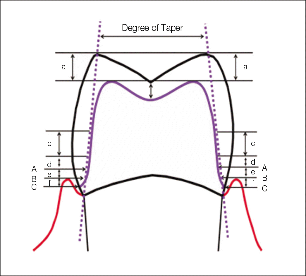 | Fig. 1.Measuring method for occlusal reduction, width & depth of margine, convergence angle. A: Measuring point of margin width, B: Artificial gingiva position, C: Margin position, a: Thickness of occlusal surface reduction (Cusp tip), b: Thickness of Occlusal surface reduction (Fossa), c: Measuring point of convergence angle (Upper part of margin: 1.5 - 3 mm), d: Upper part of measuring point of margin width (0.5 mm), e: Upper part of artificial gingiva position (0.5 mm), f: Lower part of artificial gingiva position (0.5 mm). |
Table 1.
Guideline of tooth reduction for Home position (Max. left 2nd premolar)
Table 2.
Guideline of tooth reduction for Home position (Max. left 2nd molar)
Table 3.
Average thickness of occlusal surface reduction on maxillary left 2nd premolar (Unit: mm)
Table 4.
Average thickness of occlusal surface reduction on maxillary left 2nd molar (Unit: mm)
| Measuring point | Operator's position | Average thickness of occlusal reduction (SD) | Significance |
|---|---|---|---|
| 1. Mesiobuccal cusp tip | Random P. | 1.69 (0.50) | 0.95 |
| Home P. | 1.68 (0.43) | ||
| 2. Distobuccal cusp tip | Random P. | 1.61 (0.43) | 0.01∗ |
| Home P. | 1.93 (0.43) | ||
| 3. Palatal cusp tip | Random P. | 2.05 (0.53) | 0.24 |
| Home P. | 2.00 (0.44) | ||
| 4. Distolingual cusp tip | Random P. | 1.74 (1.02) | 0.63 |
| Home P. | 1.63 (0.35) | ||
| 5. Mesial fossa | Random P. | 1.02 (0.34) | 0.09 |
| Home P. | 0.85 (0.30) | ||
| 6. Central fossa | Random P. | 0.92 (0.39) | 0.39 |
| Home P. | 0.84 (0.33) | ||
| 7. Distal fossa | Random P. | 0.85 (0.38) | 0.27 |
| Home P. | 0.94 (0.27) |
Table 5.
Average width of prepared margin on maxillary left 2nd premolar (Unit: mm)
Table 6.
Average width of prepared margin on maxillary left 2nd molar (Unit: mm)
| Measuring point | Operator's position | Average width of prepared margin (SD) | Significance |
|---|---|---|---|
| 1. MB Lineangle | Random P. | 1.55 (0.54) | 0.44 |
| Home P. | 1.84 (1.65) | ||
| 2. Buccal midline | Random P. | 1.74 (0.39) | 0.62 |
| Home P. | 1.86 (1.02) | ||
| 3. DB lineangle | Random P. | 1.54 (0.35) | 0.08 |
| Home P. | 1.33 (0.39) | ||
| 4. Distal midline | Random P. | 2.51 (0.80) | 0.25 |
| Home P. | 2.29 (0.40) | ||
| 5. DL lineangle | Random P. | 1.91 (0.45) | 0.67 |
| Home P. | 1.98 (0.50) | ||
| 6. Lingual midline | Random P. | 1.44 (0.39) | 0.07 |
| Home P. | 1.25 (0.26) | ||
| 7. ML lineangle | Random P. | 1.44 (0.29) | 0.82 |
| Home P. | 1.46 (0.33) | ||
| 8. Mesial midline | Random P. | 1.66 (0.46) | 0.01∗ |
| Home P. | 2.00 (0.34) |
Table 7.
Average depth of prepared margin on maxillary left 2nd premolar (Unit: mm)
Table 8.
Average depth of prepared margin on maxillary left 2nd molar (Unit: mm)
| Measuring point | Operator's position | Average depth of prepared margin (SD) | Significance |
|---|---|---|---|
| 1. MB Lineangle | Random P. | 0.55 (0.82) | 0.03∗ |
| Home P. | 0.17 (0.43) | ||
| 2. Buccal midline | Random P. | 1.03 (0.77) | 0.01∗ |
| Home P. | 0.54 (0.41) | ||
| 3. DB lineangle | Random P. | 1.78 (0.63) | 0.01∗ |
| Home P. | 1.34 (0.53) | ||
| 4. Distal midline | Random P. | 1.44 (0.68) | 0.01∗ |
| Home P. | 1.02 (0.40) | ||
| 5. DL lineangle | Random P. | 1.38 (0.75) | 0.01∗ |
| Home P. | 0.90 (0.42) | ||
| 6. Lingual midline | Random P. | 0.99 (0.66) | 0.02∗ |
| Home P. | 0.58 (0.39) | ||
| 7. ML lineangle | Random P. | 0.81 (0.63) | 0.04∗ |
| Home P. | 0.46 (0.33) | ||
| 8. Mesial midline | Random P. | 1.39 (0.83) | 0.03∗ |
| Home P. | 0.97 (0.46) |
Table 9.
Average convergence angle on maxillary left 2nd premolar (Unit: °)
Table 10.
Average convergence angle on maxillary left 2nd molar (Unit: °)
Table 11.
Pearson correlation of random position on maxillary left 2nd premolar
| Occlusal reduction | Margin depth | Margin width | Mesiodistal convergence angle | Buccolingual convergence angle | ||
|---|---|---|---|---|---|---|
| Occlusal reduction | P.C.C. | 1 | ||||
| Significance | ||||||
| Margin depth | P.C.C. | -0.080 | 1 | |||
| Significance | 0.436 | |||||
| Margin width | P.C.C. | 0.518∗∗ | -0.084 | 1 | ||
| Significance | 0.000 | 0.247 | ||||
| Mesiodistal convergence angle | P.C.C. | -0.090 | 0.557∗∗ | 0.160 | 1 | |
| Significance | 0.674 | 0.005 | 0.457 | |||
| Buccolingual convergence angle | P.C.C. | -0.154 | 0.510∗ | 0.206 | 0.570∗∗ | 1 |
| Significance | 0.472 | 0.11 | 0.333 | 0.004 |
Table 12.
Pearson correlation of random position on maxillary left 2nd molar
| Occlusal reduction | Margin depth | Margin width | Mesiodistal convergence angle | Buccolingual convergence angle | ||
|---|---|---|---|---|---|---|
| Occlusal reduction | P.C.C. | 1 | ||||
| Significance | ||||||
| Margin depth | P.C.C. | -0.326∗∗ | 1 | |||
| Significance | 0.000 | |||||
| Margin width | P.C.C. | -0.154∗ | 0.169∗ | 1 | ||
| Significance | 0.046 | 0.019 | ||||
| Mesiodistal convergence angle | P.C.C. | -0.330 | 0.694∗∗ | -0.080 | 1 | |
| Significance | 0.877 | 0.000 | 0.711 | |||
| Buccolingual convergence angle | P.C.C. | 0.235 | 0.382 | -0.363 | 0.444∗ | 1 |
| Significance | 0.269 | 0.065 | 0.081 | 0.030 |
Table 13.
Pearson correlation of home position on maxillary left 2nd premolar
| Occlusal reduction | Margin depth | Margin width | Mesiodistal convergence angle | Buccolingual convergence angle | ||
|---|---|---|---|---|---|---|
| Occlusal reduction | P.C.C. | 1 | ||||
| Significance | ||||||
| Margin depth | P.C.C. | -0.209∗ | 1 | |||
| Significance | 0.041 | |||||
| Margin width | P.C.C. | 0.043 | -0.092 | 1 | ||
| Significance | 0.675 | 0.204 | ||||
| Mesiodistal convergence angle | P.C.C. | 0.010 | 0.260 | 0.192 | 1 | |
| Significance | 0.962 | 0.220 | 0.368 | |||
| Buccolingual convergence angle | P.C.C. | 0.217 | 0.126 | 0.115 | 0.214 | 1 |
| Significance | 0.309 | 0.556 | 0.594 | 0.314 |
Table 14.
Pearson correlation of home position on maxillary left 2nd molar
| Occlusal reduction | Margin depth | Margin width | Mesiodistal convergence angle | Buccolingual convergence angle | ||
|---|---|---|---|---|---|---|
| Occlusal reduction | P.C.C. | 1 | ||||
| Significance | ||||||
| Margin depth | P.C.C. | -0.423∗∗ | 1 | |||
| Significance | 0.000 | |||||
| Margin width | P.C.C. | 0.059 | 0.014 | 1 | ||
| Significance | 0.450 | 0.848 | ||||
| Mesiodistal convergence angle | P.C.C. | 0.224 | 0.172 | -0.059 | 1 | |
| Significance | 0.293 | 0.421 | 0.784 | |||
| Buccolingual convergence angle | P.C.C. | 0.126 | 0.217 | -0.130 | 0.800∗∗ | 1 |
| Significance | 0.558 | 0.310 | 0.545 | 0.000 |
 XML Download