Abstract
Purpose
The purpose of this study was to colorimetrically evaluate the masking effect of different opacity of ingots on the final shade of IPS Empress Esthetic laminate veneer restorations using the CIE L∗a∗b∗ system.
Materials and methods
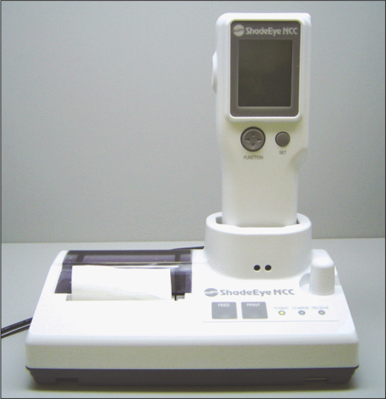
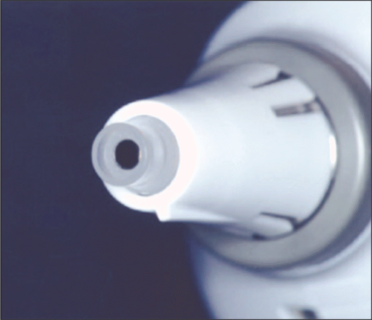
Six porcelain disks of IPS Empress Esthetic system (translucency: E 01, E 03, E 0C-1, E TC-1, E TC-2, E TC-3) were fabricated with 7 mm in diameter and 0.6 mm in thickness. Six extracted human incisors (shade: A1, A3, A4, B2, B3, C3) were used as the abutment specimens. The incisors were prepared using a diamond wheel and made with a flat labial surface on the middle 1/3. For each combination of different shades of abutments and copings, the change in color was measured with a colorimeter. CIE L∗a∗b∗ coordinates were recorded for each specimen. Color differences (ΔE) were calculated. Descriptive statistical analysis was done.
Results
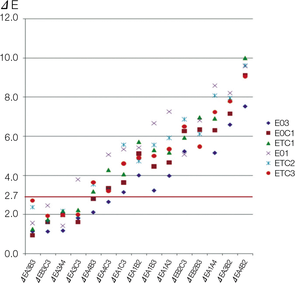
ΔE values were significantly affected by coping translucency and abutment shade (P<.05). The color differences (ΔE) of laminate veneers among abutments with A3, B3, C3, and A4 shade were mostly below 2.7 which was within the clinically acceptable range, while color differences between A4 and B2, A3 and B2, and A1 and A4 showed more than 2.7.
Go to : 
REFERENCES
1.Davis LG., Ashworth PD., Spriggs LS. Psychological effects of aesthetic dental treatment. J Dent. 1998. 26:547–54.

2.Faunce FR., Myers DR. Laminate veneer restoration of permanent incisors. J Am Dent Assoc. 1976. 93:790–2.

3.Helpin ML., Fleming JE. Laboratory technique for the laminate veneer restoration. Pediatr Dent. 1982. 4:48–50.
4.Barreto MT., Shiu A., Renner RP. Anterior porcelain laminate veneers: clinical and laboratory procedures. Quintessence Dent Technol. 1986. 10:493–9.
5.Garber DA., Goldstein RE., Feinman RA. Porcelain Laminate Veneers. Chicago, Quintessence Pub Co.;1988.
6.Fradeani M. Six-year follow-up with Empress veneers. Int J Periodontics Restorative Dent. 1998. 18:216–25.
7.Belser UC., Magne P., Magne M. Ceramic laminate veneers: continuous evolution of indications. J Esthet Dent. 1997. 9:197–207.

8.Yaman P., Qazi SR., Dennison JB., Razzoog ME. Effect of adding opaque porcelain on the final color of porcelain laminates. J Prosthet Dent. 1997. 77:136–40.

9.Antonson SA., Anusavice KJ. Contrast ratio of veneering and core ceramics as a function of thickness. Int J Prosthodont. 2001. 14:316–20.
10.Vichi A., Ferrari M., Davidson CL. Influence of ceramic and cement thickness on the masking of various types of opaque posts. J Prosthet Dent. 2000. 83:412–7.

11.Davis BK., Aquilino SA., Lund PS., Diaz-Arnold AM., Denehy GE. Colorimetric evaluation of the effect of porcelain opacity on the resultant color of porcelain veneers. Int J Prosthodont. 1992. 5:130–6.
12.Davis BK., Papcum LJ., Johnston WM. Effect of cement shade on final shade of porcelain veneers. J Dent Res. 1991. 70:297. Abstract 250.
13.Davis BK., Scott JO., Johnston WM. Effect of porcelain shades on final shade of porcelain veneers. J Dent Res. 1991. 70:475. Abstract 1671.
14.Simonsen RJ., Calamia JR. Tensile bond strength of etched porcelain [abstract]. J Dent Res. 1983. 62:297. Abstract 1099.
15.Calamia JR., Simonsen RJ. Effect of coupling agents on bond strength of etched porcelain[abstract]. J Dent Res. 1984. 63:179. Abstract 79.
16.Hasegawa A., Ikeda I., Kawaguchi S. Color and translucency of in vivo natural central incisors. J Prosthet Dent. 2000. 83:418–23.

17.Kim HE., Cho IH., Lim JH., Lim HS. Shade analysis of anterior teeth using digital shade analysis system. J Korean Acad Prosthodont. 2003. 41:565–81.
18.Shimada K., Kakehashi Y., Matsumura H., Tanoue N. In vivo quantitative evaluation of tooth color with hand-held colorimeter and custom template. J Prosthet Dent. 2004. 91:389–91.

19.Bolt RA., Bosch JJ., Coops JC. Influence of window size in small-window colour measurement, particularly of teeth. Phys Med Biol. 1994. 39:1133–42.

20.Wee AG., Monaghan P., Johnston WM. Variation in color between intended matched shade and fabricated shade of dental porcelain. J Prosthet Dent. 2002. 87:657–66.

21.Douglas RD., Steinhauer TJ., Wee AG. Intraoral determination of the tolerance of dentists for perceptibility and acceptability of shade mismatch. J Prosthet Dent. 2007. 97:200–8.

22.Seghi RR., Hewlett ER., Kim J. Visual and instrumental colorimetric assessments of small color differences on translucent dental porcelain. J Dent Res. 1989. 68:1760–4.

23.Seghi RR., Johnston WM., O' Brien WJ. Performance assessment of colorimetric devices on dental porcelains. J Dent Res. 1989. 68:1755–9.

24.O' Brien WJ., Groh CL., Boenke KM. A new, small-color-difference equation for dental shades. J Dent Res. 1990. 69:1762–4.
25.Johnston WM., Kao EC. Assessment of appearance match by visual observation and clinical colorimetry. J Dent Res. 1989. 68:819–22.

26.Preston JD. Current status of shade selection and color matching. Quintessence Int. 1985. 16:47–58.
27.Sorensen JA., Torres TJ. Improved color matching of metal-ceramic restorations. Part I: A systematic method for shade determination. J Prosthet Dent. 1987. 58:133–9.

28.Sorensen JA., Torres TJ. Improved color matching of metal ceramic restorations. Part II: Procedures for visual communication. J Prosthet Dent. 1987. 58:669–77.

30.Sproull RC. Color matching in dentistry. I. The three-dimensional nature of color. J Prosthet Dent. 1973. 29:416–24.
31.Sproull RC. Color matching in dentistry. II. Practical applications of the organization of color. J Prosthet Dent. 1973. 29:556–66.
32.Sproull RC. Color matching in dentistry. 3. Color control. J Prosthet Dent. 1974. 31:146–54.
33.Macentee M., Lakowski R. Instrumental colour measurement of vital and extracted human teeth. J Oral Rehabil. 1981. 8:203–8.

34.Tung FF., Goldstein GR., Jang S., Hittelman E. The repeatability of an intraoral dental colorimeter. J Prosthet Dent. 2002. 88:585–90.

35.Paul S., Peter A., Pietrobon N., Ha¨mmerle CH. Visual and spectrophotometric shade analysis of human teeth. J Dent Res. 2002. 81:578–82.

36.Johnston WM., Hesse NS., Davis BK., Seghi RR. Analysis of edge-losses in reflectance measurements of pigmented maxillofacial elastomer. J Dent Res. 1996. 75:752–60.

37.van der Burgt TP., ten Bosch JJ., Borsboom PC., Kortsmit WJ. A comparison of new and conventional methods for quantification of tooth color. J Prosthet Dent. 1990. 63:155–62.

38.Brewer JD., Garlapo DA., Chipps EA., Tedesco LA. Clinical discrimination between autoglazed and polished porcelain surfaces. J Prosthet Dent. 1990. 64:631–4.

39.Kim IJ., Lee YK., Lim BS., Kim CW. Effect of surface topography on the color of dental porcelain. J Mater Sci Mater Med. 2003. 14:405–9.
Go to : 
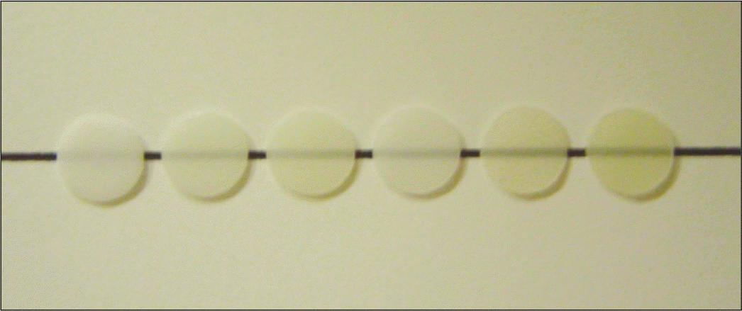 | Fig. 1.IPS Empress Esthetic® laminate veneer disk specimen (Ivoclar Vivadent) (from the left, E 03, E 0C-1, E TC-1, E 01, E TC-2, E TC-3). |
Table 1.
Mean L∗, a∗, b∗ values of the abutment teeth specimens
Table 2.
Mean L∗, a∗, b∗ values of laminate veneer specimens on various abutment teeth specimens
Table 3.
Two-way ANOVA for mean ΔE values of laminate veneers on pair of abutment teeth specimens with different shades
| DF | Sum of Squares | Mean Square | F value | Pr > F | |
|---|---|---|---|---|---|
| Laminate veneer | 5 | 26.87 | 5.374 | 16.16 | <.05 |
| Pair of abutment teeth | 14 | 446.441 | 31.888 | 95.87 | <.05 |
Table 4.
Tukey's HSD test for mean ΔE values of laminate veneer specimens
| Tukey Grouping | Mean | N | Laminate veneer | |
|---|---|---|---|---|
| A | 5.44 | 15 | E 01 | |
| A | 4.56 | 15 | E TC2 | |
| B | A | 4.53 | 15 | E TC1 |
| B | A | 4.43 | 15 | E TC3 |
| B | 4.35 | 15 | E 0C1 | |
| C | 3.68 | 15 | E 03 | |
Table 5.
Tukey's HSD test for mean ΔE values of pair of abutment teeth specime


 PDF
PDF ePub
ePub Citation
 Print
Print


 XML Download