

 PDF
PDF ePub
ePub Citation
 Print
Print


Abstract
Purpose
This study compared the marginal bone loss around dental implant that were placed in the canine areas of the mandibles and finded the survival rate of implants, marginal bone loss around implants and prosthetic complications in 10 patients treated with overdentures retained with Locator attachments.
Materials and methods
Ten patients who had received implant retained overdentures in the mandibules using two implants and Locator attachments at Daegu Catholic University Medical Center from 2004 to 2010 were included in this study. Evaluations of the survival rate of implants, marginal bone loss and prosthetic complications were performed.
Results
Implants placed in this study showed a 100% survival rate and the average annual bone loss was 1.03 mm ± 0.20 mm in the first year. The patients have verbally indicated that they are comfortable and that their overdentures function well. But, implant retained overdentures had various prosthetic complications such as male change, relining, rebasing and denture fracture.
Go to : 
REFERENCES
1.Mericske-Stern RD., Taylor TD., Belser U. Management of the edentulous patient. Clin Oral Implants Res. 2000. 11:108–25.
2.Naert I., Gizani S., Vuylsteke M., Van Steenberghe D. A 5-year prospective randomized clinical trial on the influence of splinted and unsplinted oral implants retaining a mandibular overdenture: prosthetic aspects and patient satisfaction. J Oral Rehabil. 1999. 26:195–202.

3.Raghoebar GM., Meijer HJ., Stegenga B., van' t Hof MA., van Oort RP., Vissink A. Effectiveness of three treatment modalities for the edentulous mandible. A five-year randomized clinical trial. Clin Oral Implants Res. 2000. 11:195–201.
4.Thomason JM. The McGill Consensus Statement on Overdentures. Mandibular 2-implant overdentures as first choice standard of care for edentulous patients. Eur J Prosthodont Restor Dent. 2002. 10:95–6.
5.Mericske-Stern R., Steinlin ST., Marti P., Geering AH. Peri-implant mucosal aspects of ITI implants supporting overdentures. A five-year longitudinal study. Clin Oral Implants Res. 1994. 5:9–18.
6.Kim SO. Implant Overdenture. Seoul: Myungmoon Publishing;2007. p. 115–8.
7.Buser D., Mericske-Stern R., Bernard JP., Behneke A., Behneke N., Hirt HP., Belser UC., Lang NP. Long-term evaluation of non-submerged ITI implants. Part 1: 8-year life table analysis of a prospective multi-center study with 2359 implants. Clin Oral Implants Res. 1997. 8:161–72.
8.Adell R., Lekholm U., Rockler B., Branemark PI. A 15-year study of osseointegrated implants in the treatment of the edentulous jaw. Int J Oral Surg. 1981. 10:387–416.
9.Cox JF., Zarb GA. The longitudinal clinical efficacy of os-seointegrated dental implants: a 3-year report. Int J Oral Maxillofac Implants. 1987. 2:91–100.
10.van Steenberghe D., Quirynen M., Calberson L., Demanet M. A prospective evaluation of the fate of 697 consecutive intraoral fixtures ad modem Branemark in the rehabilitation of edentulism. J Head Neck Pathol. 1987. 6:53–8.
11.Adell R., Eriksson B., Lekholm U., Branemark PI., Jemt T. Longterm follow-up study of osseointegrated implants in the treatment of totally edentulous jaws. Int J Oral Maxillofac Implants. 1990. 5:347–59.
12.Johns RB., Jemt T., Heath MR., Hutton JE., McKenna S., McNamara DC., van Steenberghe D., Taylor R., Watson RM., Herrmann I. A multicenter study of overdentures supported by Branemark implants. Int J Oral Maxillofac Implants. 1992. 7:513–22.
13.Naert I., Gizani S., Vuylsteke M., van Steenberghe D. A 5-year randomized clinical trial on the influence of splinted and unsplinted oral implants in the mandibular overdenture therapy. Part I: Peri-implant outcome. Clin Oral Implants Res. 1998. 9:170–7.
14.Mericske-Stern R. Clinical evaluation of overdenture restorations supported by osseointegrated titanium implants: a retrospective study. Int J Oral Maxillofac Implants. 1990. 5:375–83.
15.Cooper LF., Scurria MS., Lang LA., Guckes AD., Moriarty JD., Felton DA. Treatment of edentulism using Astra Tech implants and ball abutments to retain mandibular overdentures. Int J Oral Maxillofac Implants. 1999. 14:646–53.
16.Tallgren A. The continuing reduction of the residual alveolar ridges in complete denture wearers: a mixed-longitudinal study covering 25 years. J Prosthet Dent. 1972. 27:120–32.
17.Kim YS. Implant adapted occlusion. J Korean Dent Assoc. 2005. 43:406–15.
18.Wismeijer D., van Waas MA., Kalk W. Factors to consider in selecting an occlusal concept for patients with implants in the edentulous mandible. J Prosthet Dent. 1995. 74:380–4.
19.Kim SO. Implant Overdenture. Seoul: Myungmoon Publishing;2007. p. 311–22.
Go to :
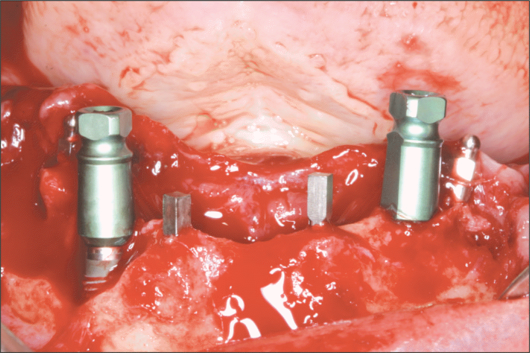 | Fig. 1.Two implants were placed in anterior mandible. 4 mini implants were placed simultaneously for immediate loading. Note bone defect around at the #43 implant. |
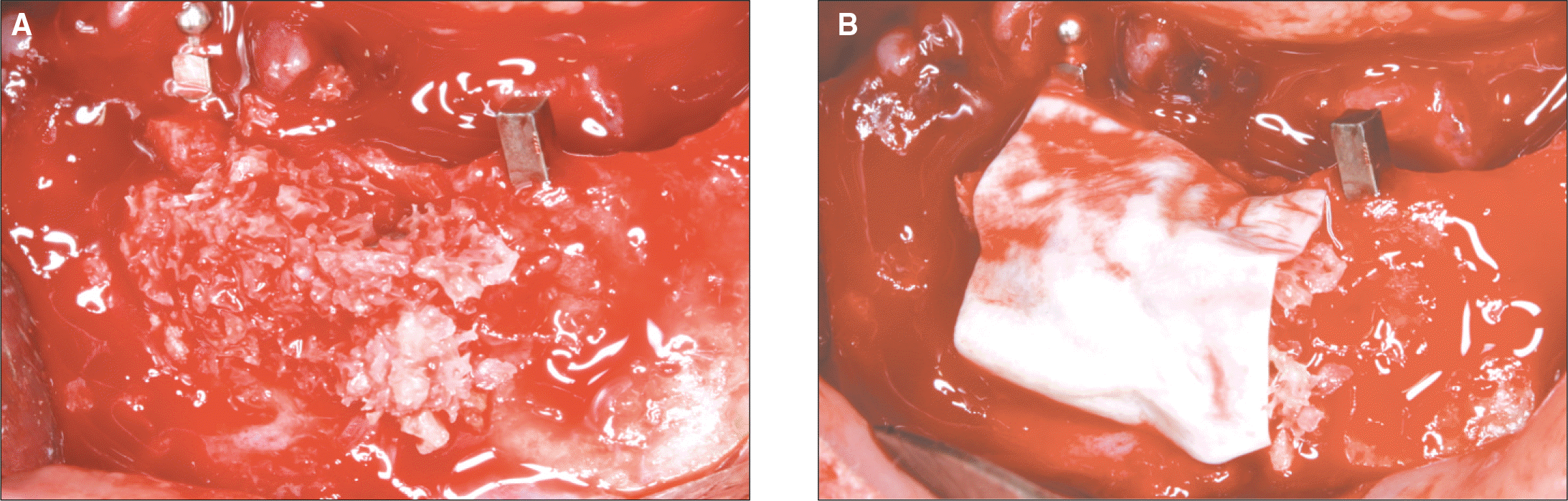 | Fig. 2.Mineral allograft was grafted in the bone defect and collagen membrane was covered over the bone. |
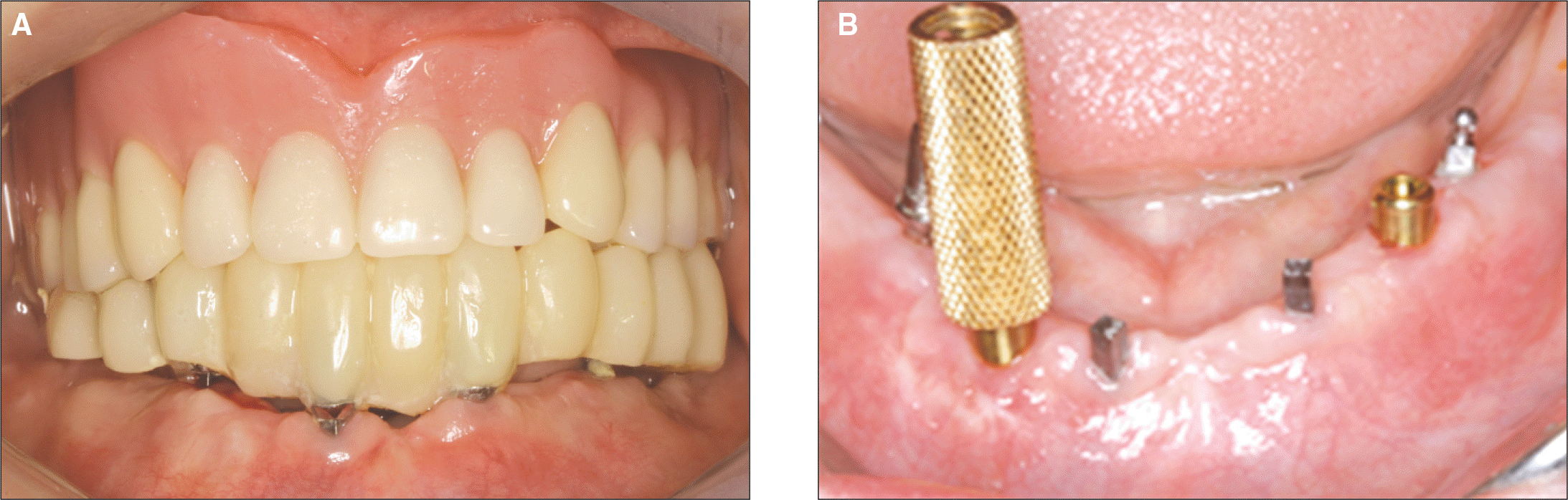 | Fig. 4.A: Fixed provisional restoration was delivered after the stitch-out, B: Lacator attachment was seated on the implant with torque of 30 Ncm after 8 months healing period. Mini implants were removed out when final denture was delivered. |
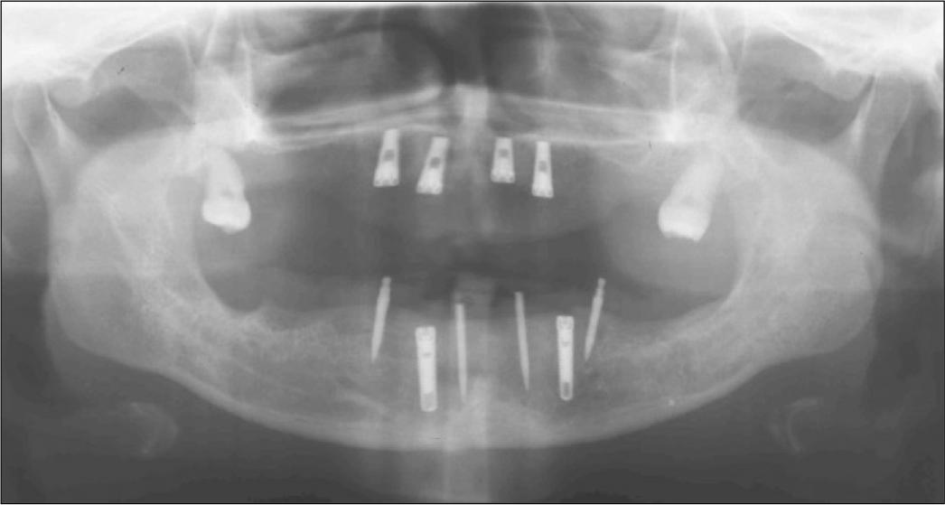
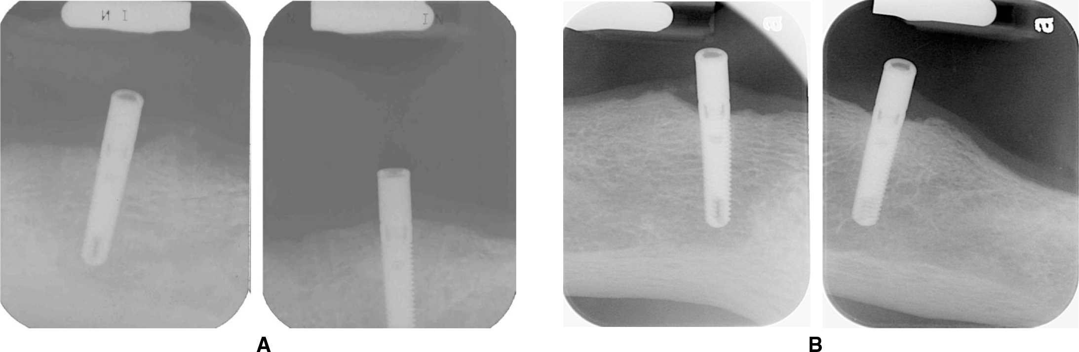
 | Fig. 5.A: Periapical radiograms after delivery of final denture, B: Periapical radiograms after 6.5 years show minimal bone loss around implants. |
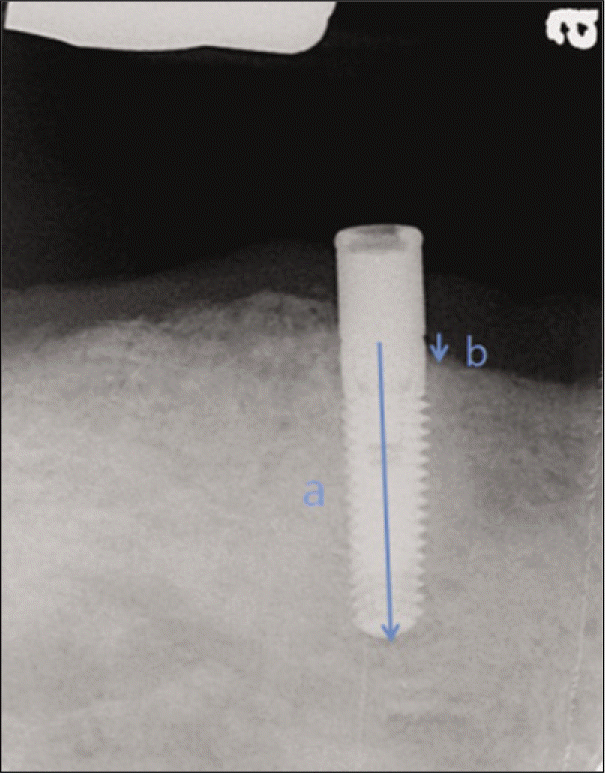
 | Fig. 6.References used to measure actual marginal bone loss. a: The distance from top level of implant platform to implant apex. b: The distance from top level of implant platform to marginal bone contact level. |
Table 1.
Datum of patients and implants
| Patient number | Gender | Age | PMH | Manufacture | Diameter | Length | GBR | Stage | Opposing jaw |
|---|---|---|---|---|---|---|---|---|---|
| 1 | F | 50 | N | TSV® | 4.7 | 13 | Autobone+ Orthoblast II® | 1' | Implant overdenture |
| 2 | M | 83 | N | Replace® | 3.5 | 13 | N | 2' | Complete denture |
| 3 | M | 62 | Dysuresia | Replace® | 4.3 | 13 | OrthoblastII® + Tutodent® | 1' | Complete denture |
| 4 | F | 65 | Hypertension, Hwa-byung | Replus® | 3.7 | 13 | Auto bone + orthoblast II® | 2' | Natural teeth |
| 5 | F | 69 | Osteoporosis | UNI® | 4.1 | 14 | N | 2' | Implant overdenture |
| 6 | F | 68 | N | TSV® | 3.7 | 13 | Autobone. + Biocera® + Tutodent® | 2' | Removable partial denture |
| 7 | M | 61 | Diabetes | TSV® | 3.7 | 13 | N | 1' | Complete denture |
| 8 | F | 68 | Osteoporosis | Dentis® | 3.7 | 12 | N | 1' | Complete denture |
| 9 | M | 41 | N | Legacy® | 3.7 | 13 | N | 1' | Implant overdenture |
| 10 | F | 51 | N | Dentis® | 3.7 | 12 | Allotis® + Orthoblast II® + Lyoplant® | 2' | Implant overdenture |
Table 2.
Number of implants placed according to implant length and diameter
| 3.5 mm | 3.7 mm | 4.1 mm | 4.3 mm | 4.7 mm | Total | |
|---|---|---|---|---|---|---|
| 12 mm | 4 | 4 | ||||
| 13 mm | 1 | 8 | 3 | 2 | 14 | |
| 14 mm | 2 | 2 | ||||
| Total | 1 | 12 | 2 | 3 | 2 |
Table 3.
Life table analysis of cumulative survival rate after the insertion
 XML Download