

 PDF
PDF ePub
ePub Citation
 Print
Print


Abstract
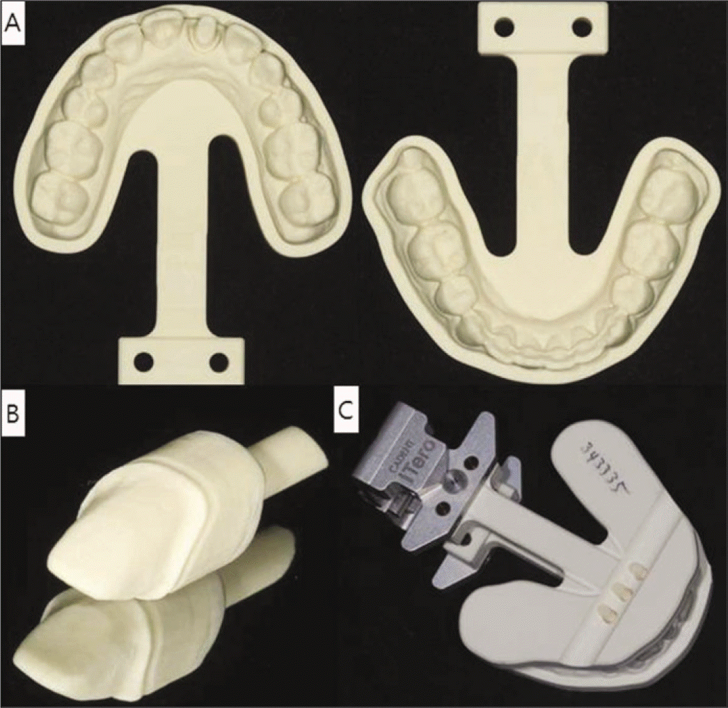
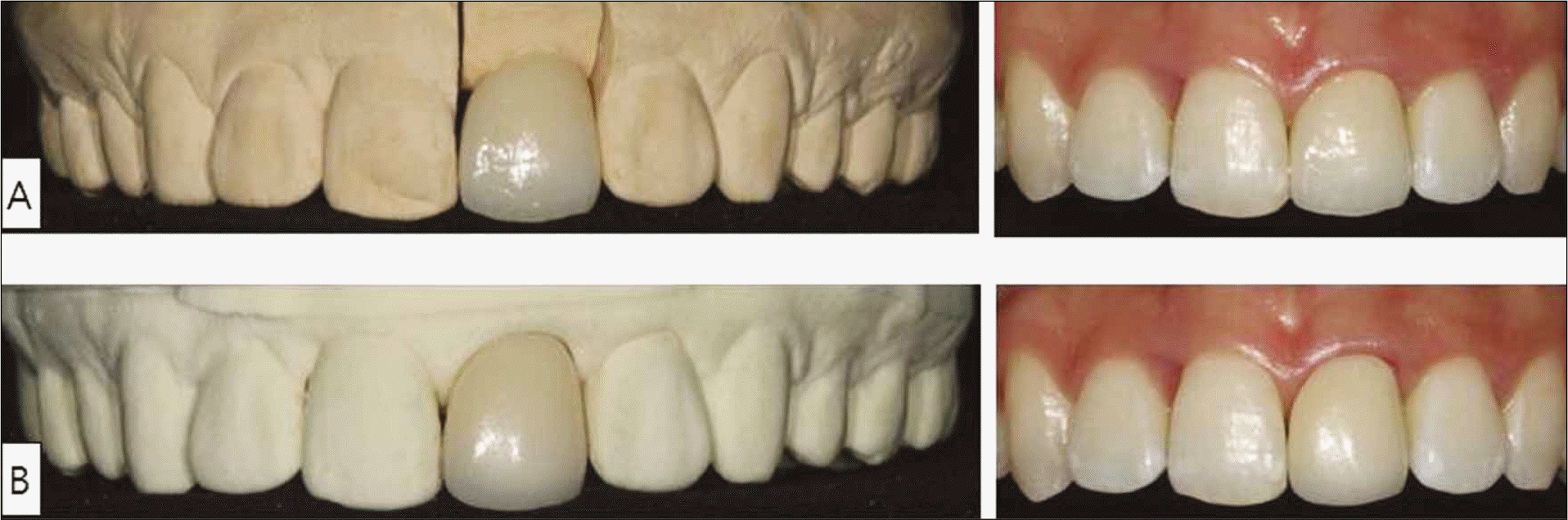
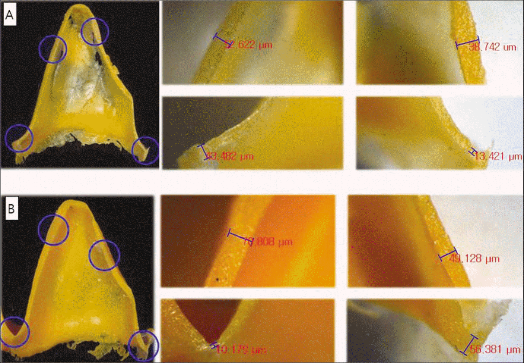
The digital impression technique has been developed for more precise restorations and convenient procedures in prosthodontic treatment compared to the conventional impression technique. Along with the utilization of CAD/CAM, the introduction of digital impression technique actualizes the digitalization of dental treatments. This case is to compare two different prostheses introducing each procedure in detail; one from CAD/CAM after taking digital impression and the other from the conventional technique. A 22-year-old female visited the clinic with the chief complaint of correction of discoloration and shape on maxillary right central incisor. Due to the trauma 8 years ago, the tooth was endodontically treated with post and resin core. The treatment was planned and the procedures were conducted under the patient's consent to treat the tooth with all ceramic crowns in two different ways for comparison. In conclusion, both prostheses presented clinically acceptable results with comparing the internal fitness of two all ceramic crowns. The prosthesis from digital impression, however, showed more errors in respect of internal fitness. (J Korean Acad Prosthodont 2011;49:263-9)
Go to : 
REFERENCES
1.Miyazaki T., Hotta Y., Kunii J., Kuriyama S., Tamaki Y. A review of dental CAD/CAM: current status and future perspectives from 20 years of experience. Dent Mater J. 2009. 28:44–56.

2.Christensen GJ. The state of fixed prosthodontic impressions: room for improvement. J Am Dent Assoc. 2005. 136:343–6.
3.Christensen GJ. Impressions are changing: deciding on conventional, digital or digital plus in-office milling. J Am Dent Assoc. 2009. 140:1301–4.
4.Cadent iTero Digital Impression System and Virtual 3-D Dentistry[Internet]. Carlstadt, NJ 07072;. 2007. Available at. www.cadent.biz.
5.Jeffrey BD. The future of impressions. Dental Economics 2007 June 1. Available at. www.dentaleconomics.com.
6.Garg AK. Cadent iTero' s digital system for dental impressions: the end of trays and putty? Dent Implantol Update. 2008. 19:1–4.
7.Henkel GL. A comparison of fixed prostheses generated from conventional vs digitally scanned dental impressions. Compend Contin Educ Dent. 2007. 28:422–4. 426-8, 430-1.
8.Beuer F., Schweiger J., Edelhoff D. Digital dentistry: an overview of recent developments for CAD/CAM generated restorations. Br Dent J. 2008. 204:505–11.
9.Christensen GJ. Marginal fit of gold inlay castings. J Prosthet Dent. 1966. 16:297–305.
10.Moon BH., Yang JH., Lee SH., Chung HY. A study on the marginal fit of all-ceramic crown using CCD camera. J Korean Acad Prosthodont. 1998. 36:273–92.
11.McLean JW., von Fraunhofer JA. The estimation of cement film thickness by an in vivo technique. Br Dent J. 1971. 131:107–11.
12.Sorensen JA. A standardized method for determination of crown margin fidelity. J Prosthet Dent. 1990. 64:18–24.
13.Molin M., Karlsson S. The fit of gold inlays and three ceramic inlay systems. A clinical and in vitro study. Acta Odontol Scand. 1993. 51:201–6.
14.Rahme HY., Tehini GE., Adib SM., Ardo AS., Rifai KT. In vitro evaluation of the "replica technique" in the measurement of the fit of Procera crowns. J Contemp Dent Pract. 2008. 9:25–32.
15.Syrek A., Reich G., Ranftl D., Klein C., Cerny B., Brodesser J. Clinical evaluation of all-ceramic crowns fabricated from intraoral digital impressions based on the principle of active wavefront sampling. J Dent. 2010. 38:553–9.
Go to :
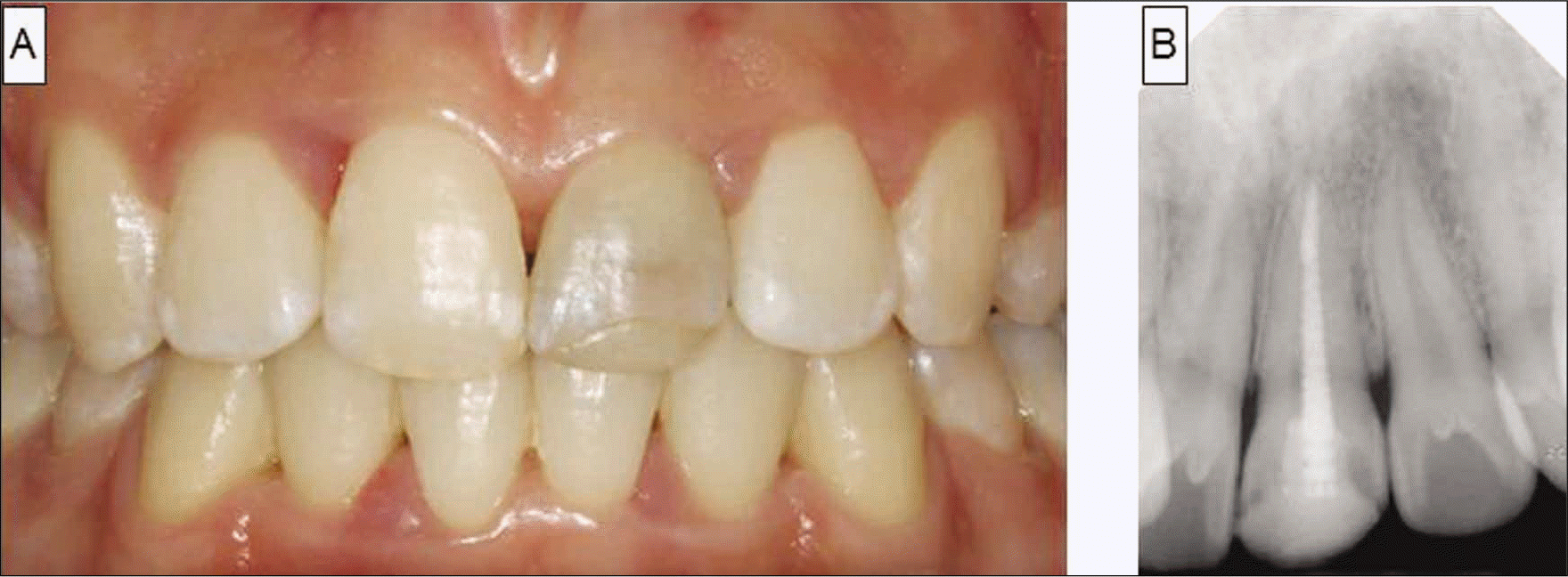 | Fig. 1.Intraoral photo and periapical radiograph at first visit. The maxillary left central incisor was discolored. (A) The tooth was treated with post and resin core after endodontic treatment. (B) |
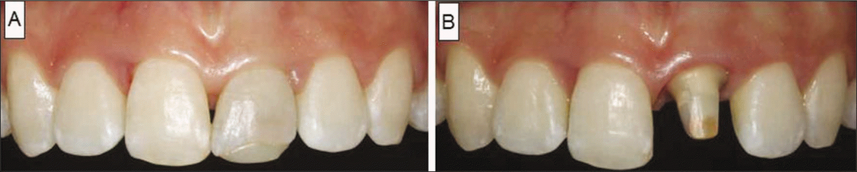 | Fig. 2.A: Before tooth preparation and gingivectomy, B: After tooth preparation and gingivectomy. |
 XML Download