Abstract
Purpose
To date most of finite element analysis assumed the presence of 100% contact between bone and implant, which is inconsistent with clinical reality. In human retrieval study bone-implant contact (BIC) ratio ranged from 20 to 80%. The objective of this study was to explore the influence of bone-implant contact pattern on bone of the interface using nonlinear 3-dimensional finite element analysis.
Materials and methods
A computer tomography-based finite element models with two types of implant (Mark III Branemark, Inplant) which placed in the maxillary 2nd premolar area were constructed. Two different degrees of bone-implant contact ratio (40, 70%) each implant design were simulated. 5 finite element models were constructed each bone-implant contact ratio and implant design, and sum of models was 40. The position of bone-implant contact was determined according to random shuffle method. Elements of bone-implant contact in group W (wholly randomized osseointegration) was randomly selected in terms of total implant length including cortical and cancellous bone, while ones in group S (segmentally randomized osseointegration) was randomly selected each 0.75 mm vertically and horizontally.
Results
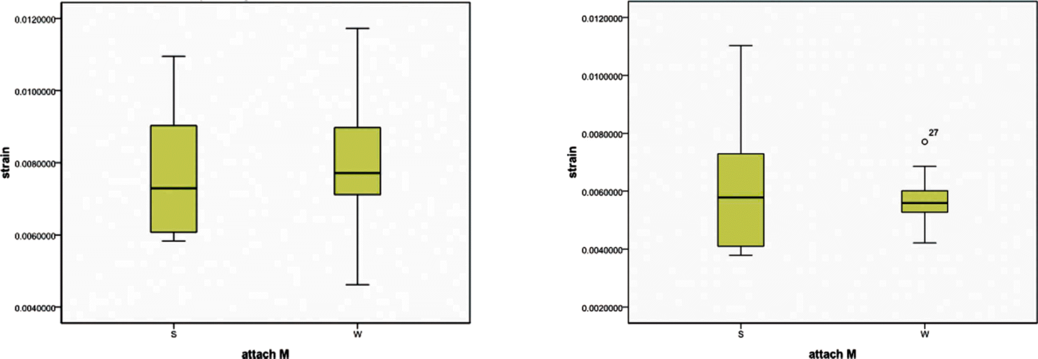
Maximum von Mises strain between group W and group S was not significantly different regardless of bone-implant contact ratio and implant design (P=.939). Peak von Mises strain of 40% BIC was significantly lower than one of 70% BIC (P=.007). There was no significant difference between Mark III Branemark and Inplantin 40% BIC, while average of peak von Mises strain for Inplant was significantly lower (4886 ± 1034 μ m/m) compared with MK III Branemark(7134 ± 1232 μ m/m) in BIC 70% (P<.0001).
Conclusion
Assuming bone-implant contact in finite element method, whether the contact elements in bone were wholly randomly or segmentally randomly selected using random shuffle method, both methods could be effective to be no significant difference regardless of sample size. (J Korean Acad Prosthodont 2011;49:214-21)
Go to : 
REFERENCES
2.Block MS., Finger IM., Fontenot MG., Kent JN. Loaded hydroxylapatite-coated and grit-blasted titanium implants in dogs. Int J Oral Maxillofac Implants. 1989. 4:219–25.
3.Coelho PG., Marin C., Granato R., Suzuki M. Histomorphologic analysis of 30 plateau root form implants retrieved after 8 to 13 years in function. A human retrieval study. J Biomed Mater Res B Appl Biomater. 2009. 91:975–9.

4.Papavasiliou G., Kamposiora P., Bayne SC., Felton DA. 3D-FEA of osseointegration percentages and patterns on implant-bone interfacial stresses. J Dent. 1997. 25:485–91.

5.Huang HL., Hsu JT., Fuh LJ., Tu MG., Ko CC., Shen YW. Bone stress and interfacial sliding analysis of implant designs on an immediately loaded maxillary implant: a nonlinear finite element study. J Dent. 2008. 36:409–17.

6.Pessoa RS., Muraru L., Ju′nior EM., Vaz LG., Sloten JV., Duyck J., Jaecques SV. Influence of implant connection type on the biomechanical environment of immediately placed implants - CT-based nonlinear, three-dimensional finite element analysis. Clin Implant Dent Relat Res. 2010. 12:219–34.

7.Lian Z., Guan H., Ivanovski S., Loo YC., Johnson NW., Zhang H. Effect of bone to implant contact percentage on bone remodelling surrounding a dental implant. Int J Oral Maxillofac Surg. 2010. 39:690–8.

8.Deng B., Tan KB., Liu GR., Lu Y. Influence of osseointegration degree and pattern on resonance frequency in the assessment of dental implant stability using finite element analysis. Int J Oral Maxillofac Implants. 2008. 23:1082–8.
9.Sennerby L., Ericson LE., Thomsen P., Lekholm U., Astrand P. Structure of the bone-titanium interface in retrieved clinical oral implants. Clin Oral Implants Res. 1991. 2:103–11.

10.Garetto LP., Chen J., Parr JA., Roberts WE. Remodeling dynamics of bone supporting rigidly fixed titanium implants: a histomorphometric comparison in four species including humans. Implant Dent. 1995. 4:235–43.

11.Degidi M., Perrotti V., Strocchi R., Piattelli A., Iezzi G. Is insertion torque correlated to bone-implant contact percentage in the early healing period? A histological and histomorphometrical evaluation of 17 human-retrieved dental implants. Clin Oral Implants Res. 2009. 20:778–81.

12.Holmes DC., Loftus JT. Influence of bone quality on stress distribution for endosseous implants. J Oral Implantol. 1997. 23:104–11.
13.Lin CL., Wang JC., Ramp LC., Liu PR. Biomechanical response of implant systems placed in the maxillary posterior region under various conditions of angulation, bone density, and loading. Int J Oral Maxillofac Implants. 2008. 23:57–64.
14.Rubin PJ., Rakotomanana RL., Leyvraz PF., Zysset PK., Curnier A., Heegaard JH. Frictional interface micromotions and anisotropic stress distribution in a femoral total hip component. J Biomech. 1993. 26:725–39.

15.Mellal A., Wiskott HW., Botsis J., Scherrer SS., Belser UC. Stimulating effect of implant loading on surrounding bone. Comparison of three numerical models and validation by in vivo data. Clin Oral Implants Res. 2004. 15:239–48.
16.Ramos A., Simões JA. Tetrahedral versus hexahedral finite elements in numerical modelling of the proximal femur. Med Eng Phys. 2006. 28:916–24.

17.Rho JY., Ashman RB., Turner CH. Young' s modulus of trabecular and cortical bone material: ultrasonic and microtensile measurements. J Biomech. 1993. 26:111–9.
18.Meyer U., Vollmer D., Bourauel C., Joos U. Sensitivity analysis of bone geometries around oral implants upon bone loading using finite element method. Comp Meth Biomech Biomed Eng. 2001. 3:553–59.
19.Van Staden RC., Guan H., Loo YC. Application of the finite element method in dental implant research. Comput Methods Biomech Biomed Engin. 2006. 9:257–70.

20.Geng JP., Tan KB., Liu GR. Application of finite element analysis in implant dentistry: a review of the literature. J Prosthet Dent. 2001. 85:585–98.

21.Hattori Y., Satoh C., Kunieda T., Endoh R., Hisamatsu H., Watanabe M. Bite forces and their resultants during forceful intercuspal clenching in humans. J Biomech. 2009. 42:1533–8.

22.Chou HY., Jagodnik JJ., Mu ¨ftu ¨ S. Predictions of bone remodeling around dental implant systems. J Biomech. 2008. 41:1365–73.

23.Merz BR., Hunenbart S., Belser UC. Mechanics of the implant-abutment connection: an 8-degree taper compared to a butt joint connection. Int J Oral Maxillofac Implants. 2000. 15:519–26.
24.Steinemann SG., Mausli PA., Szmukler-Moncler S. Betatitanium alloy for surgical implants. Froes FH, Caplan I, editors. Titanium' 92 Science and technology. Warrendale, PA: The Minerals, Metals & Materials Society;1993. p. 2689–96.
25.Slaets E., Carmeliet G., Naert I., Duyck J. Early cellular responses in cortical bone healing around unloaded titanium implants: an animal study. J Periodontol. 2006. 77:1015–24.

26.Slaets E., Carmeliet G., Naert I., Duyck J. Early trabecular bone healing around titanium implants: a histologic study in rabbits. J Periodontol. 2007. 78:510–7.

27.Sennerby L., Thomsen P., Ericson LE. A morphometric and bio-mechanic comparison of titanium implants inserted in rabbit cortical and cancellous bone. Int J Oral Maxillofac Implants. 1992. 7:62–71.
28.Lin CL., Wang JC., Ramp LC., Liu PR. Biomechanical response of implant systems placed in the maxillary posterior region under various conditions of angulation, bone density, and loading. Int J Oral Maxillofac Implants. 2008. 23:57–64.
29.Tada S., Stegaroiu R., Kitamura E., Miyakawa O., Kusakari H. Influence of implant design and bone quality on stress/strain distribution in bone around implants: a 3-dimensional finite element analysis. Int J Oral Maxillofac Implants. 2003. 18:357–68.
Go to : 
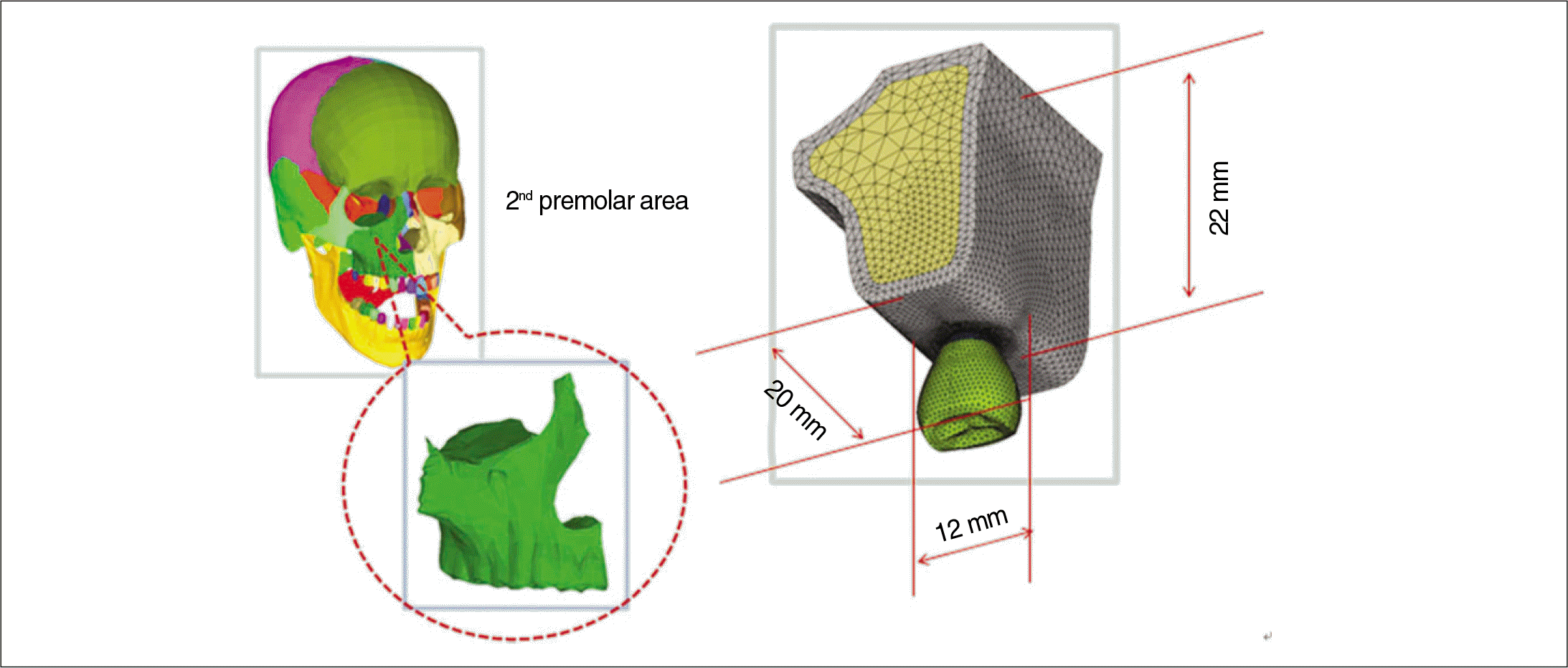 | Fig. 1.The bone model represents CT generated images of the second premolar of the human maxillary bone to provide bone geometric information. The size of the edentulous area used was approximately 20 mm mesiodistally, 12 mm buccolingully and 22 mm in the bone height. |
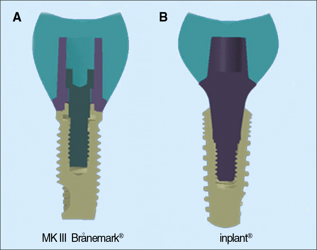 | Fig. 2.Two types of implant models employed in this study. A: MK III Bra®nemark® implant with external hex, B: Inplant® with Morse-taper. |
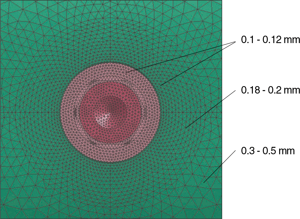 | Fig. 3.The element size near bone-implant interface was 0.1 - 0.12 mm for more reality, the one of the distant zone from the interface was 0.3 - 0.5 mm for model simplification. |
 | Fig. 5.Constraints of bone model and implant-abutment interface. A: boundary condition of bone model, B: tied condition and frictional contact between abutment and abutment screw. |
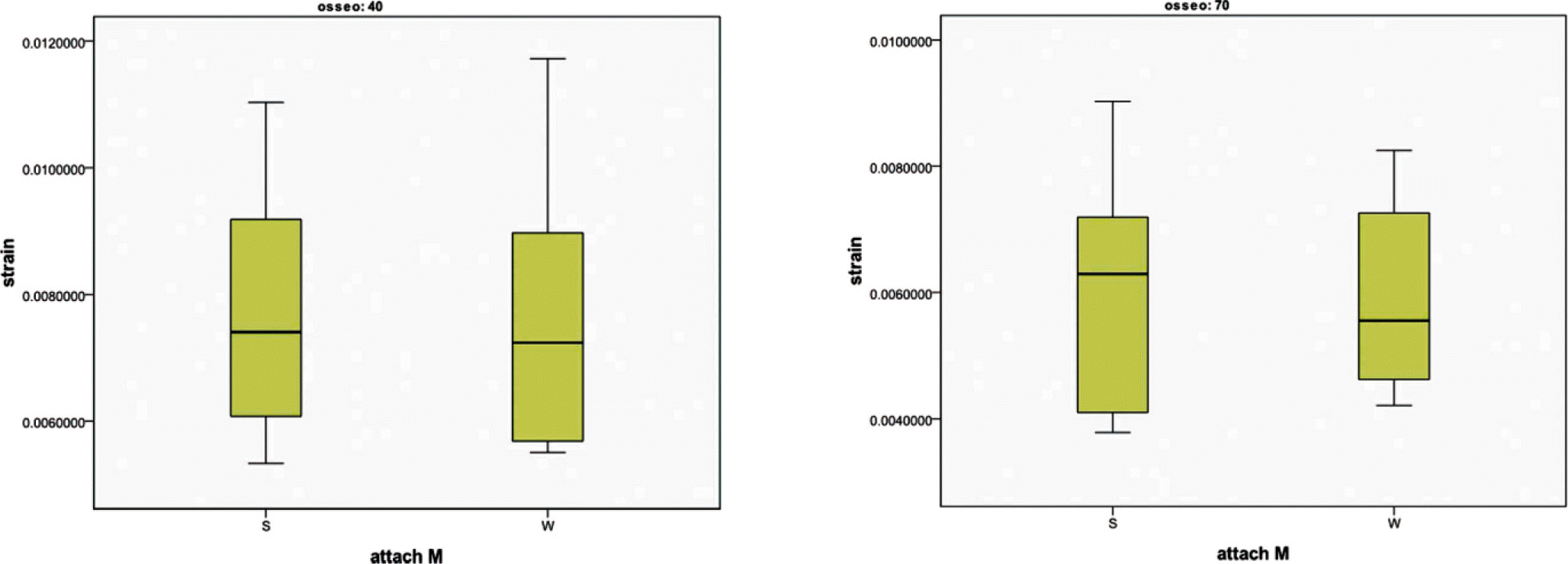 | Fig. 6.Comparison between S (segmentally randomized osseointegration) and W (wholly randomized osseointegration) group according to osseointegraton degrees (40, 70%). ‘osseo’ is osseointegration degree and ‘attach M’ is attachment method of bone-implant contact. |
 | Fig. 7.Comparison between S (segmentally randomized osseointegration) and W (wholly randomized osseointegration) group according to implant design. ‘attach M’ is attachment method of bone-implant contact. |
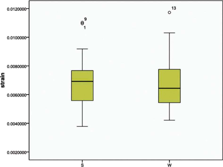 | Fig. 8.Comparison between S (segmentally randomized osseointegration) and W (wholly randomized osseointegration) group including osseointegration degree and implant design. ‘attach M’ is attachment method of bone-implant contact. |
Table 1.
Mechanical properties of bone, implant, and prosthetic materials
| Material | Young's moduls (GPa) | Possion's ratio |
|---|---|---|
| Cortical bone | 13 | 0.3 |
| Dense trabecular bone | 2.6 | 0.3 |
| Implant | 110 | 0.3 |
| Abutment | 90 | 0.3 |
| Abutment screw | 90 | 0.3 |
| Crown | 11.7 | 0.3 |
Table 2.
Averages and standard deviations of von Mises strain in terms of osseointegration degrees (40, 70%). ‘osseo’ is osseointegration degree and ‘attach M’ is attachment method of bone-implant contact
| Osseo | attach M | Number | Average | Standard deviation |
|---|---|---|---|---|
| 40 | S | 10 | .007722 | .002052 |
| W | 10 | .007606 | .002137 | |
| 70 | S | 10 | .006001 | .001800 |
| W | 10 | .006019 | .001469 |
Table 3.
Averages (Standard deviations) of von Mises strain in terms of implant design (MK III Bra®nemark®; BI, Inplant®; II). ‘attach M’ is attachment method of bone-implant contact
| Implant.design | attach M | Number | Average | SD |
|---|---|---|---|---|
| BI | S | 10 | .007585 | .001676 |
| W | 10 | .007934 | .002070 | |
| II | S | 10 | .006138 | .002263 |
| W | 10 | .005690 | .001022 |


 PDF
PDF ePub
ePub Citation
 Print
Print


 XML Download