

 PDF
PDF ePub
ePub Citation
 Print
Print


Abstract
Purpose
Resonance frequency analysis, Periotest, and removal torque (RT) test were known as the methods to assess implant stability. The results of these methods are affected by the bone condition, implant diameter and shape. The purpose of this study is to access the meaning and the correlationship of the resonance frequency analysis, Periotest and RT test in osseointegration simulated acrylic resin when the engaged bone thickness and peri-implant bone defect are changed.
Materials and methods
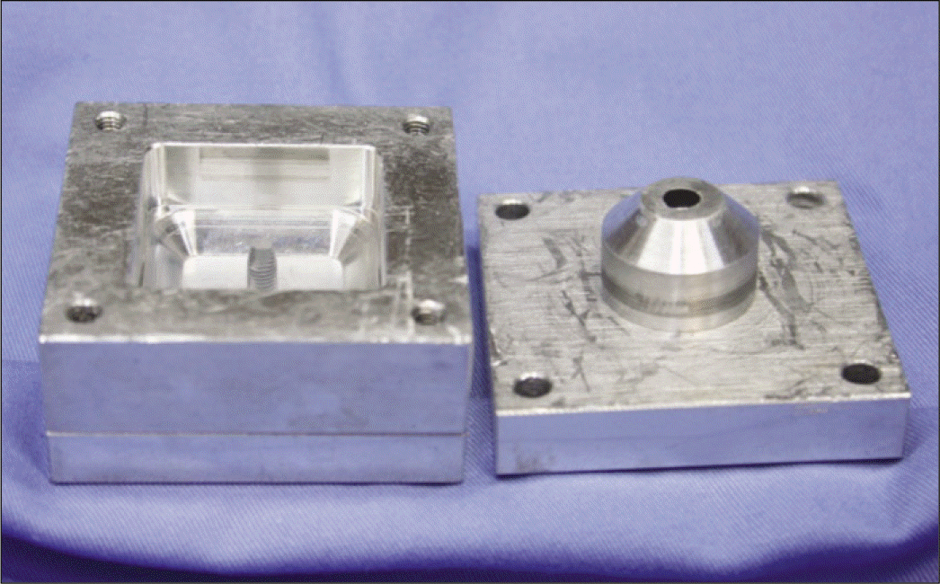
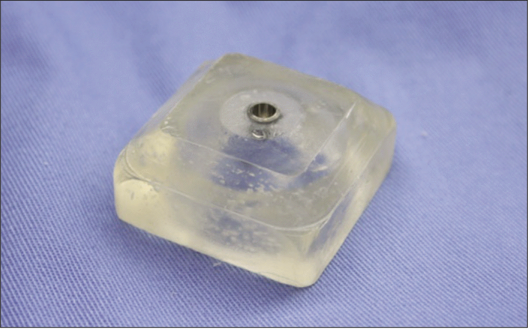
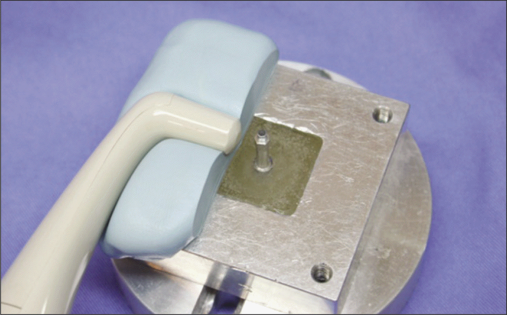
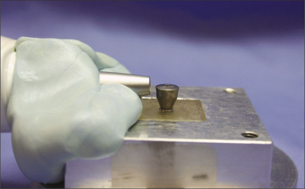
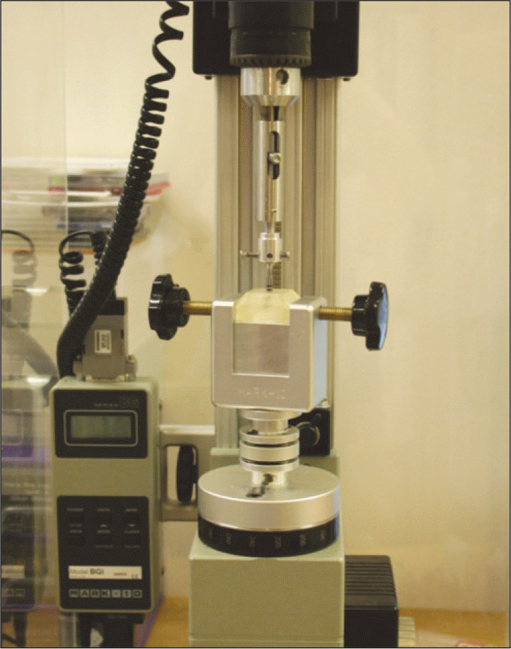
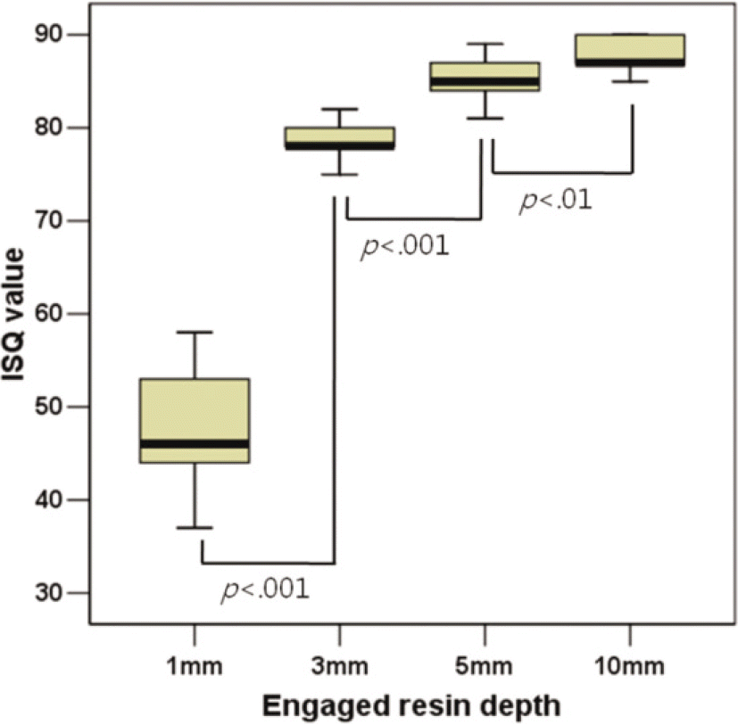
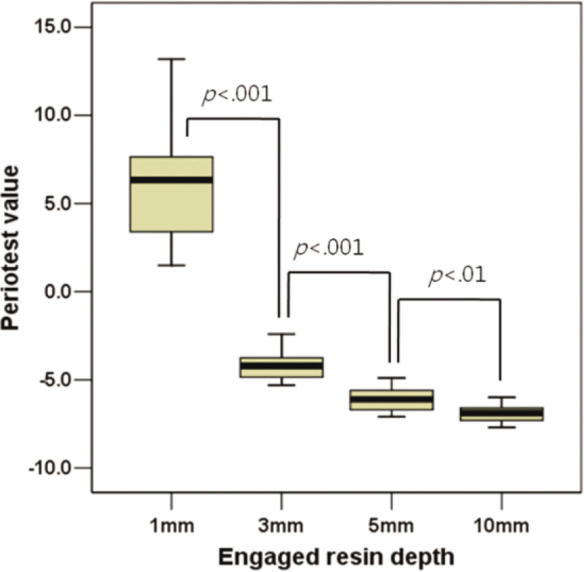
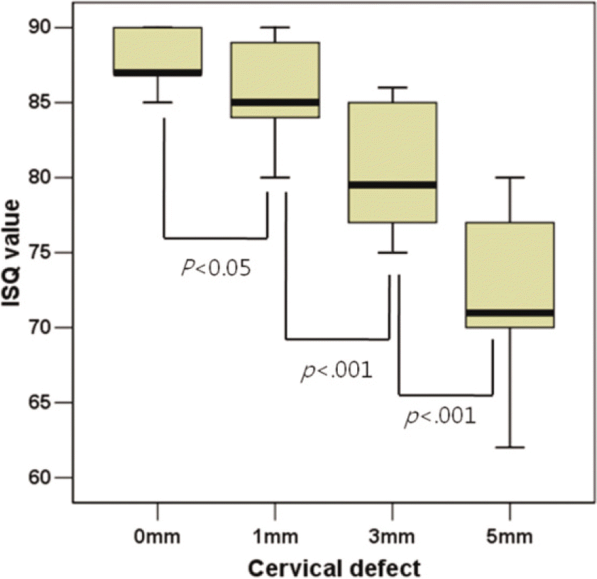
To simulate osseointegration, the fixture was fixed to an aluminum mold with a screw. Acrylic resin powder and liquid were poured into the mold for polymerization. The engaged resin thickness with implant was controlled. Simulated cortical bone thicknesses were 1, 3, 5 and 10 mm. Additional 1, 3 and 5 mm peri-implant bone defects were simulated. Three types of implants were used; 4 mm diameter implants of straight shape, 4 mm diameter implants of tapered shape and 5 mm diameter implants of tapered shape. Five fixtures per each type were tested in respective bone condition. Resonance frequency analysis and Periotest were evaluated in all bone conditions. Peak removal torque was measured at simulated cortical bone thicknesses of 1 and 3 mm. The statistical analysis was performed with the Kruskal-Wallis test, Mann-Whitney U test, and Spearman test using a 95% level of confidence.
Results
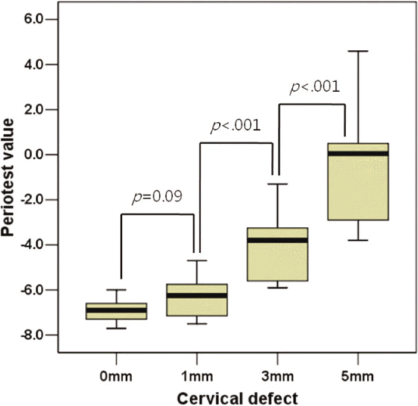
With increasing engaged bone depth, the Implant Stability Quotient (ISQ) values increased and the Periotest values (PTVs) decreased (P<.001, P<.001). With increasing peri-implant bone defect, ISQ values decreased and PTVs increased (P<.001). When the diameter of implant increased, ISQ values increased and Periotest values (PTV) decreased (P<.001). There was a strong correlation between ISQ values and PTVs (r = -0.99, P<.001). Furthermore, the peak removal torque values had weak correlations with both ISQ values and PTVs (r = 0.52, P<.001; r = -0.52, P<.001).
Conclusion
This study confirmed favorable implant stability with increasing engaged bone depth and implant diameter and decreasing peri-implant bone defect. ISQ values and PTVs showed strong correlation with each other and not with the peak removal torque values. (J Korean Acad Prosthodont 2011;49:128-37)
Go to : 
REFERENCES
1.Friberg B., Jemt T., Lekholm U. Early failures in 4,641 consecutively placed Branemark dental implants: a study from stage 1 surgery to the connection of completed prostheses. Int J Oral Maxillofac Implants. 1991. 6:142–6.
2.Meredith N. Assessment of implant stability as a prognostic determinant. Int J Prosthodont. 1998. 11:491–501.
3.Cochran DL., Schenk RK., Lussi A., Higginbottom FL., Buser D. Bone response to unloaded and loaded titanium implants with a sandblasted and acid-etched surface: a histometric study in the canine mandible. J Biomed Mater Res. 1998. 40:1–11.

4.Schulte W., Lukas D. The Periotest method. Int Dent J. 1992. 42:433–40.
5.Olive′ J., Aparicio C. Periotest method as a measure of osseointe-grated oral implant stability. Int J Oral Maxillofac Implants. 1990. 5:390–400.
6.Meredith N., Friberg B., Sennerby L., Aparicio C. Relationship between contact time measurements and PTV values when using the Periotest to measure implant stability. Int J Prosthodont. 1998. 11:269–75.
7.Aparicio C. The use of the Periotest value as the initial success criteria of an implant: 8-year report. Int J Periodontics Restorative Dent. 1997. 17:150–61.
8.Walker L., Morris HF., Ochi S. Periotest values of dental implants in the first 2 years after second-stage surgery: DICRG interim report no. 8. Dental Implant Clinical Research Group. Implant Dent. 1997. 6:207–12.
9.Truhlar RS., Morris HF., Ochi S. Stability of the bone-implant complex. Results of longitudinal testing to 60 months with the Periotest device on endosseous dental implants. Ann Periodontol. 2000. 5:42–55.
10.Meredith N., Alleyne D., Cawley P. Quantitative determination of the stability of the implant-tissue interface using resonance frequency analysis. Clin Oral Implants Res. 1996. 7:261–7.
11.Atsumi M., Park SH., Wang HL. Methods used to assess implant stability: current status. Int J Oral Maxillofac Implants. 2007. 22:743–54.
12.Meredith N., Book K., Friberg B., Jemt T., Sennerby L. Resonance frequency measurements of implant stability in vivo. A cross-sectional and longitudinal study of resonance frequency measurements on implants in the edentulous and partially dentate maxilla. Clin Oral Implants Res. 1997. 8:226–33.
13.Rasmusson L., Meredith N., Cho IH., Sennerby L. The influence of simultaneous versus delayed placement on the stability of titanium implants in onlay bone grafts. A histologic and biomechanic study in the rabbit. Int J Oral Maxillofac Surg. 1999. 28:224–31.
14.Roberts WE., Smith RK., Zilberman Y., Mozsary PG., Smith RS. Osseous adaptation to continuous loading of rigid endosseous implants. Am J Orthod. 1984. 86:95–111.
15.Johansson C., Albrektsson T. Integration of screw implants in the rabbit: a 1-year follow-up of removal torque of titanium im- plants. Int J Oral Maxillofac Implants. 1987. 2:69–75.
16.Roze′ J., Babu S., Saffarzadeh A., Gayet-Delacroix M., Hoornaert A., Layrolle P. Correlating implant stability to bone structure. Clin Oral Implants Res. 2009. 20:1140–5.
17.Gardner MP., Chong AC., Pollock AG., Wooley PH. Mechanical evaluation of large-size fourth-generation composite femur and tibia models. Ann Biomed Eng. 2010. 38:613–20.
18.Heiner AD. Structural properties of fourth-generation composite femurs and tibias. J Biomech. 2008. 41:3282–4.
19.Chong AC., Friis EA., Ballard GP., Czuwala PJ., Cooke FW. Fatigue performance of composite analogue femur constructs under high activity loading. Ann Biomed Eng. 2007. 35:1196–205.
20.Turkyilmaz I., Sennerby L., Tumer C., Yenigul M., Avci M. Stability and marginal bone level measurements of unsplinted implants used for mandibular overdentures: a 1-year randomized prospective clinical study comparing early and conventional loading protocols. Clin Oral Implants Res. 2006. 17:501–5.
21.To¨zu¨m TF., Turkyilmaz I., McGlumphy EA. Relationship between dental implant stability determined by resonance frequency analysis measurements and peri-implant vertical defects: an in vitro study. J Oral Rehabil. 2008. 35:739–44.
22.Ohta K., Takechi M., Minami M., Shigeishi H., Hiraoka M., Nishimura M., Kamata N. Influence of factors related to implant stability detected by wireless resonance frequency analysis device. J Oral Rehabil. 2010. 37:131–7.
23.To¨zu¨m TF., Bal BT., Turkyilmaz I., Gu¨lay G., Tulunoglu I. Which device is more accurate to determine the stability/mobility of dental implants? A human cadaver study. J Oral Rehabil. 2010. 37:217–24.
24.Turkyilmaz I., Sennerby L., Yilmaz B., Bilecenoglu B., Ozbek EN. Influence of defect depth on resonance frequency analysis and insertion torque values for implants placed in fresh extraction sockets: a human cadaver study. Clin Implant Dent Relat Res. 2009. 11:52–8.
25.Turkyilmaz I. A comparison between insertion torque and resonance frequency in the assessment of torque capacity and primary stability of Bra®nemark system implants. J Oral Rehabil. 2006. 33:754–9.
26.Al-Nawas B., Wagner W., Gro¨tz KA. Insertion torque and resonance frequency analysis of dental implant systems in an animal model with loaded implants. Int J Oral Maxillofac Implants. 2006. 21:726–32.
Go to :
 | Fig. 3.Schematic view of simulated bone condition with acrylic resin (1M0 : integration with 1 mm thick resin, 3M0 : integration with 3 mm thick resin, 5M0 : integration with 5 mm thick resin, 10M0 : integration with over 10 mm thick resin, 10M1 : integration with over 10 mm thick resin and simulation of 1 mm cervical bone loss, 10M3 : integration with over 10 mm thick resin and simulation of 3 mm cervical bone loss, 10M5 : integration with over 10 mm thick resin and simulation of 5 mm cervical bone loss). |
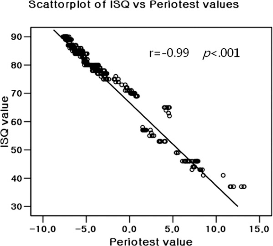 | Fig. 11.Scatterplot from 420 implant stability quotient (ISQ) values and Periotest values of all 45 implants, correlation between ISQ and PTVs. |
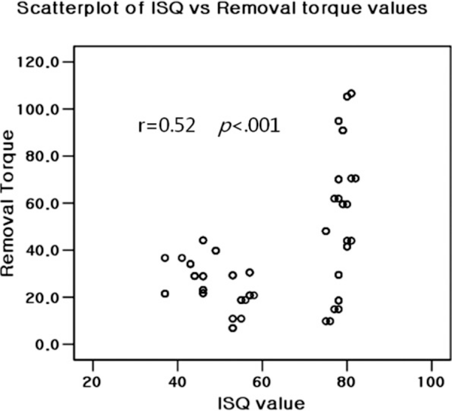
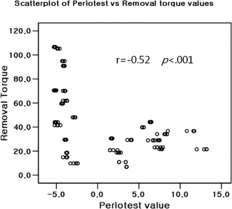
Table 1.
Implant stability quotient (ISQ), periotest value (PTV), removal torque (RT, Ncm) in part 1 experiment (mean ± SD)
Table 2.
Implant stability quotient (ISQ), periotest value (PTV) in part 2 experiment (mean ± SD)
Table 3.
Influence of diameter and shape on ISQ, Periotest Value (PTV), Removal Torque (RT) (mean ± SD)
 XML Download