

 PDF
PDF ePub
ePub Citation
 Print
Print


Abstract
Purpose
The purpose of this study was to compare the fracture strength of the zirconia ceramic crowns according to tooth position.
Material and methods
After 10 metal dies were made for each group, the zirconia ceramic crowns were fabricated using CAD/CAM system (LavaTM All-Ceramic System) and each crown was cemented on each metal die with resin cement (Rely XTM Unicem). The cemented zirconia ceramic crowns mounted on the testing jig were inclined with 30 degrees to the long axis of the tooth and the universal testing machine was used to measure the fracture strength.
Results
1. The fracture strength of the zirconia ceramic crown in the lower 1st molar (2963 N) had the highest and that in the lower central incisor (1035 N) had the lowest. 2. The fracture strength of zirconia ceramic crown was higher than that of the IPS Empress crowns in all tooth position. 3. The fracture mode of the crowns was similar. Most of fracture lines began at the loading area and extended through proximal surface perpendicular to the long axis of the crowns. 4. There were no significant differences on the fracture strength of the zirconia ceramic crowns according to tooth position except in premolar group.
Go to : 
REFERENCES
1.Southan DE., Jo ¨rgensen KD. Faulty porcelain jacket crowns. Aust Dent J. 1972. 17:436–40.

2.Anusavice KJ. Degradability of dental ceramics. Adv Dent Res. 1992. 6:82–9.
3.Blatz MB. Long-term clinical success of all-ceramic posterior restorations. Quintessence Int. 2002. 33:415–26.
4.Oh SC., Dong JK., Lu ¨thy H., Scha ¨rer P. Strength and microstructure of IPS Empress 2 glass-ceramic after different treatments. Int J Prosthodont. 2000. 13:468–72.
5.Lee HH. Dental Ceramics: Processing and clinical implications. J Korea Clin Dent. 2000. 2:106–15.
6.Song BK., Lee HH., Dong JK. Fracture Strength of the IPS Empress Crown: The effect of occlusal depth and axial inclination on upper central incisor. J Korean Acad Stomato Func Occlu. 2000. 16:237–44.
7.Shin DK., Kang HJ., Park YS., Park KS., Dong JK. Fracture Strength of the IPS Empress Crown: The effect of incisal reduction and axial inclination on upper canine. J Korean Acad Prosthodont. 2005. 43:30–40.
8.Dong JK., Oh SC., Kim SD. Fracture Strength of the IPS Empress Crown. The effect of occlusal depth and axial inclination on upper first premolar crowns. J Korean Acad Prosthodont. 1999. 37:127–33.
9.Choi TR., Lee HH., Dong JK. Fracture Strength of the IPS Empress Crown: The effect of occlusal depth and axial inclination on upper first molar. J Korean Acad Prosthodont. 2001. 39:171–83.
10.Nam YS., Dong JK. Fracture Strength of the IPS Empress Crown. The effect of incisal reduction and axial inclination on lower central incisor. J Korean Acad Stomato Func Occlu. 2003. 19:207–17.
11.Jung YC., Shin DK., Park EJ., Kim MJ., Dong JK. Fracture Strength of the IPS Empress Crown. The effect of incisal reduction depth and axial inclination on lower canine. J Korean Acad Stomato Func Occlu. 2004. 20:20–9.
12.Kim HJ., Lee HH., Nam YS., Dong JK. Fracture Strength of the IPS Empress Crown: The effect of occlusal depth and axial inclination on lower second premolar. J Korean Acad Prosthodont. 2002. 40:441–50.
13.Kim SH., Lee JH., Kim YL., Dong JK. Fracture Strength of the IPS Empress Crown: The effect of occlusal depth and axial inclination on lower first molar. J Korean Acad Prosthodont. 2003. 41:48–59.
14.Jeong HC. Fracture strength of zirconia monolithic crowns. J Korean Acad Prosthodont. 2006. 44:157–64.
15.Shin ES., Lee YS., Park WH. Comparative study in fracture strength of zirconia cores fabricated with three different CAD/CAM systems. J Korean Acad Prosthodont. 2008. 46:22–30.
16.Sundh A., Sjo ¨gren G. A comparison of fracture strength of yttrium-oxidepartially-stabilized zirconia ceramic crowns with varying core thickness, shapes and veneer ceramics. J Oral Rehabil. 2004. 31:682–8.
17.Vult von Steyern P., Ebbesson S., Holmgren J., Haag P., Nilner K. Fracture strength of two oxide ceramic crown systems after cyclic pre-loading and thermocycling. J Oral Rehabil. 2006. 33:682–9.
18.Craig RG. Restorative dental materials. St Louis: CV Mosby;1989. p. 65.
19.Lee SM., Jeong HC., Jeon YC. Fracture strength of zirconia monolithic crowns and metal-ceramic crowns after cyclic loading and thermocycling. J Korean Acad Prosthodont. 2007. 45:12–20.
20.Aboushelib MN., de Jager N., Kleverlaan CJ., Feilzer AJ. Effect of loading method on the fracture mechanics of two layered all-ceramic restorative systems. Dent Mater. 2007. 23:952–9.
21.Oilo M., Gjerdet NR., Tvinnereim HM. The firing procedure influences properties of a zirconia core ceramic. Dent Mater. 2008. 24:471–5.
22.Dong JK., Luthy H., Wohlwend A., Scha ¨rer P. Heat-pressed ceramics: Technology and strength. Int J Prosthodont. 1992. 5:9–16.
23.Studart AR., Filser F., Kocher P., Gauckler LJ. In vitro lifetime of dental ceramics under cyclic loading in water. Biomaterials. 2007. 28:2695–705.
24.Tinschert J., Natt G., Mohrbotter N., Spiekermann H., Schulze KA. Lifetime of alumina- and zirconia ceramics used for crown and bridge restorations. J Biomed Mater Res B Appl Biomater. 2007. 80:317–21.
25.Potiket N., Chiche G., Finger IM. In vitro fracture strength of teeth restored with different all-ceramic crown systems. J Prosthet Dent. 2004. 92:491–5.
Go to :
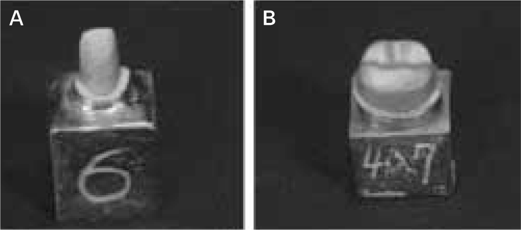 | Fig. 1.Metal dies were fabricated on upper central incisor (A) and lower 1st molar (B) with 8 degree axial inclination and 2.0 mm incisal reduction and 1.5 mm occlusal reduction. |
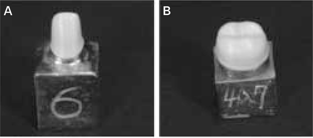 | Fig. 2.Zirconia cores were fabricated on upper central incisor (A) and lower 1st molar (B). |
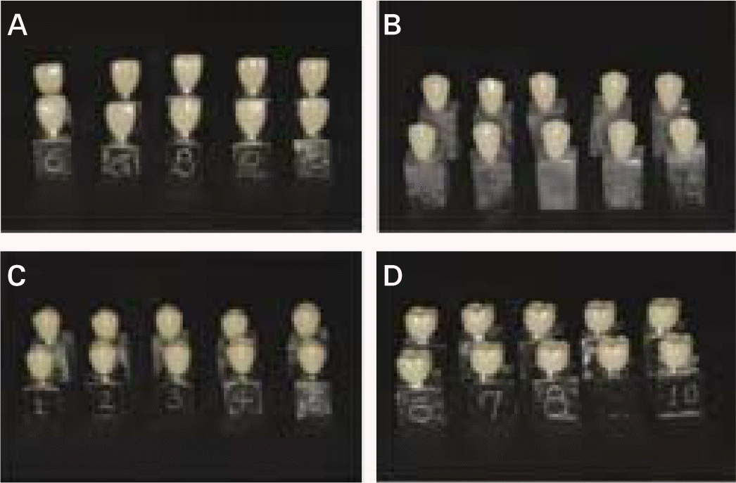 | Fig. 3.Cemented upper zirconia ceramic crowns on the metal die for fracture test (A, central incisor; B, canine; C, 1st premolar; D, 1st molar). |
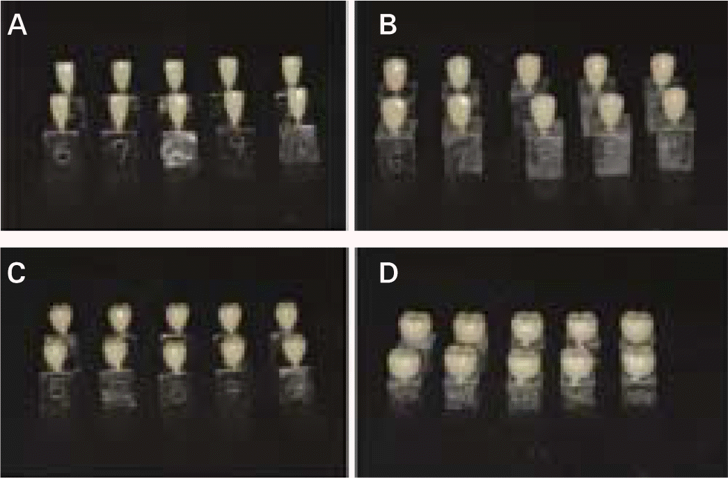 | Fig. 4.Cemented lower zirconia ceramic crowns on the metal die for fracture test (A, central incisor; B, canine; C, 2nd premolar; D, 1st molar). |
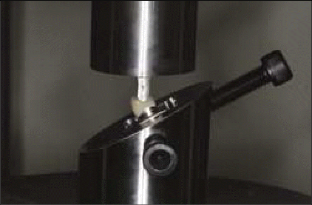 | Fig. 5.Fracture strength test of zirconia ceramic crown using the universal testing machine. |
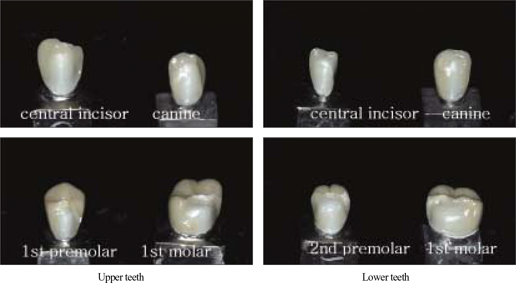
Table I.
Fracture strength of zirconia crowns on upper teeth (unit: Newton)
| Maximum | Minimum | Mean | SD | |
|---|---|---|---|---|
| Central incisor | 1525 | 955 | 1110 | 172 |
| Canine | 1881 | 972 | 1343 | 355 |
| 1st premolar | 1505 | 903 | 1101 | 194 |
| 1st molar | 3378 | 2059 | 2820 | 489 |
 XML Download