Abstract
Purpose
The objectives of the current study are to assess the accuracy of X-Ray Micro Computed Tomography (microCT) in measuring enamel thickness and to evaluate enamel thickness in maxillary incisors of Koreans.
Materials and methods
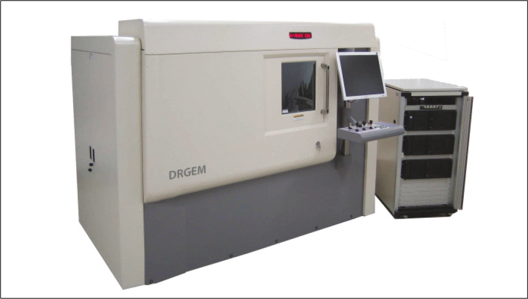
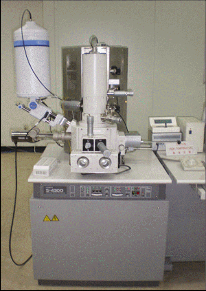
Five maxillary incisors were embedded in resin block. These teeth were longitudinally sectioned labio-lingually through the medial axis. After polishing, the teeth were scanned using a microCT (X-EYE SYSTEM; DRGEM, Seoul, Korea). On a scanning electron microscope (S-4300; Hitachi, Tokyo, Japan) (×20) and a microCT, nearly identical planes were reconstructed. In each tooth, the thickness of labial enamel was measured 1, 3 and 5 mm above the cementoenamel junction (CEJ). Thus, the accuracy of the microCT was evaluated. In addition, using 26 maxillary central incisors and 11 maxillary lateral incisors, in the medial axis and 2 mm remote areas mesially and distally from the medial axis, the thickness of labial enamel was measured 1, 3 and 5 mm above the CEJ along the long axis of the teeth.
Results
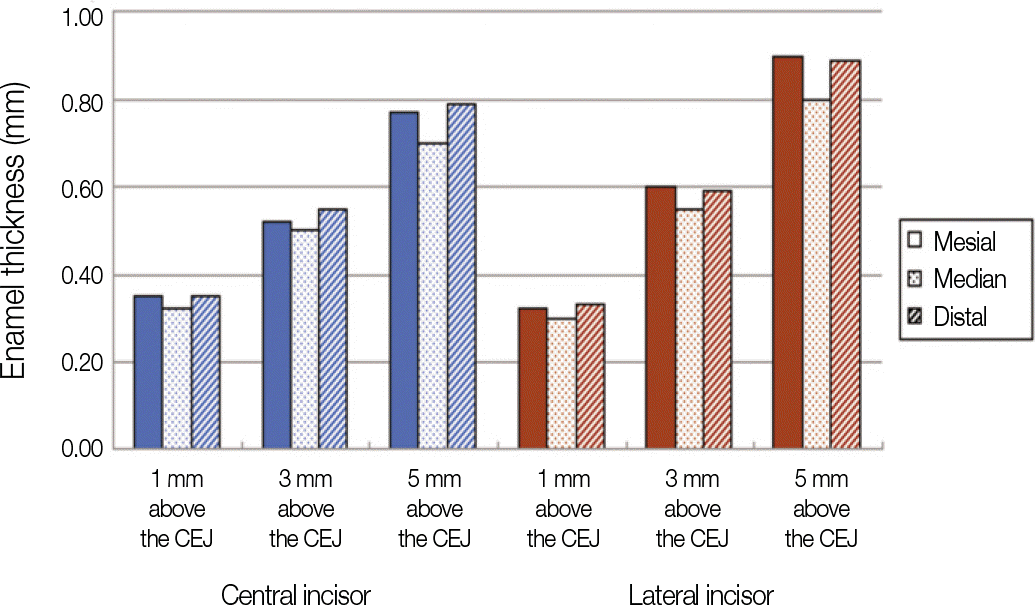
Measurements from nearly identical planes in physical and microCT sections differed by 3.81%. An independent t-test was performed and this showed that there were no significant differences in the measurements between the two methods. Mean values of labial enamel thickness in maxillary central incisors 1, 3 and 5 mm above the CEJ were 0.32 ± 0.01, 0.50 ± 0.02 and 0.70 ± 0.02 mm, respectively. Mean values of labial enamel thickness in maxillary lateral incisors 1, 3 and 5 mm above the CEJ were 0.30 ± 0.01, 0.55 ± 0.03 and 0.80 ± 0.02 mm, respectively.
Conclusion
In measuring enamel thickness, microCT is one of useful way of measurement. So according to the results of this research, when restoring a porcelain laminate veneer on maxillary incisors in Koreans, careful consideration is needed in the amount of enamel reduction. (J Korean Acad Prosthodont 2010;48:301-7)
Go to : 
REFERENCES
1.Freidman MJ. Augmenting restorative dentistry with porcelain veneers. J Am Dent Assoc. 1991. 122:29–34.
2.Shillingburg HT., Hobo S., Whitsett LD., Jacobi R., Brackett SE. Fundamentals of Fixed Prosthodontics. 3rd ed.Chicago: Quintessence;1997. p. 441.
3.Christensen GJ. Veneering of teeth. State of the art. Dent Clin North Am. 1985. 29:373–91.
5.Tjan AH., Dunn JR., Sanderson IR. Microleakage patterns of porcelain and castable ceramic laminate veneers. J Prosthet Dent. 1989. 61:276–82.

6.Ferrari M., Patroni S., Balleri P. Measurement of enamel thickness in relation to reduction for etched laminate veneers. Int J Periodontics Restorative Dent. 1992. 12:407–13.
7.Edelhoff D., Sorensen JA. Tooth structure removal associated with various preparation designs for anterior teeth. J Prosthet Dent. 2002. 87:503–9.

9.Walls AW., Steele JG., Wassell RW. Crowns and other extra-coronal restorations: porcelain laminate veneers. Br Dent J. 2002. 193:73–6. 79–82.

10.Sheets CG., Taniguchi T. Advantages and limitations in the use of porcelain veneer restorations. J Prosthet Dent. 1990. 64:406–11.

11.Hui KK., Williams B., Davis EH., Holt RD. A comparative assessment of the strengths of porcelain veneers for incisor teeth dependent on their design characteristics. Br Dent J. 1991. 171:51–5.

12.Kedici PS., Kalipcilar B., Bilir OG. Effect of glass ionomer liners on bonding strength of laminate veneers. J Prosthet Dent. 1992. 68:29–32.

13.Peumans M., Van Meerbeek B., Lambrechts P., Vanherle G. Porcelain veneers: a review of the literature. J Dent. 2000. 28:163–77.

14.Sorensen JA., Munksgaard EC. Relative gap formation of resin-cemented ceramic inlays and dentin bonding agents. J Prosthet Dent. 1996. 76:374–8.

15.Schneider PM., Messer LB., Douglas WH. The effect of enamel surface reduction in vitro on the bonding of composite resin to permanent human enamel. J Dent Res. 1981. 60:895–900.

16.Lacy AM., Wada C., Du W., Watanabe L. In vitro microleakage at the gingival margin of porcelain and resin veneers. J Prosthet Dent. 1992. 67:7–10.

17.Zaimoglu A., Karaagaçlioglu L. Microleakage in porcelain laminate veneers. J Dent. 1991. 19:369–72.
18.Zaimog ̆lu A., Karaag ̆açliog ̆lu L. Uçtaçli. Influence of porcelain ma-S terial and composite luting resin on microleakage of porcelain laminate veneers. J Oral Rehabil. 1992. 19:319–27.
19.Sim C., Neo J., Chua EK., Tan BY. The effect of dentin bonding agents on the microleakage of porcelain veneers. Dent Mater. 1994. 10:278–81.

20.Atsu SS., Aka PS., Kucukesmen HC., Kiliçarslan MA., Atakan C. Age-related changes in tooth enamel as measured by electron microscopy: implications for porcelain laminate veneers. J Prosthet Dent. 2005. 94:336–41.

21.Grine FE., Stevens NJ., Jungers WL. An evaluation of dental radiograph accuracy in the measurement of enamel thickness. Arch Oral Biol. 2001. 46:1117–25.

22.Harris EF., Hicks JD. A radiographic assessment of enamel thickness in human maxillary incisors. Arch Oral Biol. 1998. 43:825–31.

23.Stroud JL., English J., Buschang PH. Enamel thickness of the posterior dentition: its implications for nonextraction treatment. Angle Orthod. 1998. 68:141–6.
24.Stroud JL., Buschang PH., Goaz PW. Sexual dimorphism in mesiodistal dentin and enamel thickness. Dentomaxillofac Radiol. 1994. 23:169–71.

25.Scotti R., Villa L., Carossa S. Clinical applicability of the radiographic method for determining the thickness of calcified crown tissues. J Prosthet Dent. 1991. 65:65–7.

26.Barber FE., Lees S., Lobene RR. Urasonic pulse-echo measurements in teeth. Arch Oral Biol. 1969. 14:745–60.
27.Huysmans MC., Thijssen JM. Ultrasonic measurement of enamel thickness: a tool for monitoring dental erosion? J Dent. 2000. 28:187–91.

28.Schwartz GT., Thackeray JF., Reid C., van Reenan JF. Enamel thickness and the topography of the enamel-dentine junction in South African Plio-Pleistocene hominids with special reference to the Carabelli trait. J Hum Evol. 1998. 35:523–42.

29.Beynon AD., Wood BA. Variations in enamel thickness and structure in East African hominids. Am J Phys Anthropol. 1986. 70:177–93.

30.Olejniczak AJ., Grine FE. Assessment of the accuracy of dental enamel thickness measurements using microfocal X-ray computed tomography. Anat Rec A Discov Mol Cell Evol Biol. 2006. 288:263–75.

31.Piemjai M., Arksornnukit M. Compressive fracture resistance of porcelain laminates bonded to enamel or dentin with four adhesive systems. J Prosthodont. 2007. 16:457–64.

32.Nakamura T., Miyamae M., Koh N., Hino T., Maruyama T. Adhesive strength between teeth and resin cements for porcelain laminate veneer. J Osaka Univ Dent Sch. 1992. 32:21–6.
33.Dumfahrt H., Scha ¨ffer H. Porcelain laminate veneers. A retrospective evaluation after 1 to 10 years of service: Part II-Clinical results. Int J Prosthodont. 2000. 13:9–18.
34.Horn HR. Porcelain laminate veneers bonded to etched enamel. Dent Clin North Am. 1983. 27:671–84.
35.Calamia JR. Etched porcelain veneers: the current state of the art. Quintessence Int. 1985. 16:5–12.
36.Weinberg LA. Tooth preparation for porcelain laminates. N Y State Dent J. 1989. 55:25–8.
37.Chpindel P., Cristou M. Tooth preparation and fabrication of porcelain veneers using a double-layer technique. Pract Periodontics Aesthet Dent. 1994. 6:19–28.
Go to : 
 | Fig. 3.MicroCT image (left) and scanning electron micrograph (right) of the same cross-section Scale bar = 1 mm. |
 | Fig. 4.Tooth landmarks used for measurements A, Labial surface of maxillary incisor. Points are 1, 3 and 5 mm above the CEJ. B, Sagittal plane in the median axis. |
 | Fig. 5.Method of measurements in 2 mm remote axis mesially and distally from the medial axis A, Horizontal plane 1 mm above the CEJ. B, Horizontal plane 3 mm above the CEJ. C, Horizontal plane 5 mm above the CEJ. |
Table 1.
Values of measurements and percent differences between microCT and SEM (unit: mm)
Table 2.
Differences of enamel thickness between microCT and SEM (unit: mm)
| Group 1 | 1 mm above the CEJ | 3 mm above the CEJ | 5 mm above the CEJ |
|---|---|---|---|
| MicroCT | 0.39 ± 0.04 | 0.53 ± 0.07 | 0.77 ± 0.06 |
| SEM | 0.40 ± 0.05 | 0.55 ± 0.08 | 0.78 ± 0.06 |
| P-value | 0.873 | 0.835 | 0.891 |
Table 3.
Enamel thickness on the each measurement site (unit: mm)


 PDF
PDF ePub
ePub Citation
 Print
Print


 XML Download