

 PDF
PDF ePub
ePub Citation
 Print
Print


Abstract
Purpose
The present study was aimed to evaluate the level of cortical bone strain during the placement of an implant. The primary concern was to investigate if the extent of overloading area near the marginal bone could be affected by microthread fabricated at the cervical 1/3 of an implant.
Materials and methods
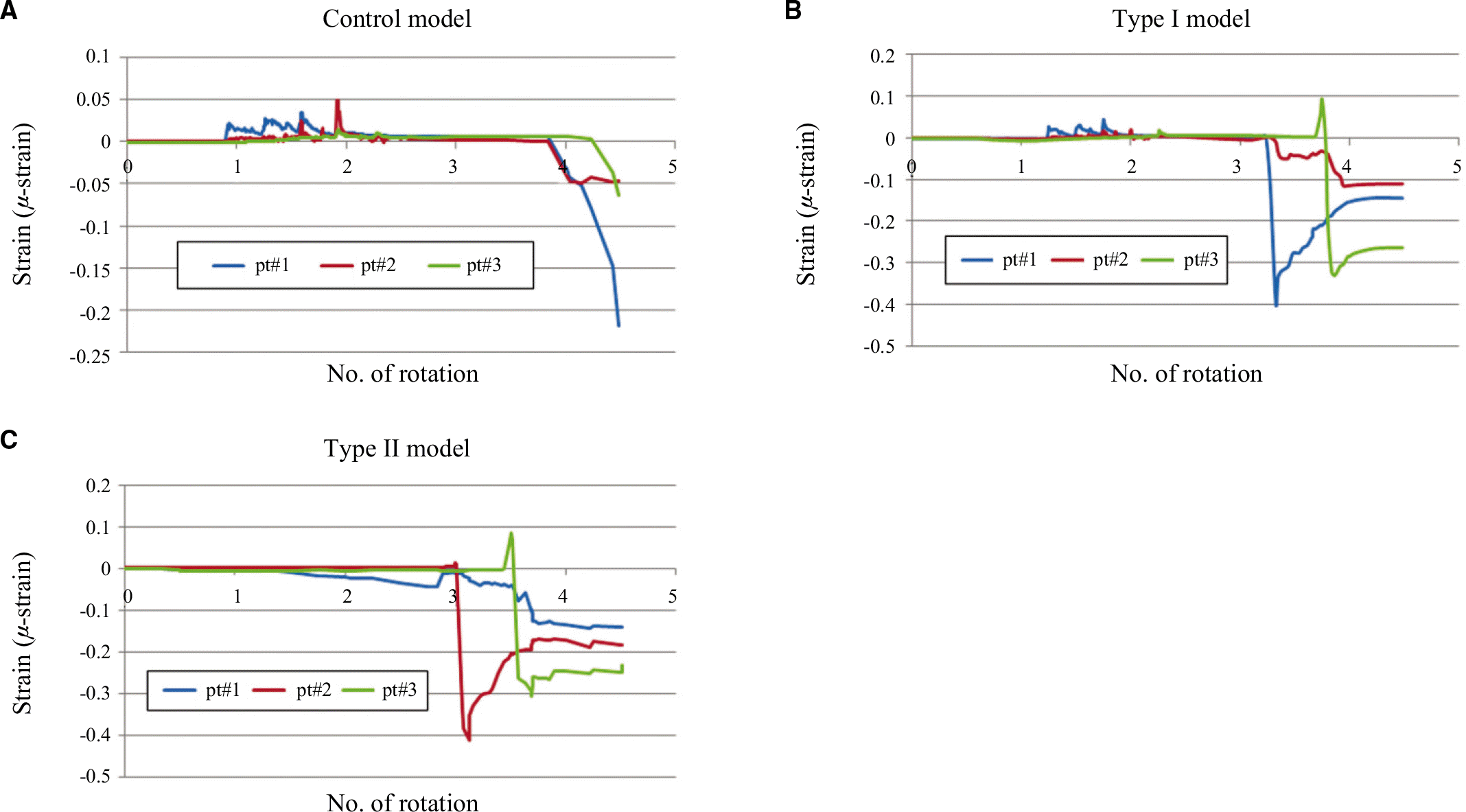
Three dimensional finite element analysis was used to simulate the insertion of 3 implants. Control model was 4.1 x 10 mm implant (Submerged model, Dentis Co, Daegu, Korea) equipped with a main thread only. Type I was with main thread and microthread, and Type II had similar thread pattern but was of tapered body. A PC-based finite element software (DEFORM 3D ver 5, SFTC, Columbus, OH, USA) was used to calculate a total of 3,600 steps of analysis, which simulated the whole insertion.
Go to : 
REFERENCES
1.Holmgren EP., Seckinger RJ., Kilgren LM., Mante F. Evaluating parameters of osseointegrated dental implants using finite element analysis-a two-dimensional comparative study examining the effects of implant diameter, implant shape, and load direction. J Oral Implantol. 1998. 24:80–8.

2.Hi mmlova ′L., Dosta ′lova ′T., Ka ′covsky ′A., Konvickova ′S. Influence of implant length and diameter on stress distribution: a finite element analysis. J Prosthet Dent. 2004. 91:20–5.
3.Chun HJ., Cheong SY., Han JH., Heo SJ., Chung JP., Rhyu IC., Choi YC., Baik HK., Ku Y., Kim MH. Evaluation of design parameters of osseointegrated dental implants using finite element analysis. J Oral Rehabil. 2002. 29:565–74.
4.Hansson S., Werke M. The implant thread as a retention element in cortical bone: the effect of thread size and thread profile: a finite element study. J Biomech. 2003. 36:1247–58.
5.Hansson S. Implant-abutment interface: biomechanical study of flat top versus conical. Clin Implant Dent Relat Res. 2000. 2:33–41.
6.Hansson S. A conical implant-abutment interface at the level of the marginal bone improves the distribution of stresses in the supporting bone. An axisymmetric finite element analysis. Clin Oral Implants Res. 2003. 14:286–93.
7.Saab XE., Griggs JA., Powers JM., Engelmeier RL. Effect of abutment angulation on the strain on the bone around an implant in the anterior maxilla: a finite element study. J Prosthet Dent. 2007. 97:85–92.
8.Petrie CS., Williams JL. Comparative evaluation of implant designs: influence of diameter, length, and taper on strains in the alveolar crest. A three-dimensional finite-element analysis. Clin Oral Implants Res. 2005. 16:486–94.
9.Hansson S. The implant neck: smooth or provided with retention elements. A biomechanical approach. Clin Oral Implants Res. 1999. 10:394–405.
10.Schrotenboer J., Tsao YP., Kinariwala V., Wang HL. Effect of mi-crothreads and platform switching on crestal bone stress levels: a finite element analysis. J Periodontol. 2008. 79:2166–72.
11.Chung JM., Jo KH., Lee CH., Yu WJ., Lee KB. Finite element analysis of peri-implant bone stress influenced by cervical module configuration of endosseous implant. J Korean Acad Prosthodont. 2009. 47:394–405.
12.Li YF. Comparative and analysis study of peri-implant bone stress around Rescue implant and standard implant using finite element method. Masters thesis, Department of Dentistry, Graduate School, Kyungpook National University, Daegu, Korea,. 2009.
13.Lee DW., Choi YS., Park KH., Kim CS., Moon IS. Effect of mi-crothread on the maintenance of marginal bone level: a 3-year prospective study. Clin Oral Implants Res. 2007. 18:465–70.
14.Engquist B., Astrand P., Dahlgren S., Engquist E., Feldmann H., Gro ¨ndahl K. Marginal bone reaction to oral implants: a prospective comparative study of Astra Tech and Bra®nemark system implants. Clin Oral Implants Res. 2002. 13:30–7.
15.Astrand P., Engquist B., Dahlgren S., Gro ¨ndahl K., Engquist E., Feldmann H. Astra Tech and Bra®nemark system implants: a 5-year prospective study of marginal bone reactions. Clin Oral Implants Res. 2004. 15:413–20.
16.Kwon MA., Kim YD., Jeong CM., Lee JY. Clinical and radiographic evaluation of implants with dual-microthread: 1-year study. J Korea Acad Periodontol. 2009. 39:27–36.
17.Rasmusson L., Kahnberg KE., Tan A. Effects of implant design and surface on bone regeneration and implant stability: an experimental study in the dog mandible. Clin Implant Dent Relat Res. 2001. 3:2–8.
18.Gotfredsen K. A 5-year prospective study of single-tooth replacements supported by the Astra Tech implant: a pilot study. Clin Implant Dent Relat Res. 2004. 6:1–8.
19.Palmer RM., Palmer PJ., Smith BJ. A 5-year prospective study of Astra single tooth implants. Clin Oral Implants Res. 2000. 11:179–82.
20.Albrektsson T., Zarb G., Worthington P., Eriksson AR. The long-term efficacy of currently used dental implants: a review and proposed criteria of success. Int J Oral Maxillofac Implants. 1986. 1:11–25.
21.Van de Velde T., Collaert B., Sennerby L., De Bruyn H. Effect of implant design on preservation of marginal bone in the mandible. Clin Implant Dent Relat Res. 2010. 12:134–41.
22.Jeong HC. Three dimensional finite element analysis of cortical bone strain induced by insertion of self tapping implant. Doctorate Thesis, Department of Dentistry, Graduate School, Kyungpook National University, Daegu, Korea,. 2008.
23.Maniatopoulos C., Pilliar RM., Smith DC. Threaded versus porous-surfaced designs for implant stabilization in bone-endodontic implant model. J Biomed Mater Res. 1986. 20:1309–33.
24.Pilliar RM., Lee JM., Maniatopoulos C. Observations on the effect of movement on boneingrowth into porous-surfaced implants. Clin Orthop Relat Res. 1986. 208:108–13.
25.Szmukler-Moncler S., Salama H., Reingewirtz Y., Dubruille JH. Timing of loading and effect of micromotion on bone-dental implant interface: review of experimental literature. J Biomed Mater Res. 1998. 43:192–203.
26.Misch CE. Contemporary Implant Dentistry. St. Louis: Mosby;1999. p. 337.
27.Han SU., Vang MS., Yang HS., Park SW., Park HO., Lim HP. Stress analysis of supporting tissues according to implant fixture diameter and residual alveolar bone width. J Korean Acad Prosthodont. 2007. 45:506–21.
28.Yu W., Jang YJ., Kyung HM. Combined influence of implant diameter and alveolar ridge width on crestal bone stress: a quantitative approach. Int J Oral Maxillofac Implants. 2009. 24:88–95.
29.Rubin CT., Lanyon LE. Regulation of bone mass by mechanical strain magnitude. Calcif Tissue Int. 1985. 37:411–7.
30.Frost HM. Wolff' s Law and bone' s structural adaptations to mechanical usage: an overview for clinicians. Angle Orthod. 1994. 64:175–88.
31.Duyck J., R � nold HJ., van Oosterwyck H., Naert I., Vander Sloten J., Ellingsen JE. The influence of static and dynamic loading on marginal bone reactions around osseointegrated implants: an animal experimental study. Clin Oral Implants Res. 2001. 12:207–18.
32.Frost HM. Bone' s mechanostat: a 2003 update. Anat Rec A Discov Mol Cell Evol Biol. 2003. 275:1081–101.
Go to :
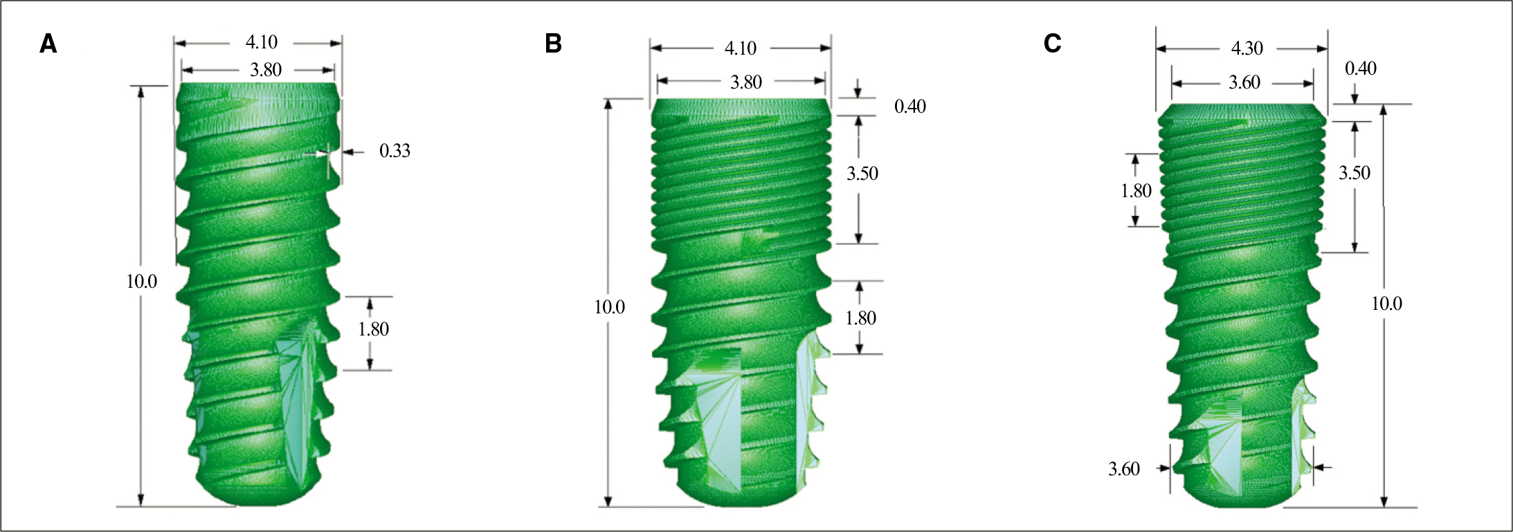 | Fig. 1.Geometry of 3 different implant systems. A: Control; straight body without microthread, B: Type I; straight body with microthread, C: Type II; tapered body with microthread. |
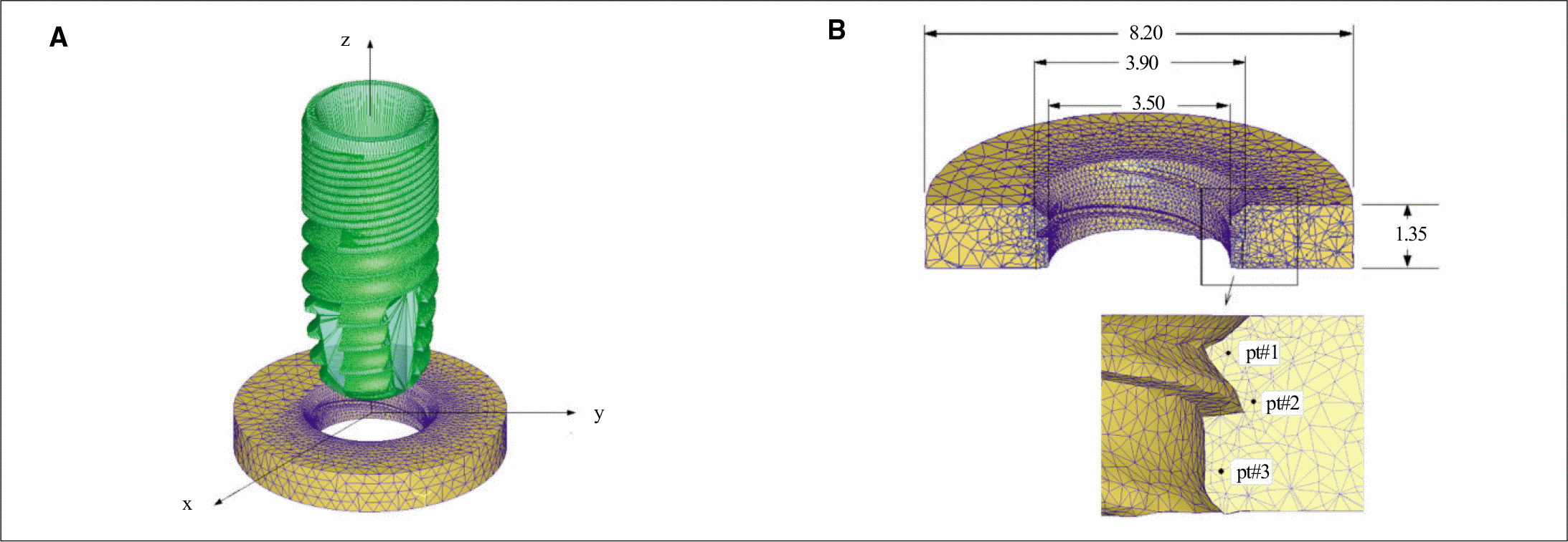 | Fig. 2.Finite element model. A: FE mesh model showing the implant prior to its placement and the axis system, B: cross sectioned cortical bone, dimensions of the threaded groove and the 3 reference points to record strain development during the implant placement. |
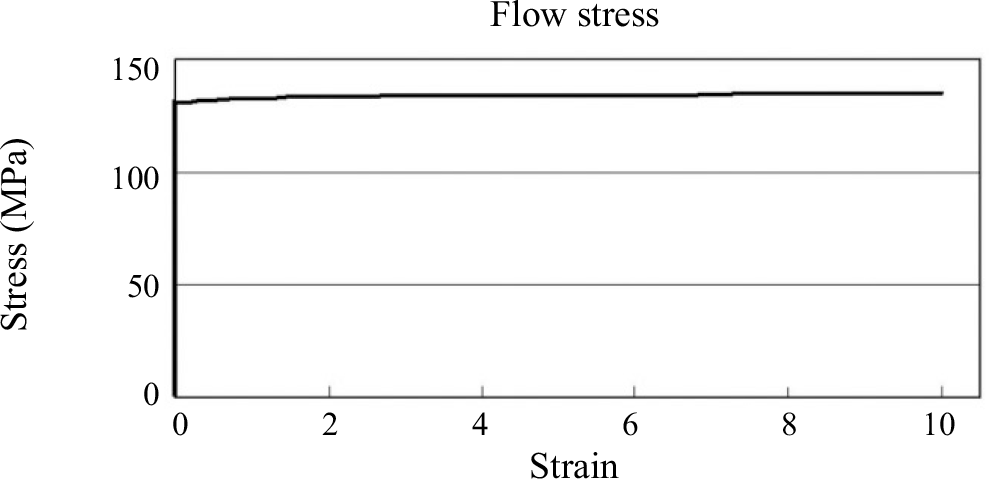 | Fig. 3.Rigid-plastic property data of cortical bone (virtually perfect plasticity was assumed for cortical bone, i.e. stress of 136 MPa was assigned at strain of 10 as compared to the yield stress, 135 MPa). |
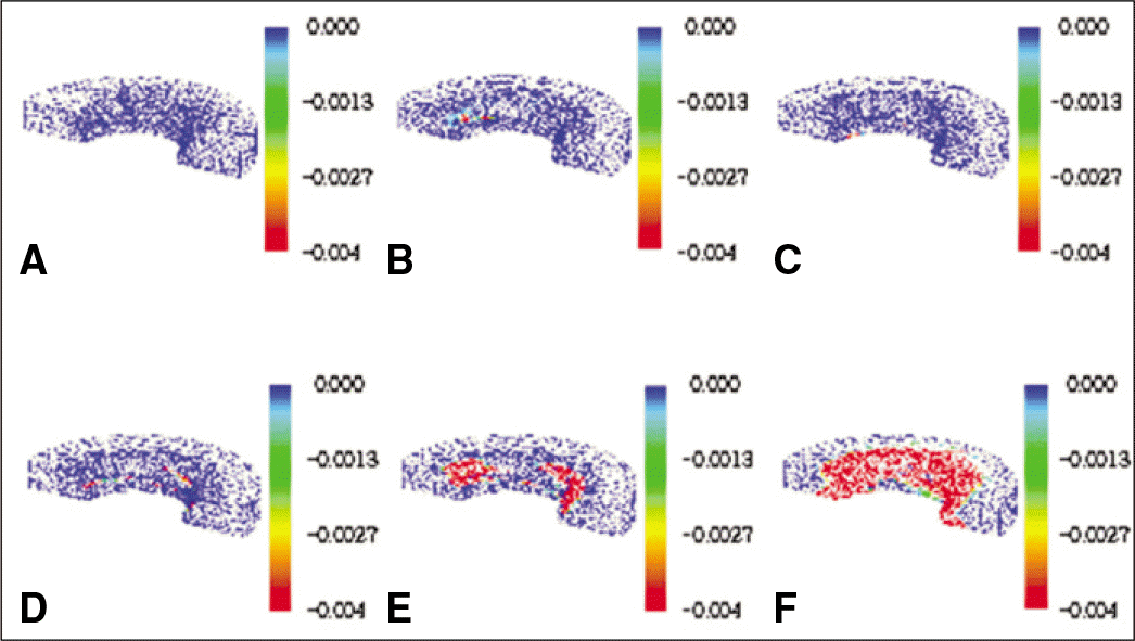
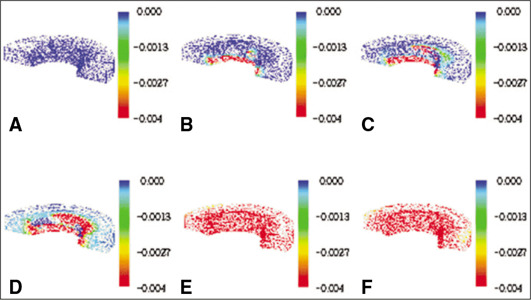
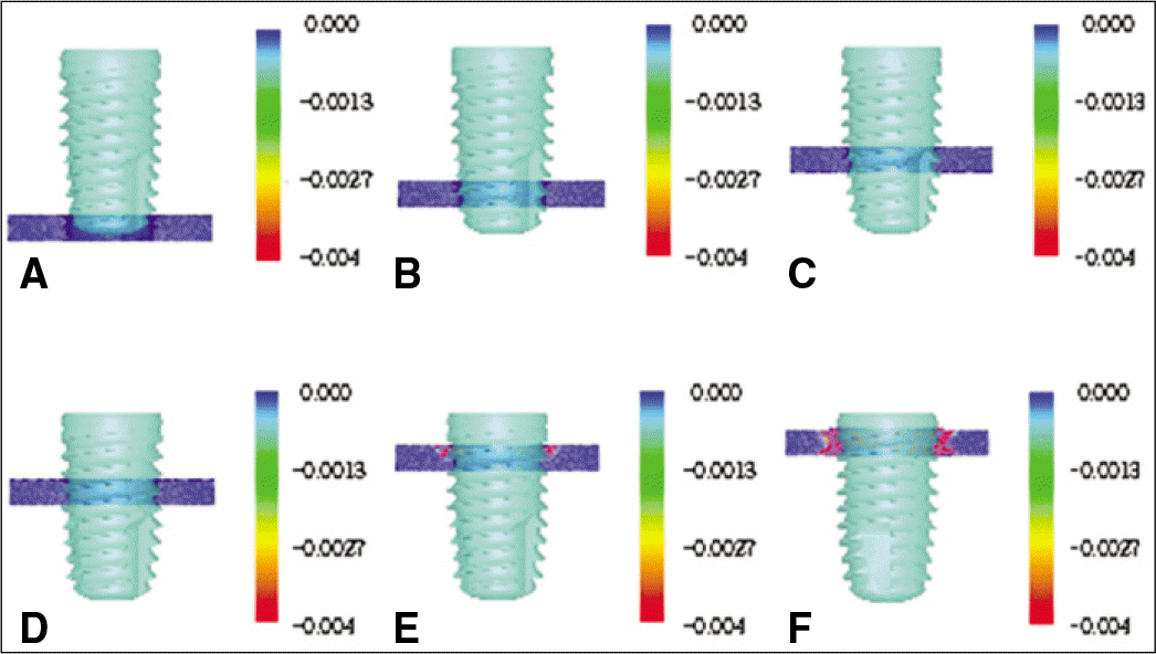 | Fig. 4.Radial strain development in the cortical bone at 6 stages of implant insertion. A: initial, B: 1 turn, C: 2 turn, D: 3 turn, E: 4 turn, F: 4.5 turn in control model implant. |
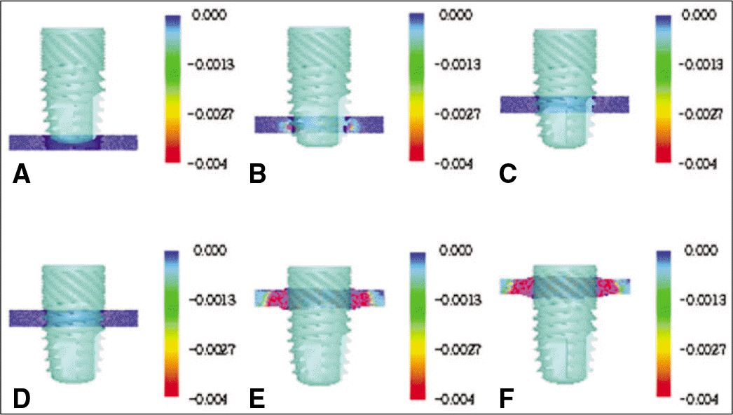
 | Fig. 5.Radial strain development in the half of cortical bone. A: initial, B: 1 turn, C: 2 turn, D: 3 turn, E: 4 turn, F: 4.5 turn in control model implant. |
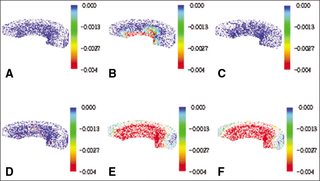
 | Fig. 6.Radial strain development in the cortical bone at 6 stages of implant insertion. A: initial, B: 1 turn, C: 2 turn, D: 3 turn, E: 4 turn, F: 4.5 turn in Type I implant. |
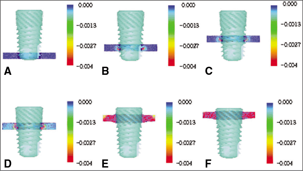
 | Fig. 7.Radial strain development in the half of cortical bone. A: initial, B: 1 turn, C: 2 turn, D: 3 turn, E: 4 turn, F: 4.5 turn in Type I implant. |
 | Fig. 8.Radial strain development in the cortical bone at 6 stages of implant insertion. A: initial, B: 1 turn, C: 2 turn, D: 3 turn, E: 4 turn, F: 4.5 turn in Type II implant. |
 | Fig. 9.Radial strain development in the half of cortical bone. A: initial, B: 1 turn, C: 2 turn, D: 3 turn, E: 4 turn, F: 4.5 turn in Type II implant. |
 XML Download