Abstract
Purpose
The purpose of this study was to investigate whether the re-osseointegration of the implants that had mechanical unscrewing possibly occurred or not. Furthermore, if it happened, the degree of re-osseointegration was evaluated by comparing with previous osseointegration.
Materials and methods
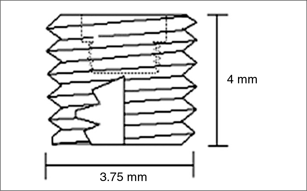
The smooth implant (commercial pure titanium 99%) specimens, whose diameter and length was 3.75 mm, 4 mm, respectively were produced. Two implants were inserted into each tibia of 7 New Zealand female white rabbits weighing at least 3.0 kg. The torque removal force for each implant after 6 weeks of implants placement was measured and included in group I . The torque removal forces were assessed after the fixtures were re-screwed to original position and the subjects were allowed to have 4 more weeks for healing and included in group II. One rabbit was sacrificed after first measurement and produced 4 slide specimens in group I, and two rabbits were sacrificed after 2nd measurement, 7 slide specimens, in group II for histomorphologic investigations. All slide specimens were assessed based on the proportion of BIC (bone-implant contact) as well as CBa (Bone area in the cortical passage) value produced by counting the screw threads embedded in the compact bones under the optical microscopic analysis (×20). Statistical analysis was conducted to evaluate the torque removal force, BIC and CBa between group I and II.
Results
As for the torque removal force, the result was 10.8 ± 3.6 Ncm for group I and 20.2 ± 9.7 Ncm for group II. Furthermore, the torque removal force of group II increased by 98.1% in average compared to group I (P < .05). On the other hand, histomorphologic analysis displayed that there was no statistical significance in BIC and CBa values between group I and the group II (P > .05), and RT/BIC and RT/CBa between group I and group II were statistically significant (P < .05).
Go to : 
REFERENCES
1.Bra � nemark PI., Hansson BO., Adell R., Breine U., Lindstro ¨m J., Halle ′n O., Ohman A. Osseointegrated implants in the treatment of the edentulous jaw. Experience from a 10-year period. Scand J Plast Reconstr Surg Suppl. 1977. 16:1–132.
2.Ivanoff CJ., Sennerby L., Lekholm U. Reintegration of mobilized titanium implants. An experimental study in rabbit tibia. Int J Oral Maxillofac Surg. 1997. 26:310–5.
3.Sennerby L., Thomsen P., Ericson LE. A morphometric and bio-mechanic comparison of titanium implants inserted in rabbit cortical and cancellous bone. Int J Oral Maxillofac Implants. 1992. 7:62–71.
4.Nobel Esthetics TM. Procedures and Products. Nobel Biocare Services, Sweden;2009. p. 103.
5.Proshtetic procedure for US implant system. Osstem, Korea;2004. p. 85.
6.Roberts WE., Smith RK., Zilberman Y., Mozsary PG., Smith RS. Osseous adaptation to continuous loading of rigid endosseous implants. Am J Orthod. 1984. 86:95–111.

7.Sennerby L., Thomsen P., Ericson LE. Early tissue response to titanium implants inserted in rabbit cortical bone. Part 1: Light microscopic observations. J Mater Sci Mater Med. 1993. 4:240–50.
8.Barzilay I., Graser GN., Iranpour B., Natiella JR., Proskin HM. Immediate implantation of pure titanium implants into extraction sockets of Macaca fascicularis. Part II: Histologic observations. Int J Oral Maxillofac Implants. 1996. 11:489–97.
Go to : 
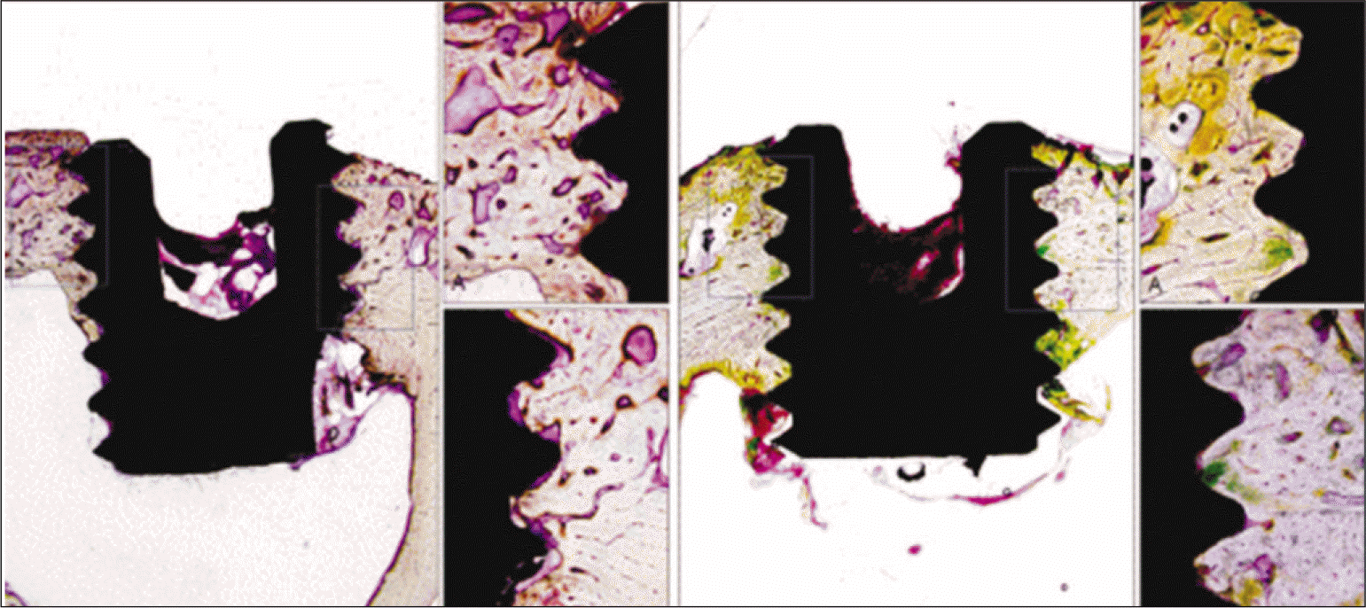
 | Fig. 3.Light microscopic findings of group I (Left) and group II (Right) (Vilanueva Bone Stain, × 20). Bone implant contact was formed at the thread parts through the cortical bone. |
Table 1.
Removal torque value (Ncm) at each group
| Group | n | Removal torque | Rate of increase (%) |
|---|---|---|---|
| I | 20 | 10.8 ± 3.6 | 98.10 |
| II | 20 | 20.2 ± 9.7 | |
| P value | 0.0015∗ |
Table 2.
BIC & CBa ratio comparison between groups
| Group | n | BIC (%) | CBa |
|---|---|---|---|
| I | 4 | 58.7 ± 9.5 | 7.73 ± 0.15 |
| II | 7 | 69.0 ± 10.5 | 7.70 ± 1.29 |
| P value | 0.145 | 0.961 |


 PDF
PDF ePub
ePub Citation
 Print
Print


 XML Download