Abstract
Purpose
This study was aimed to compare the margin and internal fitness of single anterior all-ceramic crown zirconia core made by three deferent CAD/CAM systems.
Material and methods
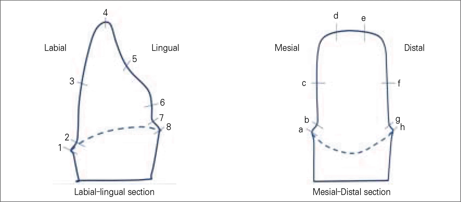
Five single zirconia cores were manufactured by three deferent CAD/CAM systems(Cerasys®system, KaVo Everest®system, LavaTMsystem). The manufactured zirconia cores were duplicated through the use of replica technique, and a replicated sample was sectioned in the center of bucolingual and mesiodistal direction to measure the marginal and internal gap. Measurement was carried out by using measuring microscope (AXIO®) and I-Solution® and analysed through the use of ANOVA.
Results
As for the mean marginal fitness of the zirconia core, it was 84.74 ± 27.57 μ m, in Cerasys®, 80.23 ± 21.07 μ m in KaVo Everest® and 96.37 ± 11.45 μ m in LavaTM, and as for the mean internal gap, it was 94.11 ± 30.07 μ m in Cerasys®, 92.31 ± 25.18 μ m in KaVo Everest®, and 94.99 ± 18.74 μ m in LavaTM. There was no significant statistically deference among the total average gap of three systems. The internal gap in KaVo Everest® seemed to be smaller than LavaTM (P < .05). The internal gap in the incisal area was larger in all of the three systems.
Conclusion
There was no difference in marginal fitness in Cerasys®, KaVo Everest® and LavaTM. As for the internal fitness, it was smaller in KaVo Everest® system than LavaTM system. In all of the three systems, there was a larger gap in incisal area. The marginal and internal gap was within the clinically allowed range in all of the three systems. (J Korean Acad Prosthodont 2010;48:135-42)
Go to : 
REFERENCES
1.Andersson M., Razzoog ME., Ode ′n A., Hegenbarth EA., Lang BR. Procera: a new way to achieve an all-ceramic crown. Quintessence Int. 1998. 29:285–96.
2.Boening KW., Wolf BH., Schmidt AE., Ka ¨stver K., Walter MH. Clinical fit of Procera Allceram crowns. J Prosthet Dent. 2000. 84:419–24.

3.Seghi PR., Sorensen JA. Relative flexural strength of six new ceramic materials. Int J Prosthodont. 1995. 8:239–46.
4.Sturdevant JR., Bayne SC., Heymann HO. Margin gap size of ceramic inlays using second-generation CAD/CAM equipment. J Esthet Dent. 1999. 11:206–14.

5.Tinschert J., Natt G., Mautsch ., Spikermann H., Anusavice KJ. Marginal fit of alumina-and zirconia-based fixed partial dentures produced by a CAD/CAM system. Oper Dent. 2001. 26:367–74.
6.Lawn BR., Deng Y., Thompson VP. Use of contact testing in the characterization and design of all-ceramic crown-like layer structures: a review. J Prosthet Dent. 2001. 86:495–510.

7.Jeon MH., Jeon YC., Jeong CM., Lim JS., Jeong HC. A study of precise fit of the CAM zirconia all-ceramic framework. J Korean Acad Prosthodont. 2005. 43:611–21.
8.Seo JY., Park IN., Lee KW. Fracture strength between different connector designs of zirconia core for posterior fixed partial dentures manufactured with CAD/CAM system. J Korean Acad Prosthodont. 2006. 44:29–39.
9.Bader J., Rozier R., McFall W Jr., Ramsey D. Effect of crown margins on periodontal conditions in regularly attending patients. J Prosthet Dent. 1991. 65:75–9.

10.Grasso J., Nalbandian J., Sanford C., Baili H. Effect of restoration quality on periodontal health. J Prosthet Dent. 1985. 53:14–9.

11.Schwartz N., Whitsett L., Berry T., Stewart J. Unserviceable crowns and fixed partial dentures: lifespan and causes for loss of serviceability. J Am Dent Assoc. 1970. 81:1395–401.

12.Felton D., Kanoy B., Bayne S., Wirthman G. Effect of in vivo crown margin discrepancies on periodontal health. J Prosthet Dent. 1991. 65:357–64.

13.Walton J., Gardner F., Agar J. A survey of crown and fixed partial denture failures: length of service and reasons for replacement. J Prosthet Dent. 1986. 56:416–21.

14.Jorgensen KD. Factors affecting the film thickness of zinc phosphate cement. Acta Odontol Scan. 1960. 18:479–90.
15.Council on dental materials and devices. Revised american national standards institute/American dental association specification No. 8 for zinc phosphate cement. J Am Dent Assoc. 1978. 96:121–3.
16.Christensen GJ. Marginal fit of gold castings. J Prosthet Dent. 1966. 16:297–305.
17.McLean JW., von Fraunhofer JA. The estimation of cement film thickness by an in vivo technique. Br Dent J. 1971. 131:107–11.
18.McLean JW., von Fraunhofer JA. Polycarboxylate cements.: Five years' experience in general practice. Br Dent J. 1972. 132:9–15.

19.Hung SH., Hung KS., Eick JD., Chappel RP. Marginal fit of porcelain-fused-to metal and two types of ceramic crown. J Prosthet Dent. 1990. 63:26–31.
20.May KB., Russell MM., Razzoog ME., Lang BR. Precision of fit: the procera Allceram crown. J Prosthet Dent. 1998. 80:394–404.

21.Hertlein G., Hoscheler S., Frank S., Suttor D. Marginal fit of CAD/CAM manufactured all ceramic prosthesis. J Dent Res. 2001. 80:42–4.
22.Kim DK., Cho IH., Lim JH., Lim HS. On the marginal fidelity of all-ceramic core using CAD/CAM system. J Korean Acad Prosthodont. 2003. 41:20–34.
23.Yang JH., Yeo IS., Lee SH., Han JS., Lee JB. Marginal fit of Celay/In-Ceram, conventional In-Ceram and Empress 2 all-ceramic single crowns. J Korean Acad Prosthodont. 2002. 40:131–9.
24.Carter JM., Sorensen SE., Johnson RR., Teitelbaum RL., Levine MS. Punch shear testing of extracted vital and endodontically treated teeth. J Biomech. 1983. 16:841–8.

25.Strawn SE., White JM., Marshall GW., Gee L., Goodis HE., Marchall SJ. Spectroscopic changes in human dentin exposed to various storage solution-short term. J Dent. 1996. 24:417–23.
26.Pera P., Bassi F., Carossa S. In vitro marginal adaptation of alumina porcelain ceramic crown. J Prosthet Dent. 1994. 72:585–90.
27.Koo JY., Lim JH., Cho IH. Marginal fidelity according to the margin types of all ceramic crowns. J Korean Acad Prosthodont. 1997. 35:445–57.
28.Sorensen JA. A standardized method for determination of crown margin. J Prosthet Dent. 1990. 64:18–24.
29.Moon BH., Yang JH Lee SH., Chung HY. A study on the marginal fit of all-ceramic crown using ccd camera. J Korean Acad Prosthodont. 1998. 36:273–92.
30.Molin M., Karlsson S. The fit of gold inlays and three ceramic inlay system. Aclinical and in vitro study. Acta Odontol Scand. 1993. 51:201–16.
31.Habib Y., Georges E., Salim M., Albert S., Khaldoun T. In vitro evaluation of the “Replica Technique” in the measurement of the fit of Procera crown. J Contemp Dent Pract. 2008. 9:25–32.
32.Davis DR. Comparison of fit of two types of all ceramic crowns. J Prosthet Dent. 1988. 59:13–6.
33.Abbate MF., Tjan A., Fox WM. Comparison of marginal fit of various ceramic crown systems. J Prosthet Dent. 1989. 61:527–31.
34.Belser UC., Mecentee MI., Richter WA. Fit of three porcelain-fusedto metal marginal designs in vivo: a scanning electron microscope study. J Prosthet Dent. 1985. 53:24–9.
35.Bindle A., Mormann WH. Marginal and internal fit of allceramic CAD/CAM crown-coping on chamfer preparations. J Oral Rehabil. 2005. 32:441–7.
36.Valderrama S., Roekel NV., Andersson M., Goodacre CJ., Munoz CA. A comparison of the marginal and internal adaptation of titanium and gold-platinum-palladium metal ceramic crowns. Int J Prosthodont. 1995. 8:29–37.
Go to : 
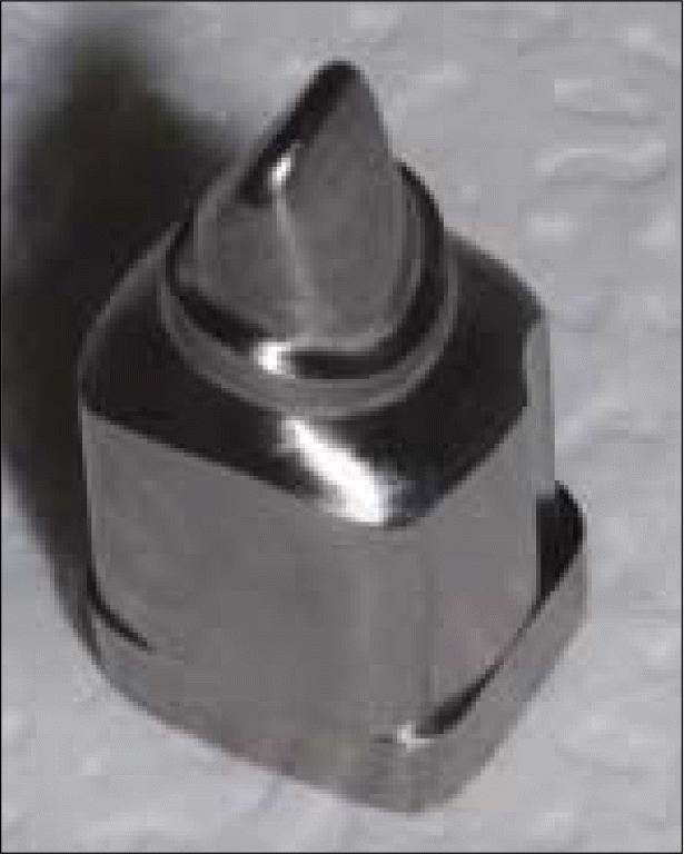
 | Fig. 1.Master models with Titanium Block made by using CAD/CAM system (Addtech Co., Seoul, Korea). |
Table I.
Mean and standard deviation (SD) at each point, and one way-ANOVA test for comparison between groups
Table II.
Two way ANOVA test for marginal and internal gap in three systems (Cerasys®, KaVo Everest®, LavaTM)
| Source | Sum of squares | DF | mean squares | F-value | P-value |
|---|---|---|---|---|---|
| Cerasys®, | |||||
| KaVo Everest® | 4031.664 | 2 | 2015.832 | 3.576 | .030 |
| LavaTM | |||||
| Marginal Av, Interval Av | 372.929 | 1 | 372.929 | 0.661 | .417 |
Table III.
Bonferroni method for post Hoc test
| System (I) | Comparing System (J) | I-J | SD | P-value | 95% confidence interval | |
|---|---|---|---|---|---|---|
| Min | Max | |||||
| Cerasys® | KaVo Everest® | 7.9931 | 3.77822 | .106 | -1.1191 | 17.1053 |
| LavaTM | -1.3863 | 3.82699 | 1.000 | -10.6161 | 7.8435 | |
| KaVo Everest® | Cerasys® | -7.9931 | 3.77822 | .106 | -17.1053 | 1.1191 |
| LavaTM | -9.3794 (∗) | 3.80331 | .043 | -18.5521 | -.2067 | |
| LavaTM | Cerasys® | 1.3863 | 3.82699 | 1.000 | -7.8435 | 10.6161 |
| KaVo Everest® | 9.3794 (∗) | 3.80331 | .043 | .2067 | 18.5521 | |


 PDF
PDF ePub
ePub Citation
 Print
Print


 XML Download