

 PDF
PDF ePub
ePub Citation
 Print
Print


Abstract
Purpose
This 3D-FEA study was performed to investigate the influence of marginal bone loss pattern around the implant to the stress distribution.
Material and methods
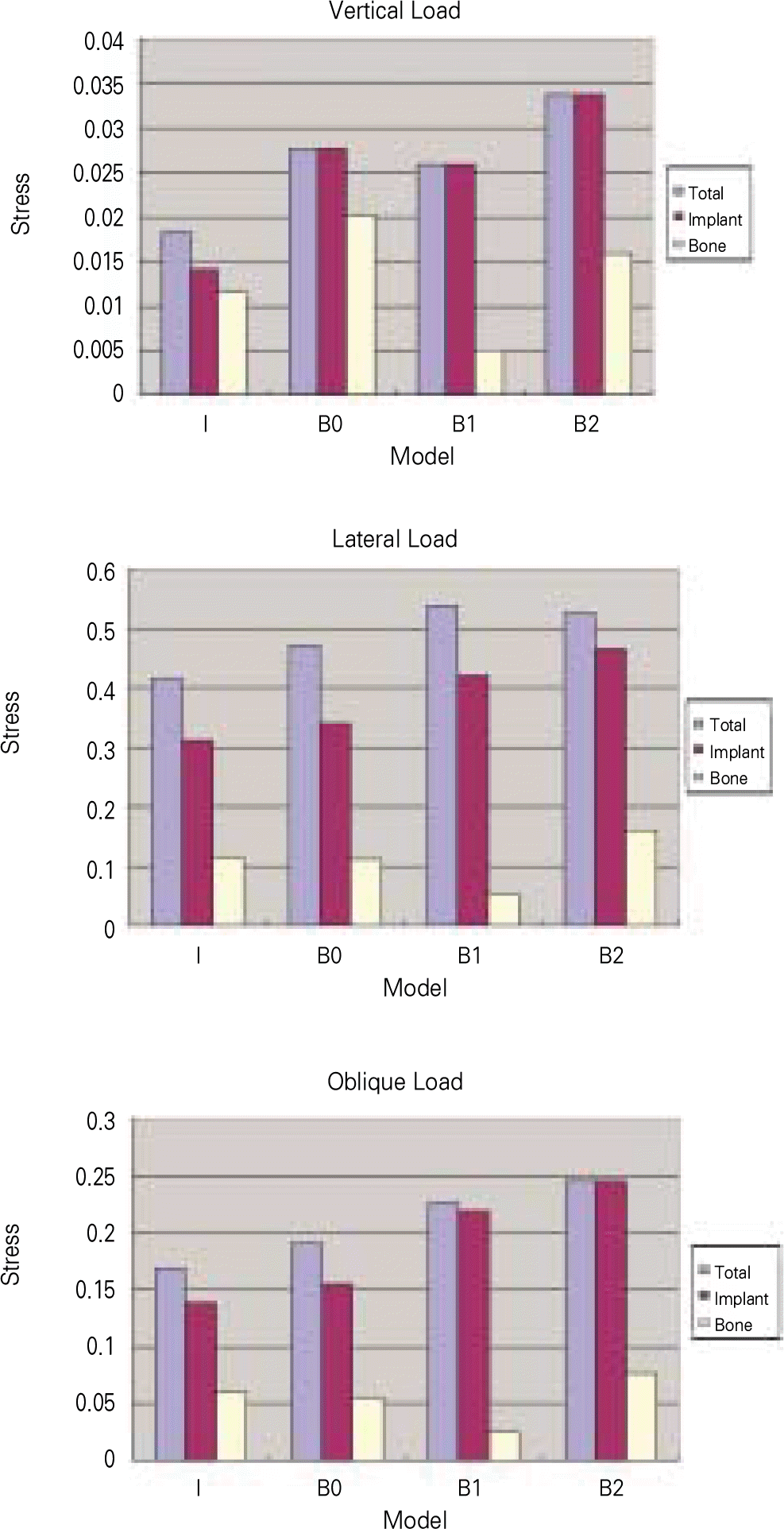
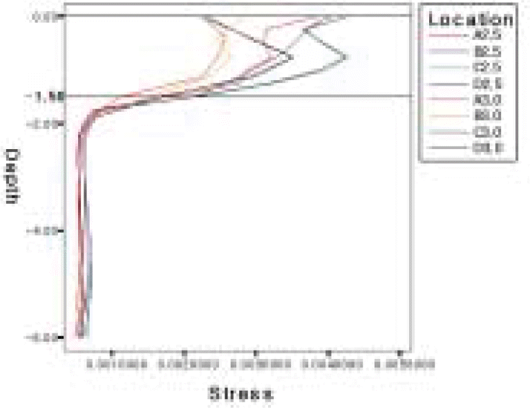
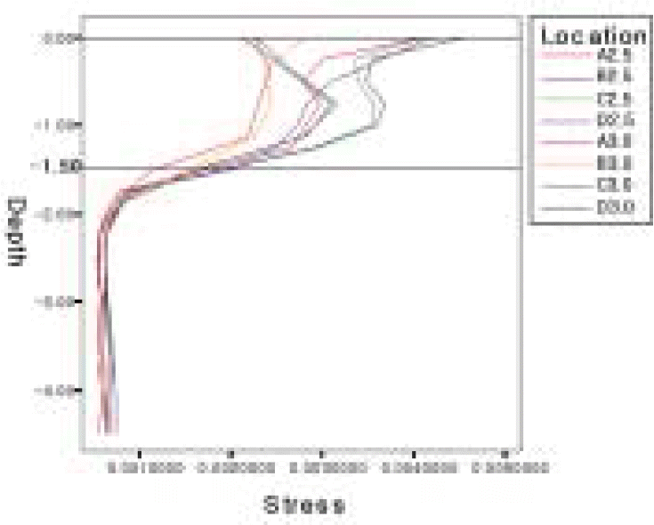
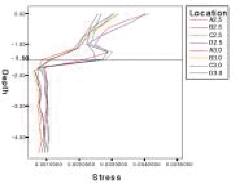
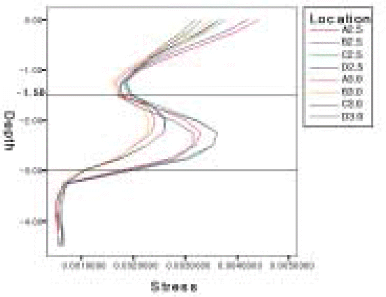
From the right second premolar to the right second molar of the mandible was modeled according to the CT data of a dentate patient. Teeth were removed and an implant (ф 4.0 × 10.0 mm) was placed in the first molar area. Twelve bone models were created: Studied bone loss conditions were horizontal bone loss and vertical bone loss, assumed bone loss patterns during biologic width formation, and pathologic vertical bone loss with or without cortification. Axial, buccolingual, and oblique force was applied independently to the center of the implant crown. The Maximum von Mises stress value and stress contour was observed and von Mises stresses at the measuring points were recorded.
Results
The stress distribution patterns were similar in the non-resorption and horizontal resorption models, but differed from those in the vertical resorption models. Models assuming biologic width formation showed altered stress distribution, and weak bone to implant at the implant neck area seams accelerates stress generation. In case of vertical bone resorption, contact of cortical bone to the implant may positively affect the stress distribution. (J Korean Acad Prosthodont 2010;48:111-21)
Go to : 
REFERENCES
1.Albrektsson T., Wennerberg A. The impact of oral implants-past and future, 1966-2042. J Can Dent Assoc. 2005. 71:327.
2.Misch CE., Suzuki JB., Misch-Dietsh FM., Bidez MW. A positive correlation between occlusal trauma and peri-implant bone loss: literature support. Implant Dent. 2005. 14:108–16.

3.Isidor F. Influence of force on peri-implant bone. Clin Oral Implants Res. 2006. 17:8–18.
4.Saadoun AP., Le Gall M., Kricheck M. Microbial infections and occlusal overload: causes of failure in osseointegrated implants. Pract Periodontics Aesthet Dent. 1993. 5:11–20.
5.Lindhe ., Berglundh T., Ericsson I., Liljenberg B., Marinello C. Experimental breakdown of peri-implant and periodontal tissues. A study in the beagle dog. Clin Oral Implants Res. 1992. 3:9–16.
6.Lang NP., Bragger U., Walther D., Beamer B., Kornman KS. Ligature-induced peri-implant infection in cynomolgus monkeys. I. Clinical and radiographic findings. Clin Oral Implants Res. 1993. 4:2–11.
7.Adell R., Lekholm U., Rockler B., Bra � nemark PI. A 15-year study of osseointegrated implants in the treatment of the edentulous jaw. Int J Oral Surg. 1981. 10:387–416.
8.Cochran DL., Hermann JS., Schenk RK., Higginbottom FL., Buser D. Biologic width around titanium implants. A histometric analysis of the implanto-gingival junction around unloaded and loaded nonsubmerged implants in the canine mandible. J Periodontol. 1997. 68:186–98.
9.Hermann JS., Cochran DL., Nummikoski PV., Buser D. Crestal bone changes around titanium implants. A radiographic evaluation of unloaded nonsubmerged and submerged implants in the canine mandible. J Periodontol. 1997. 68:1117–30.
10.Tada S., Stegaroiu R., Kitamura E., Miyakawa O., Kusakari H. Influence of implant design and bone quality on stress/strain distribution in bone around implants: a 3-dimensional finite element analysis. Int J Oral Maxillofac Implants. 2003. 18:357–68.
11.Hansson S. Surface roughness parameters as predictors of anchorage strength in bone: a critical analysis. J Biomech. 2000. 33:1297–303.
12.Ha ¨ mmerle CH., Bragger U., Bu ¨rgin W., Lang NP. The effect of subcrestal placement of the polished surface of ITI implants on marginal soft and hard tissues. Clin Oral Implants Res. 1996. 7:111–9.
13.Alomrani AN., Hermann JS., Jones AA., Buser D., Schoolfield J., Cochran DL. The effect of a machined collar on coronal hard tissue around titanium implants: a radiographic study in the canine mandible. Int J Oral Maxillofac Implants. 2005. 20:677–86.
14.Hansson S. The implant neck: smooth or provided with retention elements. A biomechanical approach. Clin Oral Implants Res. 1999. 10:394–405.
15.Schwartz-Arad D., Herzberg Ran., Levin L. Evaluation of long-term implant success. J Periodontol. 2005. 76:1623–8.
16.Albrektsson T., Zarb G., Worthington P., Eriksson AR. The long term efficacy of currently used dental implants: a review and proposed criteria of success. Int J Oral Maxillofac Implants. 1986. 1:11–25.
17.Smith DE., Zarb GA. Criteria for success of osseointegrated endosseous implants. J Prosthet Dent. 1989. 62:567–72.
18.Goodacre CJ., Kan JY., Runqcharassaeng K. Clinical complications of osseointegrated implants. J Prosthet Dent. 1999. 81:537–52.
19.Barbier L., Schepers E. Adaptive bone remodeling around oral implants under axial and nonaxial loading conditions in the dog mandible. Int J Oral Maxillofac Implants. 1997. 12:215–23.
20.Wyatt CC., Zarb GA. Bone level changes proximal to oral implants supporting fixed partial prostheses. Clin Oral Implants Res. 2002. 13:162–8.
21.Kitamura E., Stegaroiu R., Nomura S., Miyakawa O. Biomechanical aspects of marginal bone resorption around osseointegrated implants: considerations based on a three dimensional finite element analysis. Clin Oral Implants Res. 2004. 15:401–12.
22.Hermann JS., Buser D., Schenk RK., Schoolfield JD., Cochran DL. Biologic width around one- and two-piece titanium implants. A histometric evaluation of unloaded nonsubmerged and submerged implants in the canine mandible. Clin Oral Implants Res. 2001. 12:559–7.
23.Hermann JS., Buser D., Schenk RK., Higginbottom FL., Cochran DL. Biological width around titanium implants. A physiologically formed and stable dimension over time. Clin Oral Implants Res. 2000. 11:1–11.
24.Hermann JS., Buser D., Schenk RK., Cochran DL. Crestal bone changes around titanium implants. A histometric evaluation of unloaded nonsubmerged and submerged implants in the canine mandible. J Periodontol. 2000. 71:1412–24.
25.Hermann JS., Schoolfield JD., Schenk RK., Buser D., Cochran DL. Influence of the size of the microgap on crestal bone changes around titanium implants. A histometric evaluation of unloaded non-submerged implants in the canine mandible. J Periodontol. 2001. 72:1372–83.
26.Engquist B., Astrand P., Dahlgren S., Engquist E., Feldmann H., Grondahl K. Marginal bone reaction to oral implants: a prospective comparative study of Astra tech and Bra � nemark system implants. Clin Oral Implants Res. 2002. 13:30–7.
27.Lazzara RJ., Porter SS. Platform switching: a new concept in implant dentistry for controlling postrestorative crestal bone levels. Int J Periodontics Restorative Dent. 2006. 26:9–17.
28.Tarnow DP., Cho SC., Wallace SS. The effect of inter-implant distance on the height of inter-implant bone crest. J Periodontal. 2000. 71:546–9.
29.Spiekermann H., Jansen VK., Richter EJ. A 10-year follow-up study of IMZ and TPS implants in the edentulous mandible using bar-retained overdentures. Int J Oral Maxillofac Implants. 1995. 10:231–43.
30.Merickse-Stern R., Aerni D., Geering AH., Buser D. Long-term evaluation of non-submerged hollow cylinder implants. Clinical and radiographic results. Clin Oral Implants Res. 2001. 12:252–9.
31.Wiskott HW., Belser UC. Lack of integration of smooth titanium surfaces: a working hypothesis based on strains generated in the surrounding bone. Clin Oral Implants Res. 1999. 10:429–44.
32.Mellal A., Wiskott HW., Botsis J., Scherrer SS., Belser UC. Stimulating effect of implant loading on surrounding bone. Comparison of three numerical models and validation by in vivo data. Clin Oral Implants Res. 2004. 15:239–48.
33.Cha SB., Lee KB., Jo KH. Finite element approach to investigate the influence of the design configuration of the ITI solid implant on the bone stresses during the osseointegration process. J Korean Acad Prosthodont. 2006. 44:217–28.
34.Bragger U. Use of radiographs in evaluating success, stability and failure in implant dentistry. Periodontol 2000. 1998. 17:77–88.
35.Jung YC., Han CH., Lee KW. A 1-year radiographic evaluation of marginal bone around dental implants. Int J Oral Maxillofac Implants. 1996. 11:811–8.
36.Duyck J., Ronold HJ., van Oosterwyck H., Naert I., Vander Sloten J., Ellingsen JE. The influence of static and dynamic loading on marginal bone reactions around osseointegrated implants: an animal experimental study. Clin Oral Implants Res. 2001. 12:207–18.
Go to :
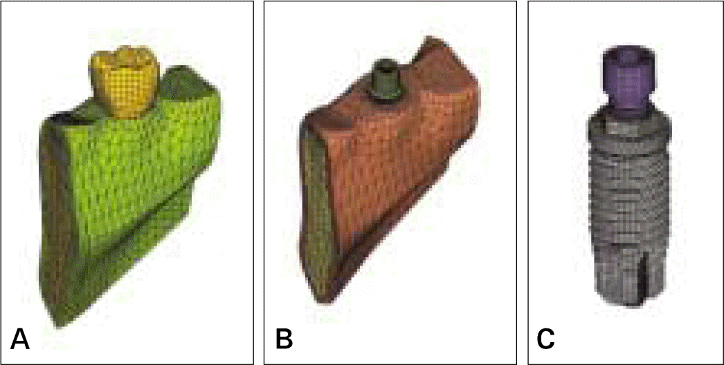 | Fig. 1.FEA models in these experiments. A, Full contour with crown; B, Bone with implant and abutment (purple: cortical bone, brown: cancellous bone); C, Implant with abutment screw. |
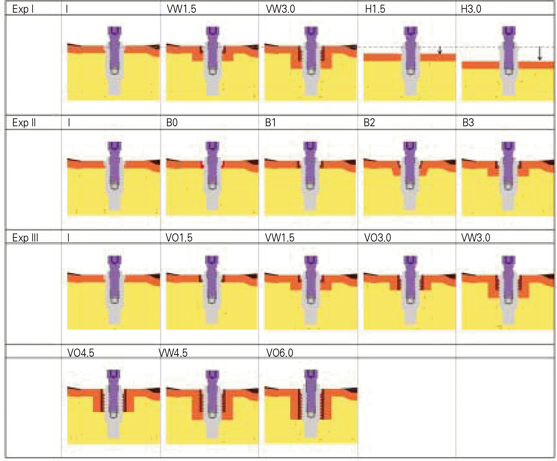 | Fig. 2.Schematic diagrams of experimental model. I: Initial state, VW1.5: 1.5 mm vertical bone loss with cortical bone contact, VW3.0: 3.0 mm vertical bone loss with cortical bone contact, H1.5: 1.5 mm horizontal bone loss, H3.0: 3.0 mm horizontal bone loss, B0: no resorption but slip mode in cortical bone, B1: loss of cortical bone contact (1.5 mm depth, 5.0 mm diameter, cylinder shape), B2: angled defect assuming stable state of implant with biologic width (1.5 mm deep, 5.0mm diameter at top), B3: vertical bone loss (1.5 mm deep, 5.0 mm diameter, cylinder shape) with cortical bone contact, VO: vertical bone loss without cortical bone contact. |
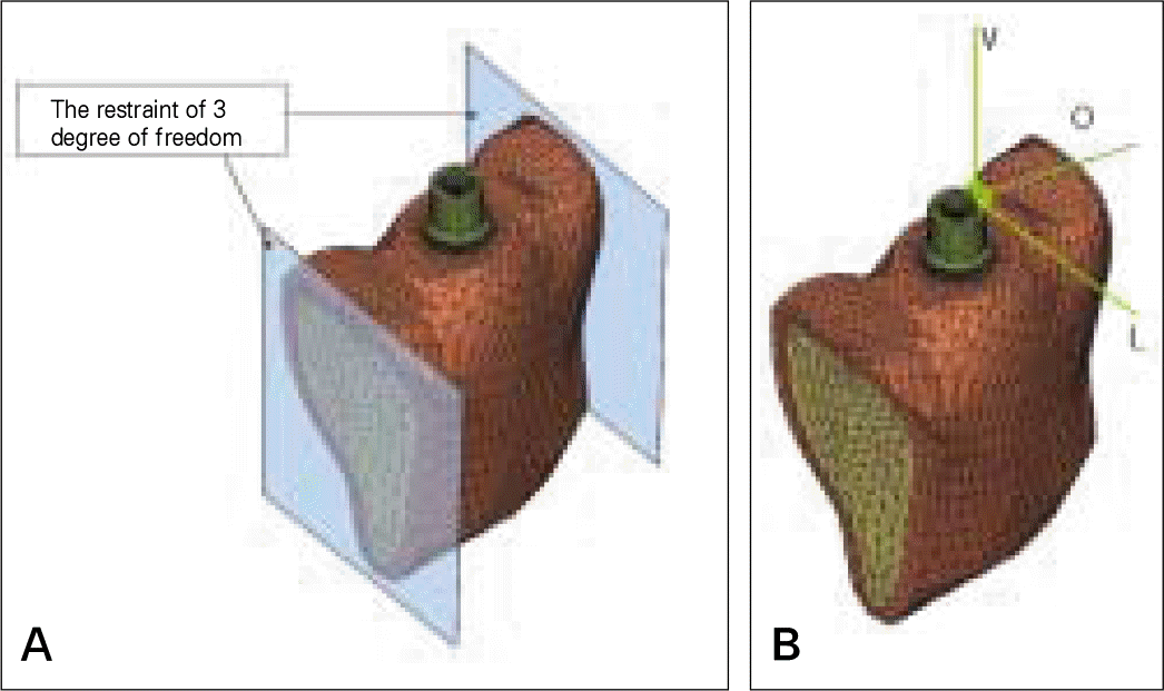 | Fig. 3.A, Boundary conditions; B, directions of forces. V: vertical load, L: lateral force, O: Oblique force (45° inclined to the long axis of implant). |
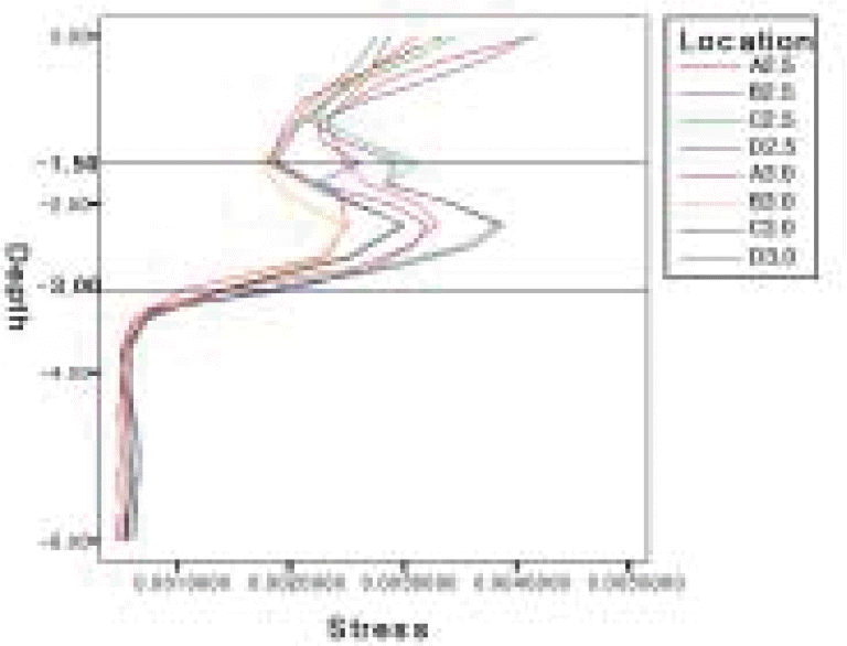
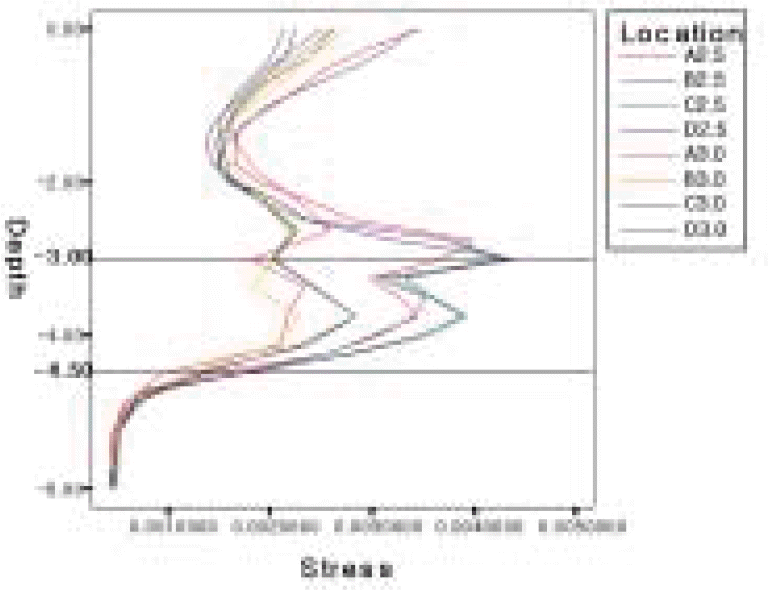
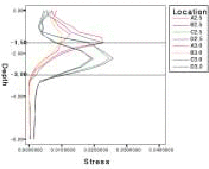
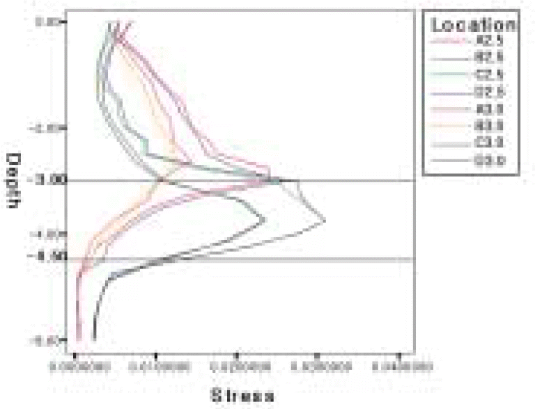
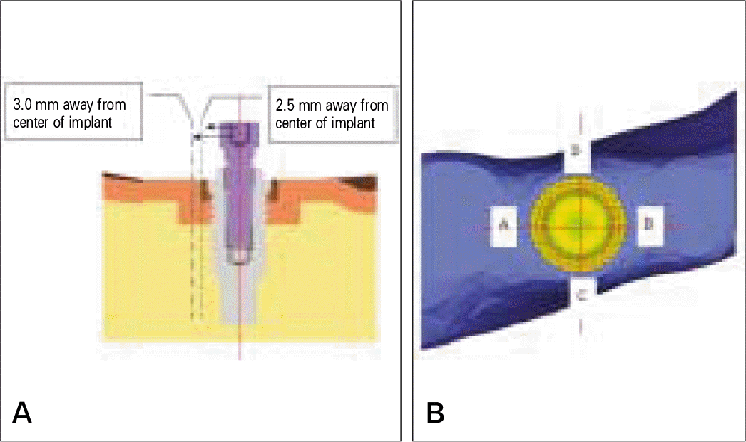 | Fig. 4.Measuring points and nomenclature of nodes. ie) A3.0 means location A in figure B and 3.0 mm away form the long axis of implant A. A: distal, B: mesial, C: buccal D: lingual. |
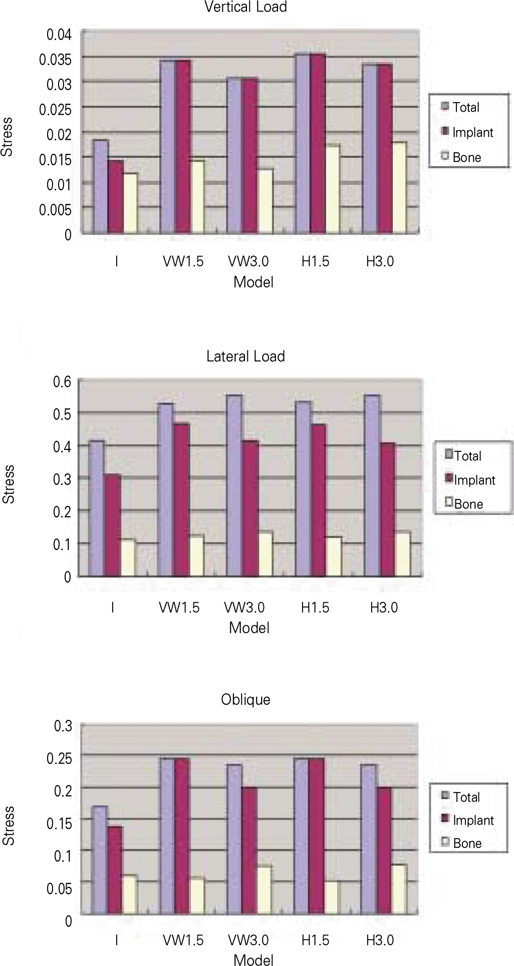
 | Fig. 6.The von Mises stress contour of Implant and bone under vertical load. The sequences of models were rearranged for the easy of comparison. |
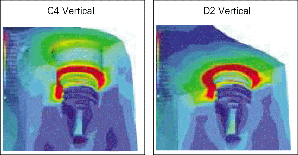
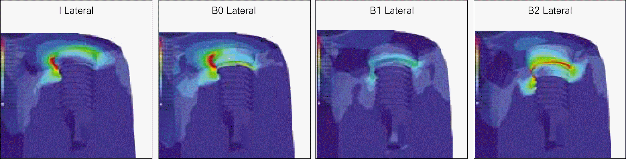 | Fig. 11.The von Mises stress contour of Implant and bone under vertical, lateral and oblique load. |
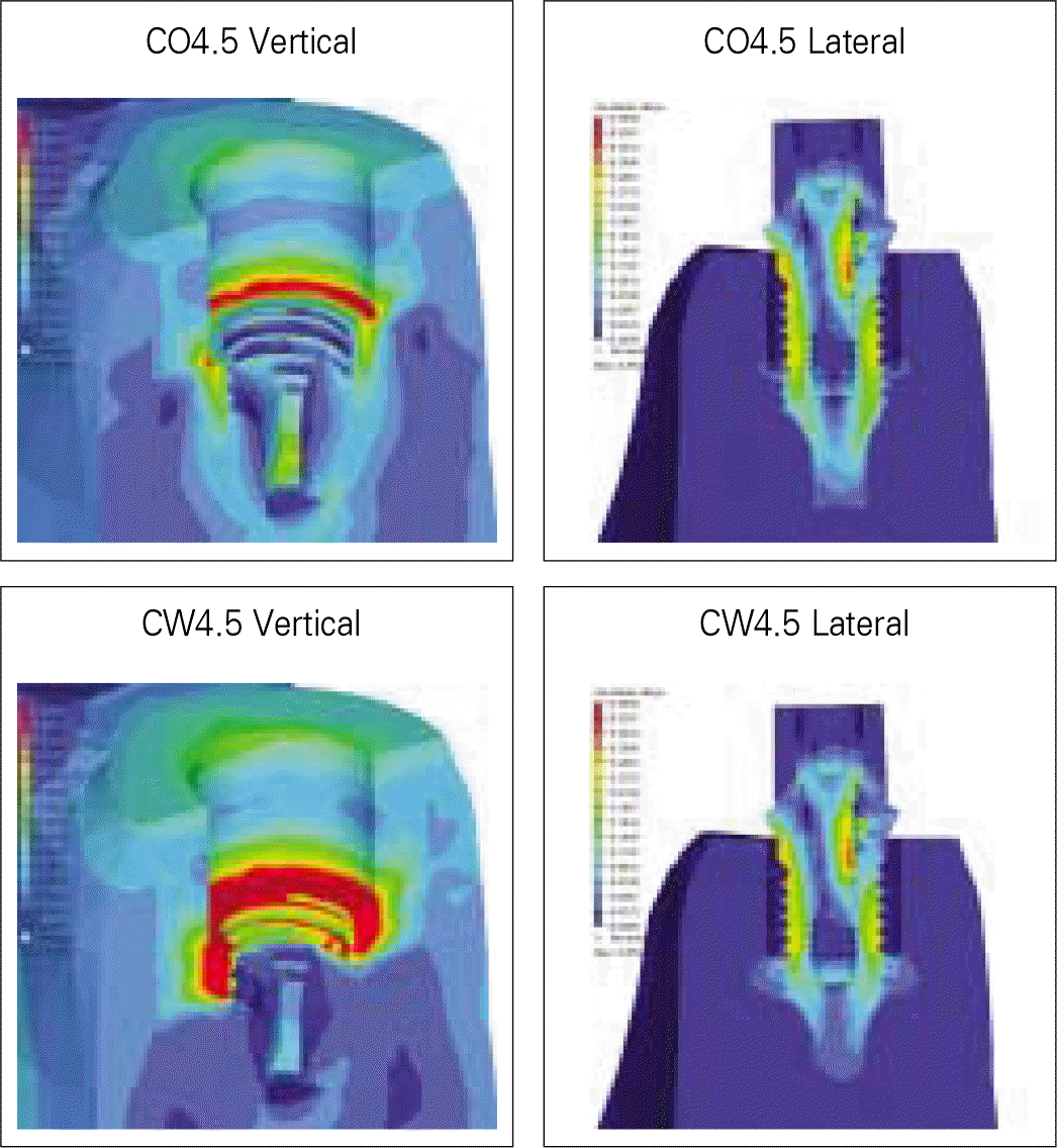 | Fig. 17.The von Mises stress contour of Implant and bone under vertical and lateral load. The results of A1, C1, C2 and C4 were presented in earlier experiments (Fig. 5, 13). |
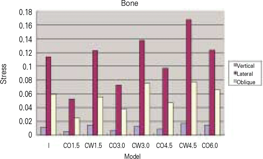
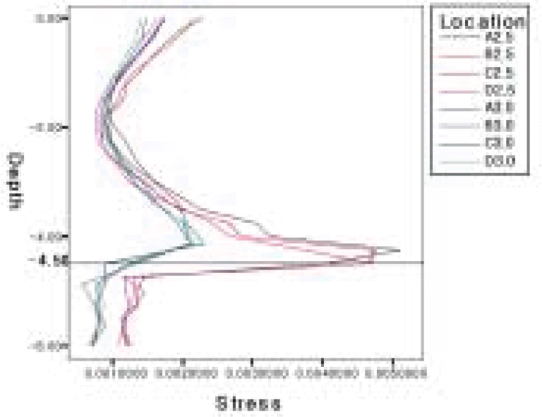
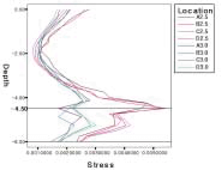
Table I.
Elements and nodes in these experiments
 XML Download