

 PDF
PDF ePub
ePub Citation
 Print
Print


Abstract
Statements of problem
Adequate bone quality and quantity were important to achieve initial stability and to prevent early failures. However there were few published data available regarding the actual effect of dimensional change in implant geometry on initial stability.
Purpose
The purpose of the current study was to investigate the influence of diameter and length changes on initial stability of implants.
Material and methods
Four types of dummy bone (D1, D2, D3 and D4) consisted of cortical and cancellous layers with different thickness were simulated. Implants which had similar surface area to each other (3.5 × 13.0-mm, 4.0 × 11.5-mm, 4.5 × 10.0-mm, 5.0 × 8.5-mm) were inserted in dummy bones. Implant stability as a function of peak insertion torque and resonance frequency values were recorded for each implant.
Results
1. Bone quality was a major influential factor to achieve initial stability (P < .05). 2. In D1, D2 and D3 dummy bones, implant stability quotient values were not significantly different to each other (P > .05), however insertion torques were increased with wider and shorter implants (P < .05). 3. In D4 dummy bone, implant stability quotient values and insertion torques were decreased with wider and shorter implants (P <. 05).
Go to : 
REFERENCES
1.Friberg B., Jemt T., Lekholm U. Early failures in 4,641 consecutively placed Bra � nemark dental implants: a study from stage 1 surgery to the connection of completed prostheses. Int J Oral Maxillofac Implants. 1991. 6:142–6.
2.Meredith N. Assessment of implant stability as a prognostic determinant. Int J Prosthodont. 1998. 11:491–501.
3.Atsumi M., Park SH., Wang HL. Methods used to assess implant stability: current status. Int J Oral Maxillofac Implants. 2007. 22:743–54.
4.O' Sullivan D., Sennerby L., Meredith N. Measurements comparing the initial stability of five designs of dental implants: a human cadaver study. Clin Implant Dent Relat Res. 2000. 2:85–92.
5.Graves SL., Jansen CE., Siddiqui AA., Beaty KD. Wide diameter implants: indications, considerations and preliminary results over a two-year period. Aust Prosthodont J. 1994. 8:31–7.
6.Gentile MA., Chuang SK., Dodson TB. Survival estimates and risk factors for failure with 6 × 5.7-mm implants. Int J Oral Maxillofac Implants. 2005. 20:930–7.
7.Ivanoff CJ., Sennerby L., Johansson C., Rangert B., Lekholm U. Influence of implant diameters on the integration of screw implants. An experimental study in rabbits. Int J Oral Maxillofac Surg. 1997. 26:141–8.
8.Friberg B., Ekestubbe A., Sennerby L. Clinical outcome of Bra � nemark System implants of various diameters: a retrospective study. Int J Oral Maxillofac Implants. 2002. 17:671–7.
9.Langer B., Langer L., Herrmann I., Jorneus L. The wide fixture: a solution for special bone situations and a rescue for the compromised implant. Part 1. Int J Oral Maxillofac Implants. 1993. 8:400–8.
10.Lazzara RJ. Criteria for implant selection: surgical and prosthetic considerations. Pract Periodontics Aesthet Dent. 1994. 6:55–62. quiz 64.
11.Matsushita Y., Kitoh M., Mizuta K., Ikeda H., Suetsugu T. Two-dimensional FEM analysis of hydroxyapatite implants: diameter effects on stress distribution. J Oral Implantol. 1990. 16:6–11.
12.Anner R., Better H., Chaushu G. The clinical effectiveness of 6 mm diameter implants. J Periodontol. 2005. 76:1013–5.

13.Krennmair G., Waldenberger O. Clinical analysis of wide-diameter frialit-2 implants. Int J Oral Maxillofac Implants. 2004. 19:710–5.
14.Ivanoff CJ., Gro ¨ndahl K., Sennerby L., Bergstro ¨m C., Lekholm U. Influence of variations in implant diameters: a 3- to 5-year retrospective clinical report. Int J Oral Maxillofac Implants. 1999. 14:173–80.
15.Friberg B., Sennerby L., Meredith N., Lekholm U. A comparison between cutting torque and resonance frequency measurements of maxillary implants. A 20-month clinical study. Int J Oral Maxillofac Surg. 1999. 28:297–303.
16.Bidez MW., Misch CE. Force transfer in implant dentistry: basic concepts and principles. J Oral Implantol. 1992. 18:264–74.
17.Bidez MW., Misch CE. Issues in bone mechanics related to oral implants. Implant Dent. 1992. 1:289–94.
18.Heidemann W., Gerlach KL., Gro ¨bel KH., Ko ¨llner HG. Influence of different pilot hole sizes on torque measurements and pullout analysis of osteosynthesis screws. J Craniomaxillofac Surg. 1998. 26:50–5.
19.Schliephake H., Sewing A., Aref A. Resonance frequency measurements of implant stability in the dog mandible: experimental comparison with histomorphometric data. Int J Oral Maxillofac Surg. 2006. 35:941–6.
20.Nkenke E., Hahn M., Weinzierl K., Radespiel-Tro ¨ger M., Neukam FW., Engelke K. Implant stability and histomor-phometry: a correlation study in human cadavers using stepped cylinder implants. Clin Oral Implants Res. 2003. 14:601–9.
21.Renouard F., Nisand D. Impact of implant length and diameter on survival rates. Clin Oral Implants Res. 2006. 17:35–51.
22.Friberg B., Sennerby L., Linden B., Gro ¨ndahl K., Lekholm U. Stability measurements of one-stage Bra � nemark implants during healing in mandibles. A clinical resonance frequency analysis study. Int J Oral Maxillofac Surg. 1999. 28:266–72.
23.Bahat O., Handelsman M. Use of wide implants and double implants in the posterior jaw: a clinical report. Int J Oral Maxillofac Implants. 1996. 11:379–86.
Go to :
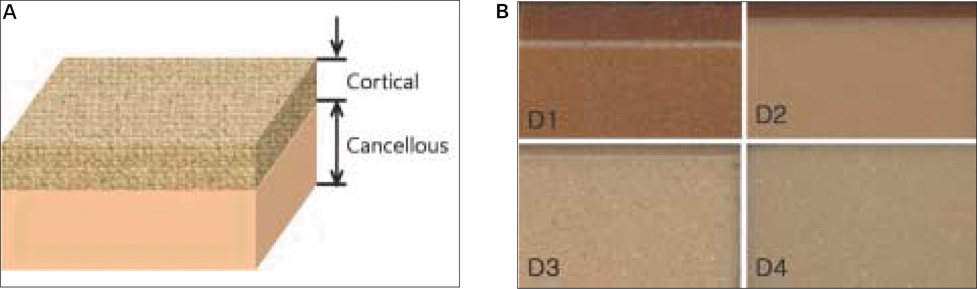
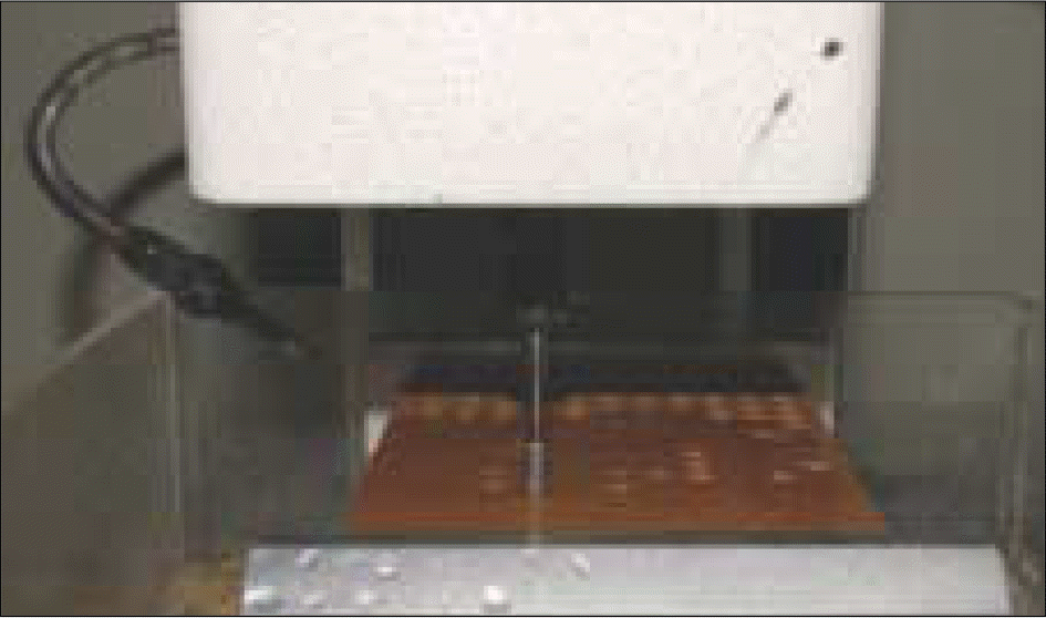
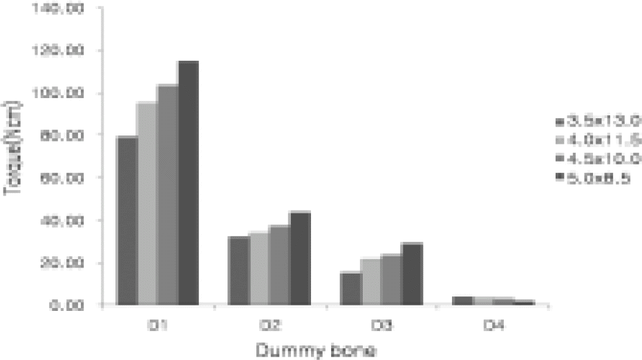
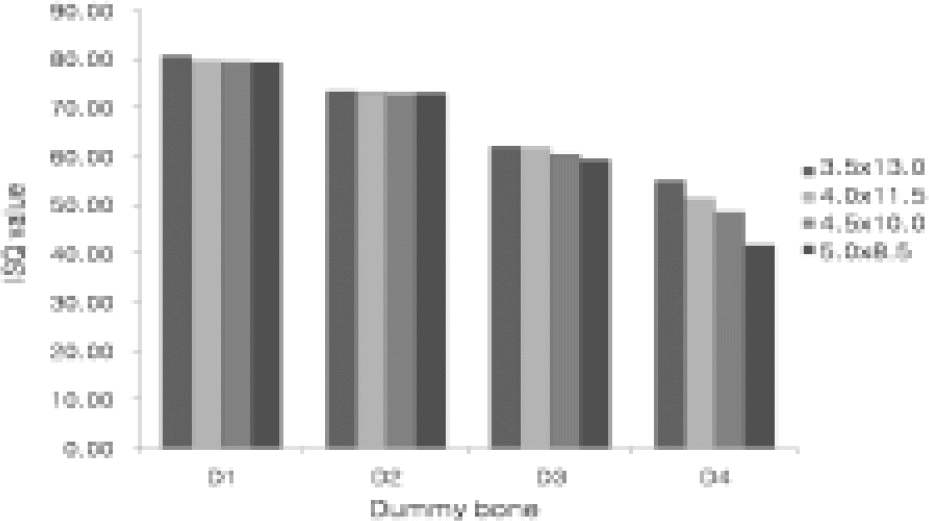
Table I.
Characteristics of dummy bone
| Type | Cortical | Cancellous | ||
|---|---|---|---|---|
| Thickness (mm) | Density (g/cc) | Thickness (mm) | Density (g/cc) | |
| D1 | 3 | 0.8 | 27 | 0.4 |
| D2 | 1.5 | 0.8 | 28.5 | 0.32 |
| D3 | 1 | 0.8 | 29 | 0.16 |
| D4 | 0 | 0.8 | 30 | 0.08 |
Table II.
Dimensions of fixtures
| Diameter (mm) | Length (mm) | Surface area (mm2) |
|---|---|---|
| 3.5 | 13.0 | 194 |
| 4.0 | 11.5 | 205 |
| 4.5 | 10.0 | 202 |
| 5.0 | 8.5 | 198 |
Table III.
Mean values and SDs of insertion torque (Ncm)
Table IV.
Mean values s and SDs of ISQ
 XML Download