

 PDF
PDF ePub
ePub Citation
 Print
Print


Abstract
Statement of problem
Insertion of endosseous implants in the atrophic maxilla is often complicated because of lack of supporting bone. Augmentation of the floor of the maxillary sinus with autogenous bone & bone substitute graft has been proven to be a reliable treatment modality, at least in the short term. The aim of this study is to evaluate the factors of implant survival rate associated with maxillary sinus lift with grafts.
Material and methods
The sinus floor was augmented with bone grafts derived from modified Caldwell-luctechnique (71 subject, 93 sinus, 180 implants), the autogenous bone or autogenous + Bio-oss. Before implant installation the width and height of the alveolar crest were increased in the first stage procedure in 10 patients while in the other 61 patients augmentation and implant installation could be performed simultaneously width and height of the alveolar crest > 4 mm) or delayed installation.
Results
In all case bone volume was sufficients for implant insertion. 14 of 180 inserted implants were lost during follow up and the healing period Patient received implant supported overdenture (5 patients) or fixed bridge (62 patients).
Conclusion
Within the limit of the result of this study, we conclude that bone grafting of the floor of the maxillary sinus floor with bone for the insertion of implants might be a reliable treatment modality and the autogenous bone graft and delayed installation method might be the factors for good results.
Go to : 
REFERENCES
1.Jemt T., Lekholm U., Adell R. Osseointegrated implants in the treatment of partially edentulous patients: a preliminary study on 876 consecutively placed fixtures. Int J Oral Maxillofac Implants. 1989. 4:211–7.
2.Ekfeldt A., Carlsson GE., Borjesson G. Clinical evaluation of single-tooth restorations supported by osseointegrated implants: a retrospective study. Int J Oral Maxillofac Implants. 1994. 9:179–83.
3.Baek JH., Kim MR. The prognosis of maxillary posterior implant installed with sinus augmentation simultaneously. Korean J Maxillofac Reconstr Surg. 2001. 10:23–30.
4.Tatum H Jr. Maxillary and sinus implant reconstructions. Dent Clin North Am. 1986. 30:207–29.
5.Boyne PJ., Marx RE., Nevins M., Triplett G., Lazaro E., Lilly LC, et al. A feasibility study evaluating rhBMP-2 absorbable collagen sponge for maxillary sinus floor augmentation. Int J Periodontics Restorative Dent. 1997. 17:11–25.
6.Grunder U., Gaberthuel T., Boitel N., Imoberdorf M., Meyenberg K., Andreoni C, et al. Evaluating the clinical performance of the Osseotite implant: defining prosthetic predictability. Compend Contin Educ Dent. 1999. 20:628–33.
7.Zarb GA., Albrektsson T. Consensus report: towards optimized treatment outcomes for dental implants. J Prosthet Dent. 1998. 80:641.
8.Jensen OT., Shulman LB., Block MS., Iacono VJ. Report of the Sinus Consensus Conference of 1996. Int J Oral Maxillofac Implants. 1998. 13:11–45.
9.Tidwell JK., Blijdorp PA., Stoelinga PJ., Brouns JB., Hinderks F. Composite grafting of the maxillary sinus for placement of endosteal implants. A preliminary report of 48 patients. Int J Oral Maxillofac Surg. 1992. 21:204–9.

10.Blomqvist JE., Alberius P., Isaksson S. Two-stage maxillary sinus reconstruction with endosseous implants: a prospective study. Int J Oral Maxillofac Implants. 1998. 13:758–66.
11.Peleg M., Mazor Z., Garg AK. Augmentation grafting of the maxillary sinus and simultaneous implant placement in patients with 3 to 5 mm of residual alveolar bone height. Int J Oral Maxillofac Implants. 1999. 14:549–56.
12.Peleg M., Chaushu G., Mazor Z., Ardekian L., Bakoon M. Radiological findings of the post-sinus lift maxillary sinus: a computerized tomography follow-up. J Periodontol. 1999. 70:1564–73.
13.Peleg M., Mazor Z., Chaushu G., Garg AK. Sinus floor augmentation with simultaneous implant placement in the severely atrophic maxilla. J Periodontol. 1998. 69:1397–403.
14.Lazzara R., Siddiqui AA., Binon P., Feldman SA., Weiner R., Phillips R, et al. Retrospective multicenter analysis of 3i endosseous dental implants placed over a five-year period. Clin Oral Implants Res. 1996. 7:73–83.
15.Lee JB., Oung YS., Shin GH., Whang BN. Clinical results of AVANA implant system. J Korean Dent Assoc. 2000. 38:23–28.
16.Valentini P., Abensur D. Maxillary sinus floor elevation for implant placement with demineralized freeze-dried bone and bovine bone (Bio-Oss): a clinical study of 20 patients. Int J Periodontics Restorative Dent. 1997. 17:232–41.
17.Moy PK., Lundgren S., Holmes RE. Maxillary sinus augmentation: histomorphometric analysis of graft materials for maxillary sinus floor augmentation. J Oral Maxillofac Surg. 1993. 51:857–62.
18.Lekholm U., Zarb G. Patient selection and preparation. Branemark PI, Zarb G, T A, editors. Tissue integrated prostheses. Chicago: Quintessence;1985.
19.Schwartz-Arad D., Samet N., Samet N., Mamlider A. Smoking and complications of endosseous dental implants. J Periodontol. 2002. 73:153–7.
20.Ivanoff C J, Gro ¨ndahl k, Sennerby L, Bergstro ¨m C, Leukholm U, Influence of variation in implant diameters: A 3-to-5- year retrospective clinical report. Int J Oral Maxillofac implants. 1999. 14:173–180.
21.Bra ¨gger U., Huber B., Lang N. Evaluation of postsurgical crest bone level adjacent to non-submerged dental implants. Clin Oral Implants Res. 1998. 9:218–24.
22.Albrektsson T., Zarb G., Warthington P., Eriksson AR. The long term efficacy of currently uesd dental implants: a review and proposed criteria of sucess. Int J Oral Maxillofac Implants. 1986. 1:11–25.
23.Jensen OT., Shulman LB., Block MS., Iacono VJ. Report of the Sinus Conference of 1996. Int J Oral Maxillofac Implants. 1998. 13:5–45.
24.Kent JN., Block MS. Simultaneous maxillary sinus floor bone grafting and placement of hydroxylapatite-coated implants. J Oral Maxillofac Surg. 1989. 47:238–42.
25.Albrektsson TIF. Consensus report in Session V. Lang N, Karring T, editors. Proceedings of the 1st European Workshop on Periodontology. London: Quintessence;p. 365–69.
26.Albrektsson T., Zarb GA. Current interpretations of the osseointegrated response: clinical significance. Int J Prosthodont. 1993. 6:95–105.
27.Misch CE., Suzuki JB., Misch-Dietsh FM., Bidez MW. A positive correlation between occlusal trauma and peri-implant bone loss: literature support. Implant Dent. 2005. 14:108–16.
Go to :
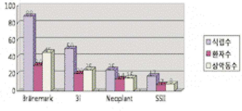
Table I.
Life table analysis of total implants
| Period | No of implants | failed | CSR (%) |
|---|---|---|---|
| Installation - Restoration | 180 | 13 | 92.70% |
| Restoration - 1 year | 167 | 0 | 92.70% |
| 1 year - 2 years | 167 | 1 | 92% |
| 2 years - 3 years | 166 | 0 | 92% |
Table II.
Survival rate according to the gender and age
Table III.
Survival rate according to the implant type
| Neoplant | Osseotite | Brarnemark | SSll | Sum | |
|---|---|---|---|---|---|
| No of Implants | 17 50 | 88 17 | 180 | ||
| Survival | 19 / 21(90%) | 45 / 50 (90%) | 83 / 88 (94.1%) | 19 / 21 (90%) | 166 / 180 (92%) |
Table IV.
Survival rate according to the graft material
| Autogenous only | Autogenous + Bio-Oss | Sum | |
|---|---|---|---|
| Patient's Number | 40 | 31 | 71 |
| Sinus graft | 45 | 48 | 93 |
| No of Implant | 95 | 85 | 180 |
| Survival (%) | 94 (99%) | 72 (85%) | 166 (92%) |
Table V.
Survival rate according to the donor site
| Iliac bone | Mandible | Maxilla | Sum | |
|---|---|---|---|---|
| Patient's Number | 26 | 25 | 20 | 71 |
| No of Implants | 62 | 66 | 52 | 180 |
| Survival (%) | 55 (90%) | 63 (94%) | 48 (90%) | 166 (92%) |
Table VI.
Survival rate according to the lengths, diameter of implants
Table VII.
Survival rate according to the timing of implants installation
| Simultaneous | delayed | Sum | |
|---|---|---|---|
| Patient's Number | 54 | 17 | 71 |
| No of Implants | 110 | 70 | 180 |
| Survival (%) | 96 (85%) | 70 (100%) | 166 (92%) |
Table VIII.
Survival rate according to the type of edentulous area
| full edentulous | partial edentulous | Sum | |
|---|---|---|---|
| No of implants | 48 | 132 | 180 |
| Survival (%) | 38 (80%) | 128 (97%) | 166 (92%) |
 XML Download