

 PDF
PDF ePub
ePub Citation
 Print
Print


Abstract
Statement of problem
Microleakage at the occlusal and gingival margin of Class V cavities restored with composite resin has traditionally been considered an obstacle to successful restoration.
Purpose
The aim of this study was to assess the effectiveness of three different surface sealants(Fortify, Permaseal and Biscover LV) on the marginal sealing of Class V light-activated composite resin restorations(Z250).
Material and methods
Forty noncarious human premolars and molars extracted within a three-month period were selected. Class V cavities with the occlusal margin in enamel and gingival margin in cementum were prepared in both buccal and lingual surfaces. The teeth, randomly assigned in four groups with twenty cavities in each group, were restored with composite resin after applying an adhesive system(Clearfil SE bond). After the finishing and polishing procedures, the restorations were covered with a specific surface sealants, except for the control samples, which were not sealed. After placing restorations, the specimens were thermocycled, and immersed in a 2% methylene blue solution for twenty four hours and sectioned longitudinally. The marginal microleakage was evaluated at the occlusal and gingival interfaces using a microscope and compared among the four groups using ANOVA test and Wilcoxon Rank-Sum test(α =0.05).
Results
Statistical analysis showed that there was significantly less leakage when the surface sealants were used than there was in control group (P < .05). There were no significant differences of microleakage at occlusal and gingival margins among groups. There were no significant differences between microleakage of occlusal and gingival margins in each group. Fortify was not statistically different from control group at the gingival margin (P > .05).
Conclusion
Application of surface sealants was an effective method of surface coating in reducing microleakage at occlusal and gingival margins of Class V composite resin restorations. However, it is certain that some microleakage still occurred despite the application of surface sealants, especially gingival margins.
Go to : 
REFERENCES
1.Munro GA., Hilton TJ., Hermesch CB. In vitro microleakage of etched and rebonded class 5 composite resin restorations. Oper Dent. 1996. 21:203–8.
2.Tjan AH., Tan DE. Microleakage at gingival margins of class V composite resin restorations rebonded with various low viscosity resin systems. Quintessence Int. 1991. 22:565–73.
3.Erhardt MC., Magalha ̃es CS., Serra MC. The effect of re-bonding on microleakage of class V aesthetic restorations. Oper Dent. 2002. 27:396–402.
4.Pameijer CH., Wendt SL Jr. Microleakage of “surface-sealing” materials. Am J Dent. 1995. 8:43–6.
5.May KN Jr., Swift EJ Jr., Wilder AD Jr., Futrell SC. Effect of a surface sealant on microleakage of Class V restorations. Am J Dent. 1996. 9:133–6.
6.Torstenson B., Oden A. Effects of bonding agent types and incremental techniques on minimizing contraction gaps around resin composite. Dent Mater. 1989. 5:218–23.
7.Sidhu SK., Henderson LJ. In vitro marginal microleakage of cervical composite restorations lined with a light-cured glass-ionomer. Oper Dent. 1992. 17:7–12.
8.Kubo S., Yokota H., Yokota H., Hayashi Y. Effect of low viscosity resin based composite on the microleakage of cervical restorations. Am J Dent. 2003. 16:244–8.
9.Goracci G., Mori G., Martinis LC. Curing light intensity and marginal leakage of resin composite restorations. Quintessence Int. 1996. 27:355–62.
10.Ramos RP., Chimello DT., Chinelatti MA., Dibb RGP., Mondelli J. Effect of three surface sealant on marginal sealing of class V composite resin restorations. Oper Dent. 2000. 25:448–53.
11.Reid JS., Saunders WP., Chen YY. The effect of bonding agent and fissure sealant on microleakage of composite resin restorations. Quintessence Int. 1991. 22:295–8.
12.Torstenson B., Bra ¨nnstro ¨m M., Mattsson B. A new method for sealing composite resin contraction gaps in lined cavities. J Dent Res. 1985. 64:450–3.
13.Irie M., Tjandrawinata R., Suzuki K. Effect of delayed polishing periods on interfacial gap formation of class V restorations. Oper Dent. 2003. 28:552–9.
14.Owens BM., Johnson WW. Effect of new generation surface sealants on the marginal permeability of Class V resin composite restorations. Oper Dent. 2006. 31:481–8.

15.Kawai K., Leinfelder KF. Effect of surface penetrating sealant on composite wear. Dent Mater. 1993. 9:108–13.
16.Ramos RP., Chinelatti MA., Chimello DT., Dibb RG. Assessing microleakage in resin composite restorations rebonded with a surface sealant and three low?viscosity resin systems. Quintessence Int. 2002. 33:450–6.
17.McCourt JW., Eick JD. Penetration of fissure sealants into contraction gaps of bulk packed auto-cured composite resin. J Pedod. 1988. 12:167–75.
18.Estafan D., Dussetschleger FL., Miuo LE., Kondamani J. Class V lesions restored with flowable composite and added surface sealing resin. Gen Dent. 2000. 48:78–80.
19.Dickinson GL., Leinfelder KF. Assessing the long-term effect of a surface penetrating sealant. J Am Dent Assoc. 1993. 124:68–72.
20.Gordon M., Plasschaert AJ., Stark MM. Microleakage of several tooth-colored restorative materials in cervical cavities. A comparative study in vitro. Dent Mater. 1986. 2:228–3l.
21.Park SE., Weber HP., Ishikawa-Nagai S. Self-bonding polymers as surface coatings of restorative resins to prevent staining. J Clin Dent. 2006. 17:134–7.
22.Hakimeh S., Vaidyanathan J., Houpt ML. Microleakage of compomer Class V restorations: effect of load cycling, thermal cycling, and cavity shape differences. J Prosthet Dent. 2000. 83:194–203.
23.Sarac D., Sarac YS., Kulunk S., Ural C., Kulunk S., Ural C., Kulunk T. The effect of polishing techniques on the surface roughness and color change of composite resins. J Prosthet Dent. 2006. 96:33–40.
24.Yang JS., Kim MH., Her S., Kim JG., Baik BJ. The effect of low-viscosity resin systems on marginal leakage of composite resin restorations. Korean J pedodontics. 1997. 24:460–74.
25.Hegdahl T., Gjerdet NR. Contraction stresses of composite resin filling materials. Acta Odontol Scand. 1977. 35:191–5.
26.Hansen EK., Asmussen E. Marginal adaptation of posterior resins: Effect of dentin-bonding agent and hygroscopic expansion. Dent Mater. 1989. 5:122–6.
27.Shortall AC. Microleakage, marginal adaptation and composite resin restorations. Br Dent J. 1982. 153:223–7.
28.Smith LA., O' Brien JA., Retief DH., Bradley EL. Microleakage of two dentinal bonding restorative systems. J Dent Res. 1988. 67:309–12.
29.Mandras RS., Retief DH., Russell CM. The effect of thermal and occlusal stresses of the microleakage of the Scotchbond 2 dentinal bonding system. Dent Mater. 1991. 7:63–7.
30.Rossomando KJ., Wendt SL Jr. Thermocycling and dwell times in microleakage evaluation for bonded restorations. Dent Mater. 1995. 11:47–51.
31.Wendt SL., Mclnnes PM., Dickinson GL. The effect of ther-mocycling in microleakage analysis. Dent Mater. 1992. 8:181–4.
32.De ′jou J., Sindres V., Camps J. Influence of criteria on the result of in vitro evaluation of microleakage. Dent Mater. 1996. 12:342–9.
33.Takeuchi CY., Orbegoso Flores VH., Palma Dibb RG., Panzeri H., Lara EH., Dinelli W. Assessing the surface roughness of a posterior resin composite: effect of surface sealing. Oper Dent. 2003. 28:281–6.
34.Dickinson GL., Leinfelder KF. Mazer RB, Russell CM. Effect of a surface penetrating sealant on wear rate of posterior composite resins. J Am Dent Assoc. 1990. 121:251–5.
35.Kim SW., Cho YB., Hong CU. Effect of surface penetrating sealant on the microleakage of cervical restorations. Korean J Conser Dent. 2001. 26:64–76.
36.Cho YG., Choi HY. Effect of Biscover on the microleakage of composite resin restorations. Korean J Conser Dent. 2005. 30:355–62.
37.Harper RH., Schnell RJ., Swartz ML., Phillips RW. In vivo measurements of thermal diffusion through restorations of various materials. J Prosthet Dent. 1980. 43:180–5.
38.Suh BI. Oxygen-inhibited layer in adhesion dentistry. J Esthet Restor Dent. 2004. 16:316–23.
Go to :
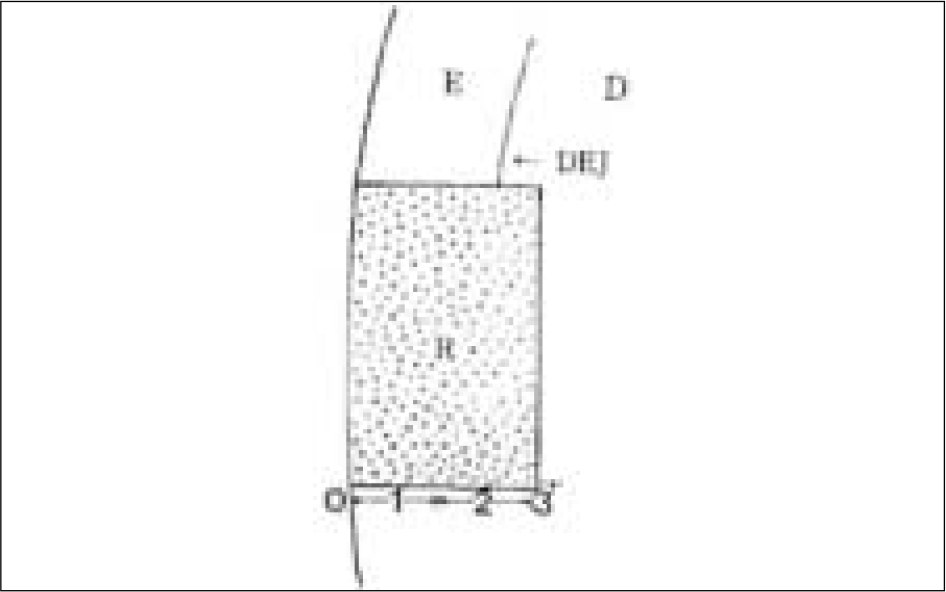 | Fig. 1.Degree of dye penetration (E: Enamel, D: Dentin, R: Composite resin, DEJ: Dentino-enamel junction) |
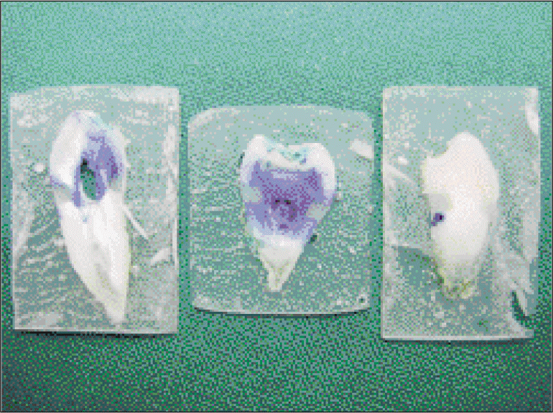
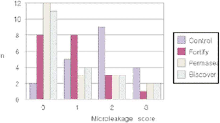
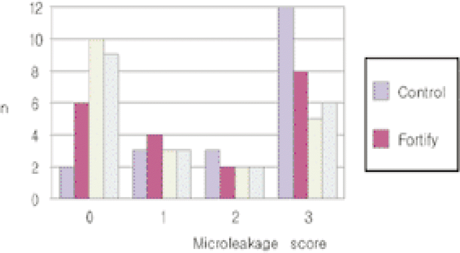
Table I.
Materials used in this study
Table II.
Ingredients of materials used in this study
Table III.
Degree of dye penetration
Table IV.
Distribution of microleakage scores
 XML Download