

 PDF
PDF ePub
ePub Citation
 Print
Print


Abstract
Statement of problem
The increasing demand for esthetic restorations has been required developing new materials for tooth colored restoration. Ceromer (Ceramic Optimized Polymer) has some advantages over porcelain, and has gained increasing popularity in restorative dentistry. However, there is little information on the dimensional changes in a clinical restoration in moist conditions.
Purpose
This study examined the dimensional changes in Ceromer restorations with a clinical crown shape that were fabricated in a clinical manner.
Material and methods
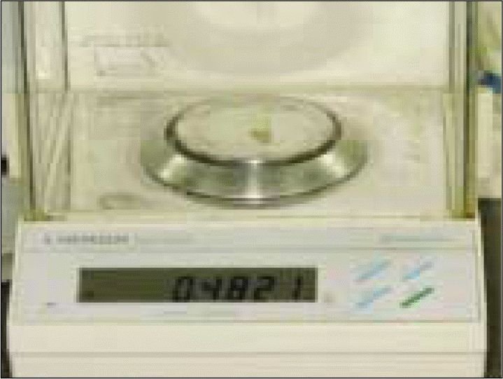
The crowns for the maxillary central incisor were fabricated with two Ceromers (BelleGlass® and Targis®) using a similar clinical restoration manufacturing technique. A total of twenty specimens were prepared and immersed in distilled water at room temperature to allow for water absorption. The weight, height and width were measured at 24, 72 and 168 hours. The accumulated ratios of the changes were calculated and evaluated using a paired t-test and an independent independent t-test.
Results
The dimensions and weight increased with increasing soaking time. Targis® showed significant differences in height and weight between 24 hours and the other times (P < .05). BelleGlass® showed significant differences in width and weight between 24 hours and the other times. The two materials showed different changing patterns of the dimensions but there were no statistically significant differences between them.
Go to : 
REFERENCES
1.Kim SJ., Shin SW., Han JS., Suh KW. Marginal fitness and marginal leakage of fiber-reinforced composite crowns depending upon luting cements. J Korean Acad Prosthodont. 2000. 38:618–30.
2.Chang HW., Lee JH., Lim HS., Cho IH. A study on the marginal fidelity and the fracture strength of ceromers. J Korean Acad Prosthodon. 2005. 43:438–52.
3.Trushkowsky RD. Ceramic optimized polyer: the nest generation of esthetic restorations-Part 1. Compend Contin Educ Dent. 1997. 18:1101–6.
4.Draughn RA., Bowen RL., Moffa JP. Composite materials. Reese JA, Valega M, editors. Restorative dental materials-an overview,. vol. 1. London: FDI;1985. p. 75–107.
5.∅ysæd H., Ruyter IE. Composites for use in posterior teeth: mechanical properties tested under dry and wet conditions, J Biomed Mater Res. 1986. 20:261–71.
6.Calais JG., So ¨ derholm KJ. Influence of filler type and water exposure on flexural strength of experimental composite resins. J Dent Res. 1988. 67:836–40.

7.Mohsen NM., Craig RG. Hydrolytic stability of silanated zirconia silica-urathane. J Oral Rehab. 1995. 22:213–20.
8.So ¨derholm KJ. Degradation of glass filler in experimental composites. J Dent Res. 1981. 60:1867–75.
9.Martin N., Jedynakiewicz NM., Fisher AC. Hygroscopic expansion and solubility of composite restoratives. Dent Materials. 2003. 19:77–86.
10.McCabe JF., Rusby S. Water absorption, dimensional change and radial pressure in resin matrix dental restorative materials. Biomaterials. 2004. 25:4001–7.
11.van Noort R. Chapter 2.2 Resin composites and polyacid-modified resin composites. Introduction to dental materials. 2nded. Mosby, Edinburgh;2002. p. 96–123.
12.Hirasawa T., Hirano S., Hirabayashi S., Harashima I., Aizawa M. Initial dimensional change of composites in dry and wet conditions. J Dent Res. 1983. 62:28–31.
13.Musanje L., Barvell BW. Aspects of water sorption from the air, water and artificial saliva in resin composite restorative materials. Dental Materials. 2003. 19:414–22.
14.Segura A., Donly KJ. In vitro posterior composite polymerization recovery following hygroscopic expansion. J Oral Rehab. 1993. 20:495–9.
15.Gohring TN., Gallo L., Luthy H. Effect of water storage, thermocycling, the incorporation and site of placement of glass-fibers on the flexural strength of veneering composite. Dent Mater. 2005. 21:761–72.
16.Feilzer AJ., de Gee AJ., Davidson CL. Relaxation of polymerization recontraction shear stress by hygroscopic expansion. J Dent Res. 1990. 69:36–9.
17.Feilzer AJ., Kakaboura AI., de Gee AJ., Davidson CL. The influence of water sorption on the development of setting shrinkage stress in traditional and resin-modified glass ionomer cements. Dent Mater. 1995. 11:186–90.
18.Keyf F., Yalc¸in F. The weight change of various light-cured restorative Materials stored in water. J Contemp Dent Pract. 2005. 6:72–9.
Go to :
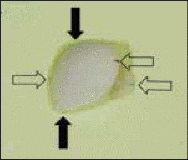 | Fig. 1.Marked reference points. Middle of incisal edge, lowest part of cervical margin and two proximal contact points were selected. Black arrows indicate reference points for mesiodistal width, White arrows indicate reference points for crown height. |
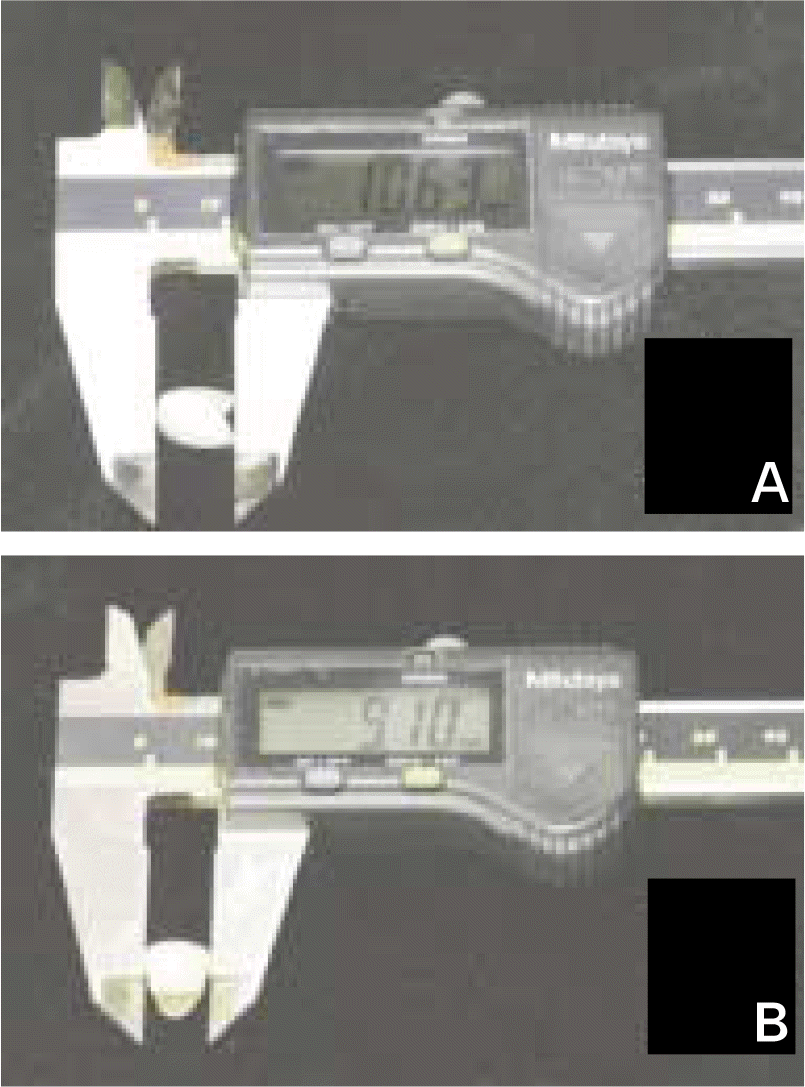 | Fig. 2.Dimension of samples were measured with electrical caliper: A. Measuring height, B. Measuring width. |
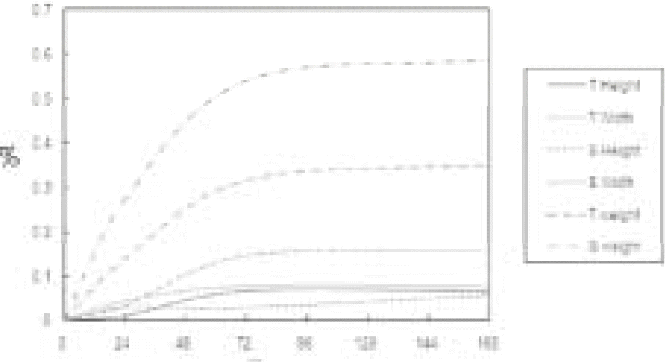 | Fig. 4.Cumulated rate of dimensional changes depending on time. T: Targis®, B: BelleGlass®. |
Table I.
Mean and standard deviation of accumulated change rate
| Material | 0 - 24 h | 0 - 72 h | 0 - 168 h | ||||
|---|---|---|---|---|---|---|---|
| Mean | SD | Mean | SD | Mean | SD | ||
| Targis® | Height (%) | 0.010∗ | 0.069 | 0.065 | 0.063 | 0.065 | 0.063 |
| Width (%) | 0.044 | 0.077 | 0.078 | 0.091 | 0.078 | 0.091 | |
| Weight (%) | 0.139∗ | 0.139 | 0.316 | 0.162 | 0.351 | 0.169 | |
| BelleGlass® | Height (%) | 0.029 | 0.047 | 0.029 | 0.065 | 0.058 | 0.081 |
| Width (%) | 0.034∗ | 0.093 | 0.147 | 0.151 | 0.159 | 0.162 | |
| Weight (%) | 0.273∗ | 0.296 | 0.542 | 0.367 | 0.589 | 0.365 | |
 XML Download