Abstract
PURPOSE
The application of computer-aided technology to implant dentistry has created new opportunities for treatment planning, surgery and prosthodontic treatment, but the correct selection and combination of available methods may be challenging in times. Hence, the purpose of this case report is to present a combination of several computer-aided tools as approaches to manage complicated implant case.
MATERIAL AND METHODS
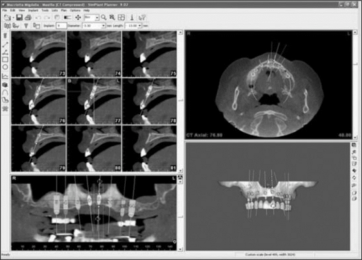
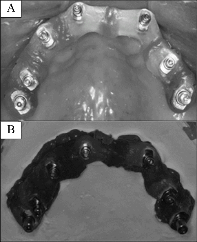
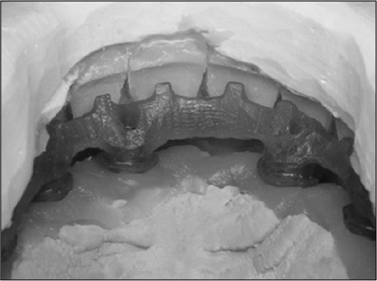
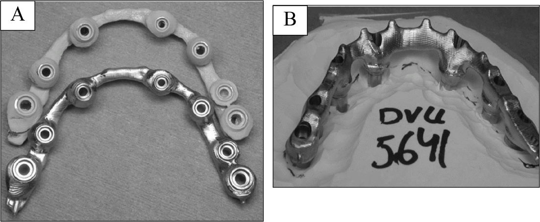
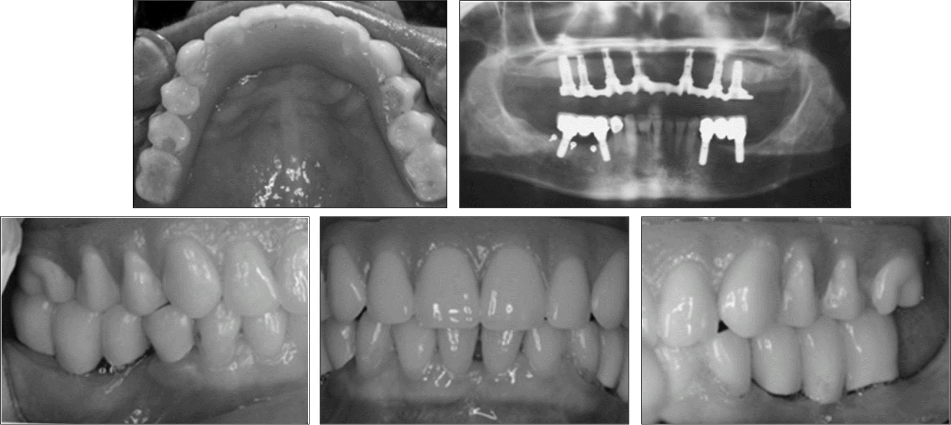
A 47 year-old female patient with severe dental anxiety, high expectations, financial restrictions and poor compliance presented for a fixed rehabilitation. A CT scan with a radiographic template obtained with software (SimPlant, Materialize, Leuven, Belgium) was used for treatment planning. The surgical plan was created and converted into a stereolithographic model of the maxilla with bone-supported surgical templates (SurgiGuide, Materialise, Leuven, Belgium), that allowed for the precise placement of 7 implants in a severely resorbed edentulous maxilla. After successful osseointegration, an accurate scan model served as the basis for the fabrication of a one-piece milled titanium framework using the Procera (Nobel Biocare, Gothenburg, Sweden) technology. The final rehabilitation of the edentulous maxilla was rendered in the form of a screw-retained maxillary metal-reinforced resin-based complete prosthesis.
RESULTS
Despite challenging circumstances, 7 implants could be placed without bone augmentation in a severely resorbed maxilla using the SimPlant software for pre-implant analysis and the SurgiGuide-system as the surgical template. The patient was successfully restored with a fixed full arch restoration, utilizing the Procera system for the fabrication of a milled titanium framework.
REFERENCES
1.McGarry TJ., Nimmo A., Skiba JF., Ahlstrom RH., Smith CR., Koumjian JH., Arbree NS. Classification system for partial edentulism. J Prosthodont. 2002. 11:181–93.

2.Bra ° nemark PI. Osseointegration and its experimental background. J Prosthet Dent. 1983. 50:399–410.
3.Jemt T., Ba ¨ ck T., Petersson A. Precision of CNC-milled titanium frameworks for implant treatment in the edentulous jaw. Int J Prosthodont. 1999. 12:209–15.
4.Shor A., Goto Y., Schuler RF. Rehabilitation of the edentulous mandible with a fixed implant-supported prosthesis. Pract Proced Aesthet Dent. 2004. 16:729–36.
5.Jemt T. Failures and complications in 391 consecutively inserted fixed prostheses supported by Bra ° nemark implants in edentulous jaws: a study of treatment from the time of prosthesis placement to the first annual checkup. Int J Oral Maxillofac Implants. 1991. 6:270–6.
6.Tan KB., Rubenstein JE., Nicholls JI., Yuodelis RA. Three-dimensional analysis of the casting accuracy of one-piece, osseointegrated implant-retained prostheses. Int J Prosthodont. 1993. 6:346–63.
7.Kan JY., Rungcharassaeng K., Bohsali K., Goodacre CJ., Lang BR. Clinical methods for evaluating implant framework fit. J Prosthet Dent. 1999. 81:7–13.

8.Ortorp A., Jemt T., Ba ¨ ck T., Ja ¨ levik T. Comparisons of precision of fit between cast and CNC-milled titanium implant frameworks for the edentulous mandible. Int J Prosthodont. 2003. 16:194–200.
9.Ortorp A., Jemt T. Clinical experiences of computer numeric control-milled titanium frameworks supported by implants in the edentulous jaw: a 5-year prospective study. Clin Implant Dent Relat Res. 2004. 6:199–209.


 PDF
PDF ePub
ePub Citation
 Print
Print



 XML Download