

 PDF
PDF ePub
ePub Citation
 Print
Print


Abstract
Construction of an obturator for rehabilitation of a patient who underwent a maxillectomy is vital. Routinely a constructed obturator includes denture portion. A patient who may present anatomical limitations due to surgical or radiotherapy complications often challenges the clinician. Purpose: This clinical report describes a patient with severe trismus after surgical resection and radiotherapy treatment of a tumor in the upper left maxilla. Conclusion: This report describes the concepts of using a rotational path insertion for an obturator and a separately constructed maxillary denture. The stability and retention of the obturator were obtained from anatomical features. Where as these were achieved through magnetic attachments and the remaining edentulous ridge to ensure esthetic and function of the prosthesis.
Go to : 
REFERENCES
1.Aramany MA. Basic principles of obturator design for partially edentulous patients. Part II: Design principles. 1978 [classical article]. J Prosthet Dent. 2001. 86:562–8.
2.Brown KE. Peripheral consideration in improving obturator retention. J Prosthet Dent. 1968. 20:176–81.

3.Kademani D., Bell RB., Schmidt BL., Blanchaert R., Fernandes R., Lambert P., Tucker WM. Oral and maxillofacial surgeons treating oral cancer: a preliminary report from the American Association of Oral and Maxillofacial Surgeons Task Force on Oral Cancer. J Oral Maxillofac Surg. 2008. 66:2151–7.
4.Sciubba JJ., Goldenberg D. Oral complications of radiotherapy. Lancet Oncol. 2006. 7:175–83.
5.Wang CJ., Huang EY., Hsu HC., Chen HC., Fang FM., Hsiung CY. The degree and time-course assessment of radiation-induced trismus occurring after radiotherapy for nasopharyngeal cancer. Laryngoscope. 2005. 115:1458–60.
6.Dijkstra PU., Kalk WW., Roodenburg JL. Trismus in head and neck oncology: a systematic review. Oral Oncol. 2004. 40:879–89.
7.Boucher LJ., Heupel EM. Prosthetic restoration of a maxilla and associated structures. J Prosthet Dent. 1966. 16:154–68.
8.Javid N. The use of magnets in a maxillofacial prosthesis. J Prosthet Dent. 1971. 25:334–41.
9.Tokuhisa M., Matsushita Y., Koyano K. In vitro study of a mandibular implant overdenture retained with ball, magnet, or bar attachments: comparison of load transfer and denture stability. Int J Prosthodont. 2003. 16:128–34.
10.Gillings BR., Samant A. Overdentures with magnetic attachments. Dent Clin North Am. 1990. 34:683–709.
11.Thean HP., Khor SK., Loh PL. Viability of magnetic denture retainers: a 3-year case report. Quintessence Int. 2001. 32:517–20.
12.Beumer J., Curtis D., Firtell D. Restoration of acquiredhard palatal defects: etiology, disability and rehabilitation. In: Maxillofacial rehabilitation: prosthodontics and surgical considerations. Beumer J III, Curis TA, Marunick MT. St. Louis: Medico Dental Media Int;1996. p. 225–84.
Go to :
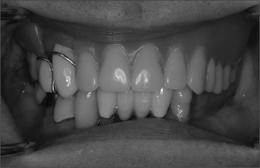
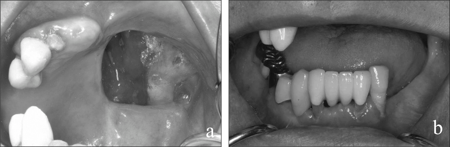 | Fig. 1. aThe occlusal view of the healed maxilla after a subtotal maxillectomy and radiotherapy. b. Limited maximum intercanine distance even after use of tongblades to improve mouth opening due to trismus. |
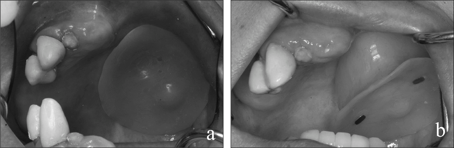 | Fig. 2 . aIntraorally fully seated obturator. b. The obturator at mid of its medio-anterior rotational insertion path. |
 XML Download