

 PDF
PDF ePub
ePub Citation
 Print
Print


INTRODUCTION
Pretransplant human leukocyte antigen (HLA) crossmatch (XM) is performed in solid organ transplantation patients to detect the presence of HLA alloantibodies reacting with donor HLA antigens. Complement-dependent cytotoxicity (CDC) XM is widely used, and it is well known that positive result in T-cell CDC XM against donor is associated with hyperacute rejection in renal transplantation. More recently, flow cytometric XM (flow XM) is increasingly used, which is a more sensitive method than CDC XM. Whereas CDC XM detects both immunoglobulin G (IgG) and IgM antibodies, flow XM detects IgG antibodies by using antihuman IgG fluorochrome conjugate.
In Korea, organ sharing among transplant candidates awaiting transplants from deceased donors is operated by the Korean Network for Organ Sharing (KONOS). At present, there are 36 hospital-based organ procurement organizations (HOPOs), and 21 laboratories are performing KONOS XMs using frozen-stored sera from solid organ transplant candidates. Majority of KONOS XMs are performed for kidney and/or pancreas transplants, and occasionally KONOS XMs are performed for heart or lung transplants. Sera from transplant candidates are renewed once a year (recently changed to every 2 years) and distributed to the laboratories performing KONOS XMs. Pretransplant KONOS T-cell XMs are performed between lymphocytes of deceased donors and stored sera of selected candidates (usually 15~25 per donor) by using (1) standard National Institute of Health (NIH) method, and (2) at least one of more sensitive methods. For the more sensitive methods, most of the HOPO laboratories are using antihuman globulin (AHG) CDC method, and a few laboratories are using flow cytometric method. Current KONOS policy for organ allocation is that negative result in pretransplant T-cell XM is a prerequisite for kidney and pancreas allocation.
We found that transplant candidates showing NIH-positive and AHG-negative (NIH+/AHG−) results in KONOS T-cell XMs are repeatedly excluded from organ allocation. In our experience, we have accumulated laboratory evidence that such XM results are mostly due to IgM type autoantibodies. However, laboratory procedures for verification of IgM autoantibodies cannot be performed for KONOS XMs. In other words, patient's autocontrol XM (patient's cell+patient's serum) cannot be included and test for dithiothreitol (DTT)-treated serum is not feasible in KONOS XM tests. IgM autoantibodies have been reported to be not associated with adverse transplantation outcomes(12). However, because IgM autoantibodies react with both patient's own lymphocytes and random donor lymphocytes(3), patients with these antibodies show positive cytotoxicity XM results with multiple donors, and they are repeatedly excluded from organ allocation. The aims of the study were to prove that these patients do not have donor-specific HLA antibodies (DSAs) and to change the current organ allocation policy of the KONOS.
MATERIALS AND METHODS
1. Study population
The study population was from KONOS XMs performed at the Seoul National University Hospital between January 2010 and June 2012, comprising 1,668 tests. From the study population, we selected probable false-positive (NIH+/AHG−) results as the study group, and probable true-positive (NIH−/AHG+ or NIH+/AHG+) results as the positive control group. For the positive control group, only the KONOS XM results of transplant candidates registered from our hospital were selected, for whom panel reactive antibody (PRA) data were available from electronic medical records review.
Further characteristics of sera showing NIH+/AHG− results in KONOS XMs are not available, such as the results of autocontrol XM, flow XM, etc. Thus, to delineate the nature of the sera showing such XM results, we analyzed additional XM characteristics of the sera of our hospital patients showing NIH+/AHG− results in preliminary or final XMs performed for organ transplantations between 2010 and 2011. The present study was approved by the Institutional Review Board of Seoul National University Hospital (number H-1207-156-422).
2. HLA crossmatch tests
T-cell CDC XMs using standard NIH and AHG methods were performed as previously described(4). For cell and serum incubation, various temperatures (4℃, 22℃ [i.e., room temperature], or 37℃) are used, and for complement incubation, 22℃ is used(4). We used room temperature for cell and serum incubation for both NIH and AHG methods. For KONOS XMs, only T-cell CDC XMs were performed using serum dilutions of 1:1, 1:2, and 1:4 in duplicate; autocontrol could not be included and DTT treatment procedure to reduce (fragment) IgM antibodies was not performed. Different from KONOS XM tests, routine XM tests for our hospital patients included both T-cell CDC XMs and T and B cell flow XMs. For T-cell CDC XMs, serum dilutions from 1:1 to 1:32 in duplicate were tested and autocontrol was routinely included to detect the presence of autoantibodies. For some of the cases showing positive reactions in autocontrol XMs, DTT-treated (5 mM in final concentration) sera were additionally tested to verify the presence of IgM autoantibodies. T and B cell flow XMs were performed by using 3-color immunofluorescence staining of pronase-treated lymphocytes as previously described(5).
3. HLA antibody tests
HLA antibody tests were performed using Luminex method. For the study group showing probable false-positive (NIH+/AHG−) results, prospective PRA screen test was performed and screen-positive samples were subjected to single antigen test to identify the class I antibody specificities (Table 1). For the positive control group showing probable true-positive (NIH−/AHG+ or NIH+/AHG+) results, PRA data were retrospectively reviewed from electronic medical records in our hospital. For these patients, class I antibody identification data were available either from single antigen test or PRA identification test. PRA screening test was performed using LIFECODES LifeScreen Deluxe kit (Gen-Probe, Stamford, CT, USA) according to manufacturer's instruction. For antibody identification, Luminex single antigen class I test (Gen-Probe) or class I ID test (Gen-Probe) were used. HLA types of the patients and donors were reviewed either from KONOS data or from electronic medical records and the presence of DSAs was determined from donor's HLA antigens and patient's HLA antibody specificities.
4. Statistical analysis
HLA XM or PRA positive rates between different groups were compared using Pearson chi-square test or Fisher exact test as appropriate. SPSS for Windows version 12.0 (SPSS Inc., Chicago, IL, USA) was used for statistical analysis. A two-tailed P-value of <0.05 was considered statistically significant.
RESULTS
1. Results of KONOS T-cell CDC XM tests
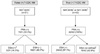
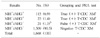
Results of KONOS T-cell CDC XM tests during the study period are shown in Table 1. Positive T-cell CDC XM results were divided into probable false-positive (NIH+/AHG−) and true-positive groups (NIH−/AHG+ or NIH+/AHG+), and their PRA and DSA results are depicted in Fig. 1. NIH+/AHG− results were detected in 21 out of 1,668 tests, comprising 1.3% of all KONOS XM tests. These 21 results were obtained from 16 different patients, and the NIH titer (1:1~1:4 tested) in these patients were 1:1 in 11 cases, 1:2 in five cases, and 1:4 in five cases. Among these 21 cases, seven (33.3%) were positive for PRA and only three (14.3%) were positive for DSA. All three cases with DSA positivity revealed weak to moderate antibody strength (sum of median fluorescence intensity values of 2,806~6,645), which was not expected to give positive CDC XM result, and the NIH+/AHG− results were not considered to be due to DSAs.
Among 138 cases of probable true-positive group (NIH−/AHG+ or NIH+/AHG+), retrospective PRA data from electronic medical records were available in 42 cases. PRA positive rate in this group was 100% (42/42), which was significantly higher than that in NIH+/AHG− group (P=3.11×10⁻⁹). Of the 42 PRA-positive cases, DSA was defined in 24 (57.1%) and could not be defined in the remaining 18 cases (42.9%) because of high PRA values (mean, 92% [range; 42%~100%]), complicating accurate identification of antibody specificities. Considering their high PRA values, many of them are expected to be positive for DSA.
2. NIH+/AHG− T-cell CDC XM results in hospital patients tested
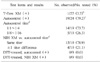
To delineate the characteristics of sera showing NIH+/AHG− XM results, we have reviewed laboratory records from our hospital patients showing such results in preliminary or final XMs performed for organ transplantations, and they are summarized in Table 2. NIH+/AHG− results were observed in 1.0% (27/2,841) of the total T-cell CDC XMs performed during the study period of 2 years for our hospital patients, which was not significantly different from the rate observed in KONOS XMs (vs. 1.3%, P=0.33). These 27 results were obtained from 20 different patients, and four patients showed NIH+/AHG− results against two or more(2-4) donors and at variable time intervals tested, up to >4 months apart. T-flow XM was positive in only one of the 27 cases (3.7%), and the presence of both autoantibody and weak alloantibody (CDC-negative and flow-positive) was suspected in this patient. Autocontrol (XM between patient's cell and patient's serum) was positive in 19 of 24 cases (79.2%) tested, and negative in five cases. All of the five autocontrol-negative cases showed negative T-flow XM results, and these patients might have IgM type donor-specific alloantibodies. The autocontrol XM was of low titer (1:1~1:4) in most cases (14/19, 73.7%), and higher titer (1:8~1:16) was observed less frequently. The titer of test XM (between donor's cell and patient's serum) and that of autocontrol XM was the same in most cases (15/19, 78.9%) and showed 1 titer difference in the rest of cases. After DTT treatment, positive reactions in autocontrol and test XM tests converted to negative in all of the nine cases tested, and the presence of IgM type autoantibodies were verified.
DISCUSSION
We have shown that NIH+/AHG− results were not rare (1.3%) in KONOS T-cell CDC XM tests and most (>85%) of these results were not due to DSAs (Table 1, Fig 1). DSA-positive cases (3/21, 14.3%) also showed antibody strength (median fluorescence intensity values) of weak to moderate intensity, which was not strong enough to be related with positive CDC XMs. Similar rate (1.0%) of NIH+/AHG− results was observed in routine XM tests performed in our laboratory for our hospital patients. Analysis of additional XM characteristics in these cases revealed that majority of them are T-flow XM negative and autocontrol XM positive, and show negative conversion of NIH-positive reactions in test and autocontrol XMs on DTT treatment of the test sera (Table 2). These findings indicate that majority of the NIH+/AHG− results are due to IgM type autoantibodies. However, in some of the cases (5/24, 20.8%), autocontrol XM was negative and the presence of IgM type donor-specific alloantibodies was suspected. Thus, NIH+/AHG− XM results are considered to be mainly due to IgM autoantibodies, and occasionally due to IgM alloantibodies.
The mechanism underlying NIH+/AHG− results in T-cell CDC XM is not well addressed in the literature. In this study, it has been shown that NIH+/AHG− XM results are due to the presence of IgM type autoantibodies or alloantibodies. IgM antibodies tend to have lower affinity than IgG antibodies(6), and might be washed off from binding to cell surface antigens during multiple (usually three times) washing steps before addition of complement in AHG XM procedure, resulting in negative result. On the other hand, IgM antibodies would give positive reactions in NIH XM, because washing step is not included in this procedure. As described in the methods, we carried out cell and serum incubation at room temperature for T-cell CDC XMs, and this incubation temperature is used by majority (>70%) of the histocompatibility laboratories in Korea (7,8). One of our colleagues using 37℃ incubation for cell and serum reaction suggested that they have not observed NIH+/AHG− XM results (personal communication). It is possible that autoantibodies are not binding to autoantigens at 37° warm temperature. This has to be further verified in future studies. If 37° incubation prevents false-positive NIH XM results due to IgM autoantibodies or alloantibodies, 37℃ warm incubation has better be used for KONOS XMs, in which further workup including autocontrol XM or repeated testing on DTT-treated sera cannot be carried out.
Although some controversies exist, IgM autoantibodies are generally known to have no adverse effects on kidney graft outcome(12). In addition, IgM donor-specific HLA alloantibodies have also been shown to exert no adverse effects on long-term graft survival in kidney transplantation(9). Thus, transplant candidates showing NIH+/AHG− results in KONOS XMs should not be excluded from organ allocation, because such XM results are due to IgM autoantibodies or alloantibodies as evidenced in the present study. Because IgM autoantibodies react with both patient's own lymphocytes and most of allogeneic lymphocytes(3), patients with these antibodies show positive XM results with multiple donors and we also have found such patients in this study. Many investigators have shown the presence of IgM antibodies by using DTT treatment(11011). Although further workup for verification of autoantibodies or IgM antibodies can be carried out by including autocontrol XMs and additional testing of DTT-treated sera in routine XM tests for hospital patients, these procedures are not feasible in KONOS XM tests.
We found that transplant candidates, whose KONOS T-cell XM tests were reported as NIH-positive and AHG-negative were repeatedly excluded from allocation of deceased donor organs. Since 2010, we changed the reporting policy in those samples showing such results. We reported NIH-negative and AHG-negative, with a comment that the test actually showed NIH-positive and AHG-negative results, suggesting the presence of autoantibody. Thus, KONOS could allocate organs to these patients. However, majority of the laboratories performing KONOS XM tests are reporting the actual test results (NIH-positive, AHG-negative), although the presence of autoantibody is suspected and these transplant candidates are excluded from organ allocation. Based on the results of the present study, we argue against the current KONOS organ allocation policy and it has to be changed in that T-NIH positive and T-AHG or T-flow negative cases may be allocated organs from deceased donors. For cases showing T-NIH positive and T-AHG or T-flow negative results in KONOS preliminary XM tests, transplant centers had better have a chance of performing final XM test including verification of IgM type autoantibody (autocontrol and XM test using sera with and without DTT treatment). With the final XM results and the DSA information from PRA test results, they can determine whether they will proceed on a transplantation. If KONOS cannot change the allocation policy, laboratories performing KONOS XM tests had better agree on reporting these results as NIH-negative and AHG-negative with comments for probable presence of autoantibody.
CONCLUSION
In this study, we have shown that NIH+/AHG− results are not rare in KONOS T-cell CDC XM tests. Most of these results are due to IgM autoantibodies and not related to donor- specific alloantibodies to be concerned in organ transplantation. KONOS organ allocation policy has to be changed so that transplant candidates with such XM results are not excluded from organ allocation.
 XML Download