Abstract
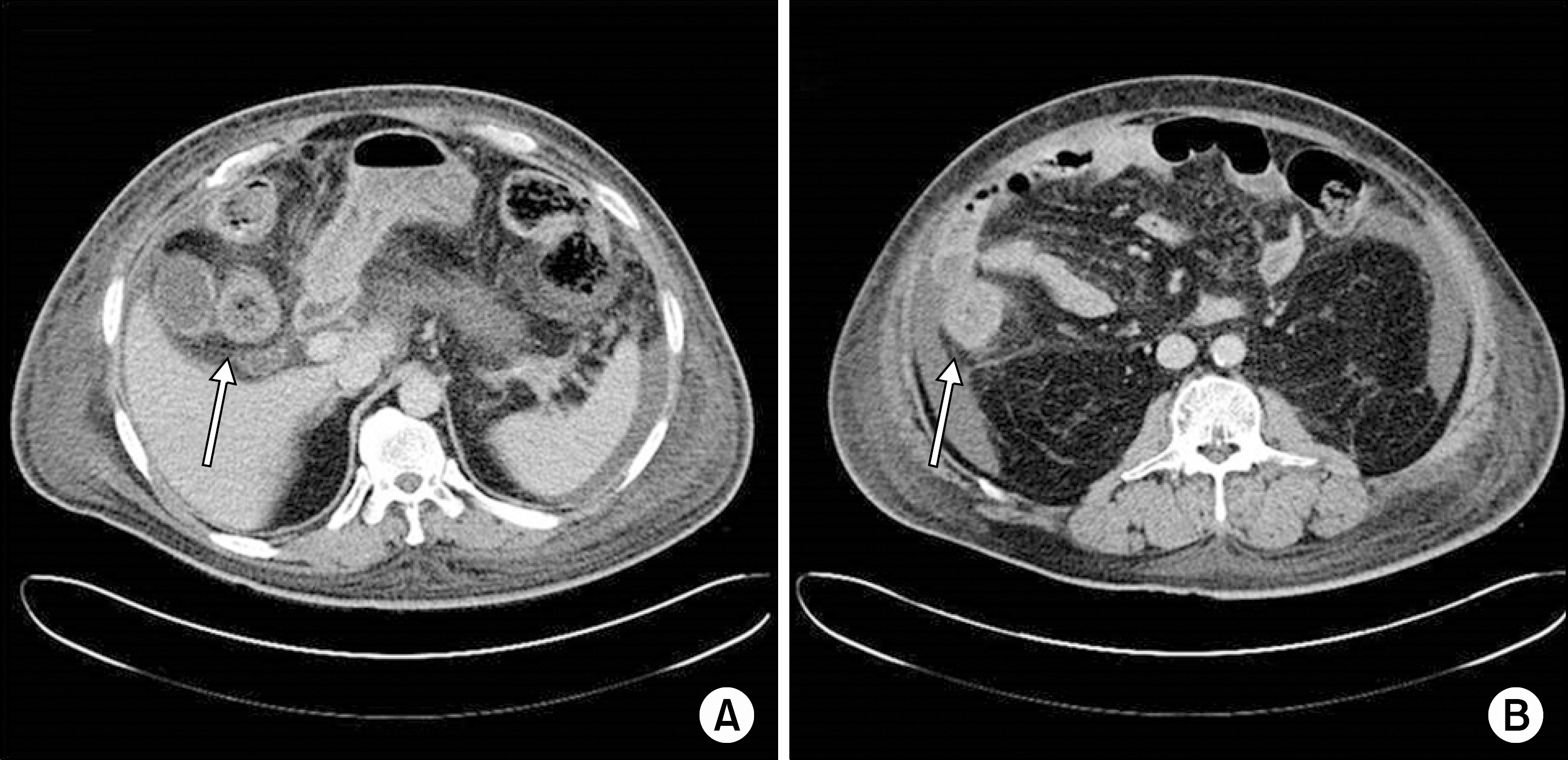
Mucormycosis is an extremely rare but potentially life-threatening fungal infection. Mucormycosis of the gastrointestinal tract manifests with features similar to ischemic colitis. A 48-year-old man with end-stage renal disease due to diabetic nephropathy underwent deceased donor kidney transplantation. He complained of abdominal pain and distension on postoperative day 17. A computed tomography (CT) scan revealed symmetrical wall thickening of the ascending colon, which was consistent with ischemic colitis. However, a follow-up CT scan showed a localized wall-off colon perforation in the hepatic flexure and segmental mural gas in the ascending colon. Microscopic examination obtained from a surgical specimen demonstrated numerous fungal hyphae and spores in the mucosa and submucosa. A total colectomy was performed, but the patient died 36 days later due to multiple organ failure, despite antifungal agents. Clinicians should be informed about fungal infection, such as colonic mucormycosis mimicking ischemic colitis, in kidney transplant patients with diabetes mellitus, and treatment should be initiated at the earliest.
Go to : 
REFERENCES
1). Neofytos D., Treadway S., Ostrander D., Alonso CD., Dierberg KL., Nussenblatt V, et al. Epidemiology, outcomes, and mortality predictors of invasive mold infections among transplant recipients: a 10-year, single-center experience. Transpl Infect Dis. 2013. 15:233–42.

2). Lanternier F., Sun HY., Ribaud P., Singh N., Kontoyiannis DP., Lortholary O. Mucormycosis in organ and stem cell transplant recipients. Clin Infect Dis. 2012. 54:1629–36.

3). Hosseini M., Lee J. Gastrointestinal mucormycosis mimicking ischemic colitis in a patient with systemic lupus erythematosus. Am J Gastroenterol. 1998. 93:1360–2.

4). Zhan HX., Lv Y., Zhang Y., Liu C., Wang B., Jiang YY, et al. Hepatic and renal artery rupture due to Aspergillus and Mucor mixed infection after combined liver and kidney transplantation: a case report. Transplant Proc. 2008. 40:1771–3.

5). Lee DG., Choi JH., Choi SM., Yoo JH., Kim YJ., Min CK, et al. Two cases of disseminated mucormycosis in patients following allogeneic bone marrow transplantation. J Korean Med Sci. 2002. 17:403–6.

6). Suh IW., Park CS., Lee MS., Lee JH., Chang MS., Woo JH, et al. Hepatic and small bowel mucormycosis after chemotherapy in a patient with acute lymphocytic leukemia. J Korean Med Sci. 2000. 15:351–4.

7). Huh JM., Jung GO., Kwon CH., Joh JW., Kim SJ., Lee SK. Invasive gastrointestinal and cutaneous mucormycosis in deceased donor small bowel transplantation: case report and review of literature. J Korean Soc Transplant. 2009. 23:172–6. (허정민, 정금오, 권준혁, 조재원, 김성주, 이석구. 단장증후군 환아에서 사체소장이식 후 Mucormycosis 감염 경험 1예. 대한이식학회지 2009;23: 172-6.).

8). Chkhotua A., Yussim A., Tovar A., Weinberger M., Sobolev V., Bar-Nathan N, et al. Mucormycosis of the renal allograft: case report and review of the literature. Transpl Int. 2001. 14:438–41.

9). Almyroudis NG., Sutton DA., Linden P., Rinaldi MG., Fung J., Kusne S. Zygomycosis in solid organ transplant recipients in a tertiary transplant center and review of the literature. Am J Transplant. 2006. 6:2365–74.

10). Spellberg B., Edwards J Jr., Ibrahim A. Novel perspectives on mucormycosis: pathophysiology, presentation, and management. Clin Microbiol Rev. 2005. 18:556–69.

11). Fogarty C., Regennitter F., Viozzi CF. Invasive fungal infection of the maxilla following dental extractions in a patient with chronic obstructive pulmonary disease. J Can Dent Assoc. 2006. 72:149–52.
12). Severo CB., Guazzelli LS., Severo LC. Chapter 7: zygomycosis. J Bras Pneumol. 2010. 36:134–41.
Go to : 
 | Fig. 1.Abdominopelvic computed tomography scan. Ascites and (A) mild to (B) moderate symmetrical wall thickening (arrows) was observed on the ascending colon. |
 | Fig. 2.Follow-up abdominopelvic computed tomography scan revealed (A) localized wall-off colon perforation in the hepatic flexure (hollow arrow) and (B, C) segmental mural gas in the ascending colon (arrows) and (D) symmetrical wall thickening (arrow) was observed in the descending colon. |
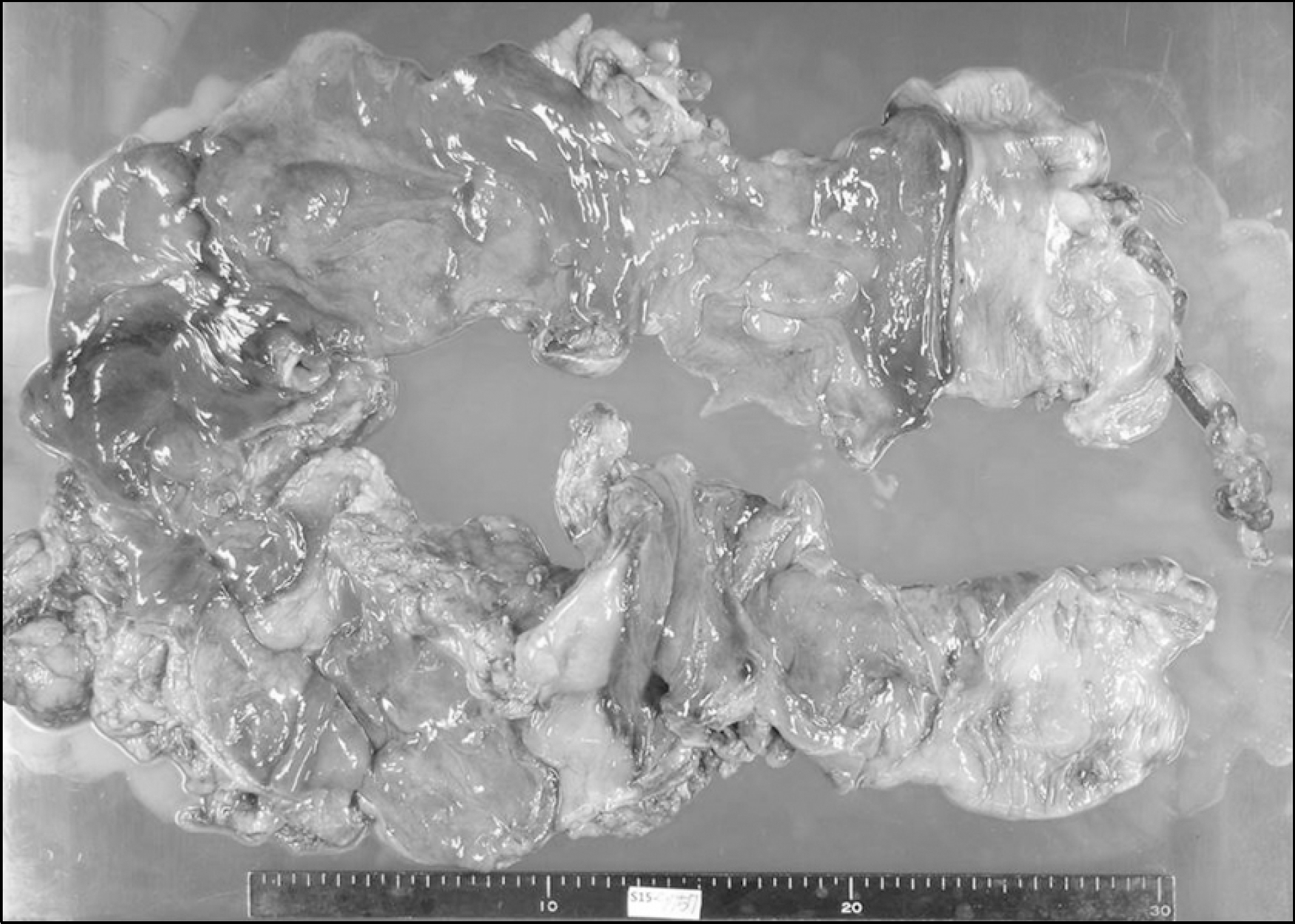
 | Fig. 3.Gross photograph of the total colectomy specimen showed necrotizing colitis from the distal transverse colon to the descending colon. |
 | Fig. 4.Colon pathologic findings. (A) Numerous fungal hyphae and spores are noted in the mucosa and submucosa (arrows) (HE stain, ×200). (B, C, D) Fungal organisms are characterized by large non-septated hyphae with acute angle branching (B: HE stain, ×400; C: PAS, ×400; D: Gomori methenamine silver, ×400). |


 PDF
PDF ePub
ePub Citation
 Print
Print


 XML Download