Abstract
Following liver transplantation, a few reports have documented hepatic venous outflow obstruction (HVOO) after inferior vena cava (IVC) stenting for the treatment of IVC stenosis. However, HVOO occurred early after IVC stenting and was mostly associated with living donor liver transplantation. Here, we report a case of HVOO that occurred 31 months after IVC stenting in a man who received deceased donor liver transplantation (DDLT) using a modified piggyback (PB) technique. The cause of HVOO was unclear, but one possible explanation is that the balloon-expandable IVC stent might have compressed the IVC chamber on the donor liver side, which would have changed the outflow hemodynamics, resulting in intimal hyperplasia. Therefore, simultaneous hepatic venous stenting with IVC stent placement could help prevent HVOO in patients receiving DDLT with the modified PB technique.
REFERENCES
1). Lerut JP., Molle G., Donataccio M., De Kock M., Ciccarelli O., Laterre PF, et al. Cavocaval liver transplantation without venovenous bypass and without temporary portocaval shunting: the ideal technique for adult liver grafting? Transpl Int. 1997. 10:171–9.

2). Jovine E., Mazziotti A., Grazi GL., Ercolani G., Masetti M., Morganti M, et al. Piggyback versus conventional technique in liver transplantation: report of a randomized trial. Transpl Int. 1997. 10:109–12.

3). Belghiti J., Panis Y., Sauvanet A., Gayet B., Fekete F. A new technique of side to side caval anastomosis during orthotopic hepatic transplantation without inferior vena caval occlusion. Surg Gynecol Obstet. 1992. 175:270–2.
4). Weeks SM., Gerber DA., Jaques PF., Sandhu J., Johnson MW., Fair JH, et al. Primary Gianturco stent placement for inferior vena cava abnormalities following liver transplantation. J Vasc Interv Radiol. 2000. 11(2 Pt 1):177–87.

5). Lee JM., Ko GY., Sung KB., Gwon DI., Yoon HK., Lee SG. Long-term efficacy of stent placement for treating inferior vena cava stenosis following liver transplantation. Liver Transpl. 2010. 16:513–9.

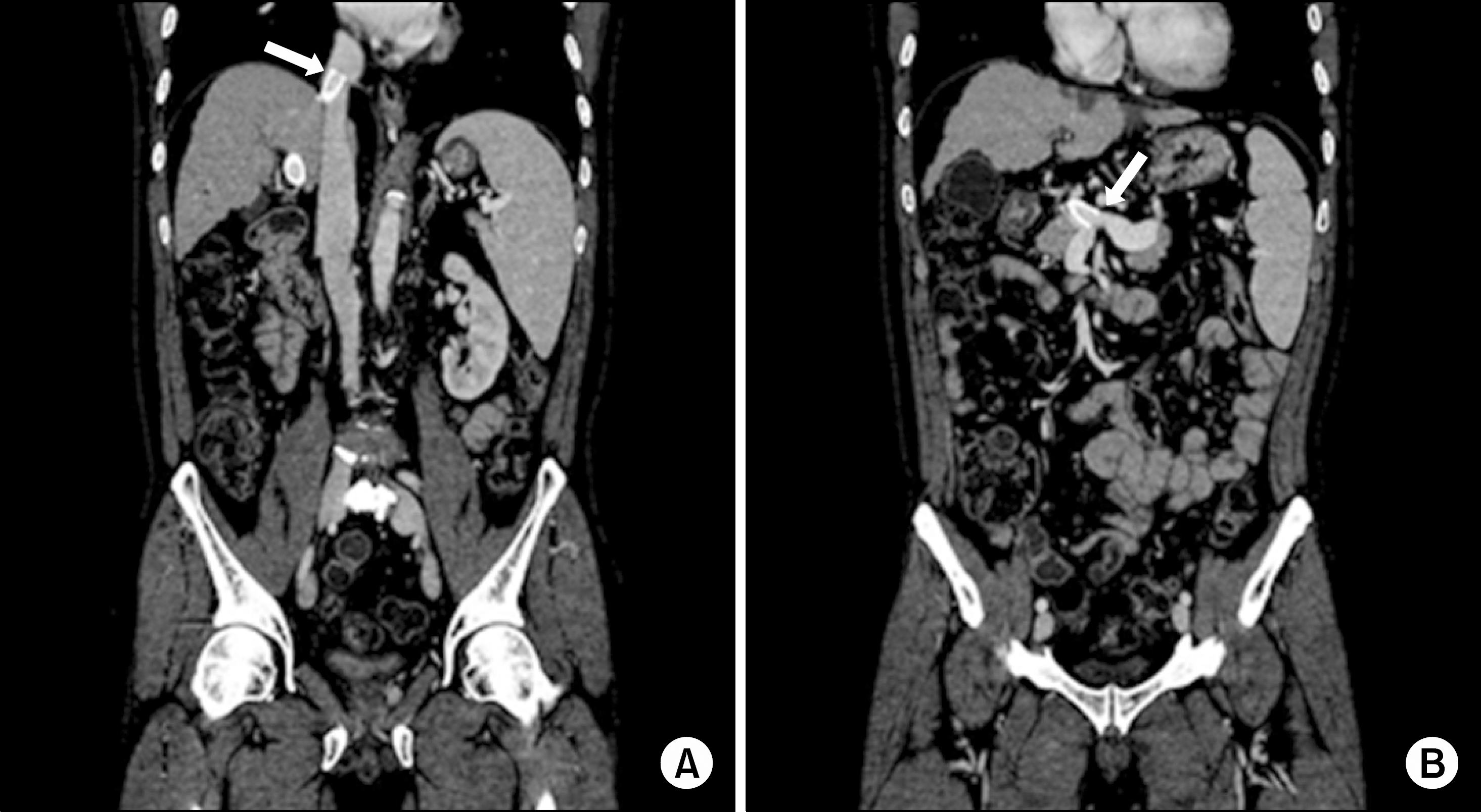
Fig. 1.
Abdominal computed tomography scan of the patient performed prior to receiving deceased donor liver transplantation. (A) The cranial end of the transjugular intrahepatic portosystemic shunt (TIPS) stent was located just below the right atrium, across the suprahepatic vena cava (arrow). (B) The caudal end of TIPS stent was placed at the junction of the inferior mesenteric and splenic veins (arrow).

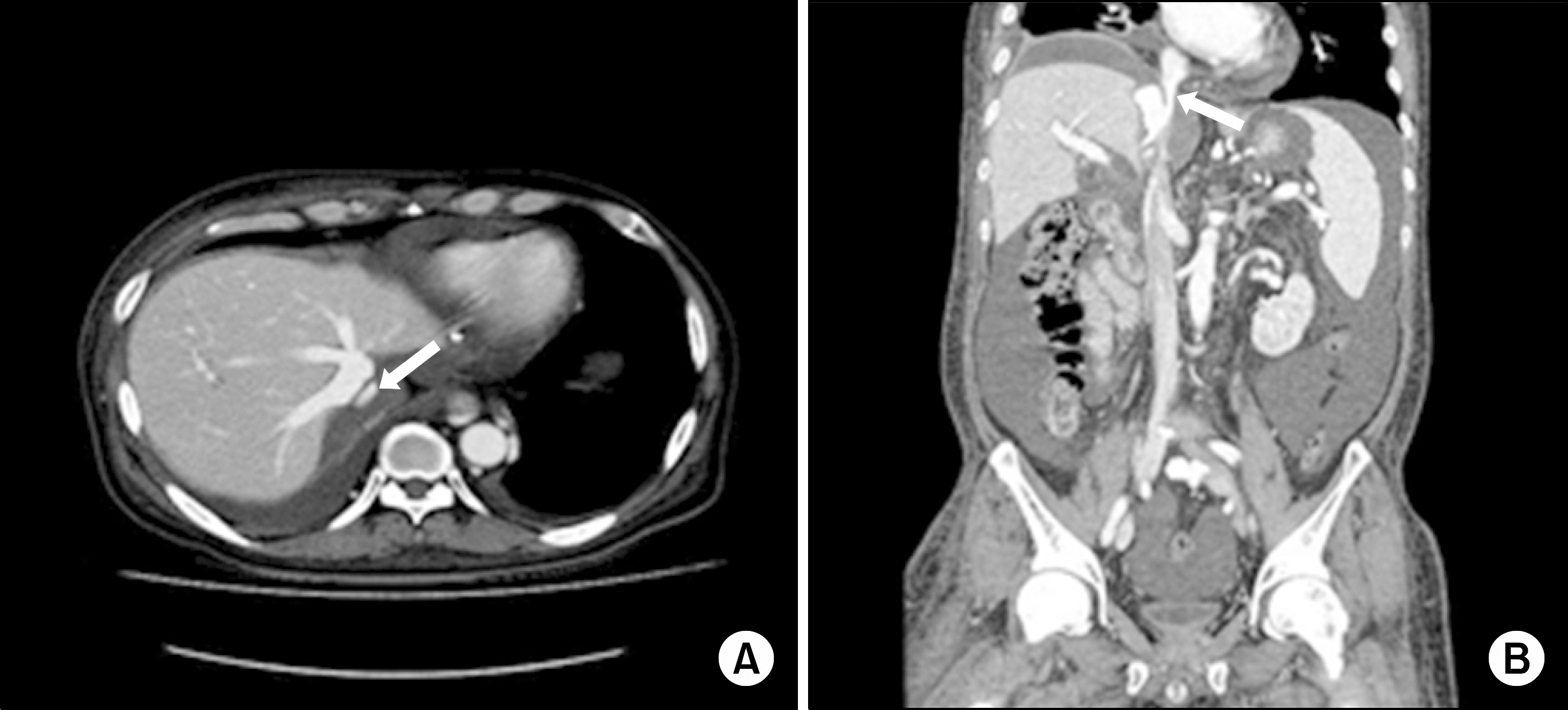
Fig. 2.
This abdominal computed tomography scan of the patient performed 6 weeks after deceased donor liver transplantation shows stenosis of the suprahepatic vena cava (arrows). (A) Axial view. (B) Coronal view.

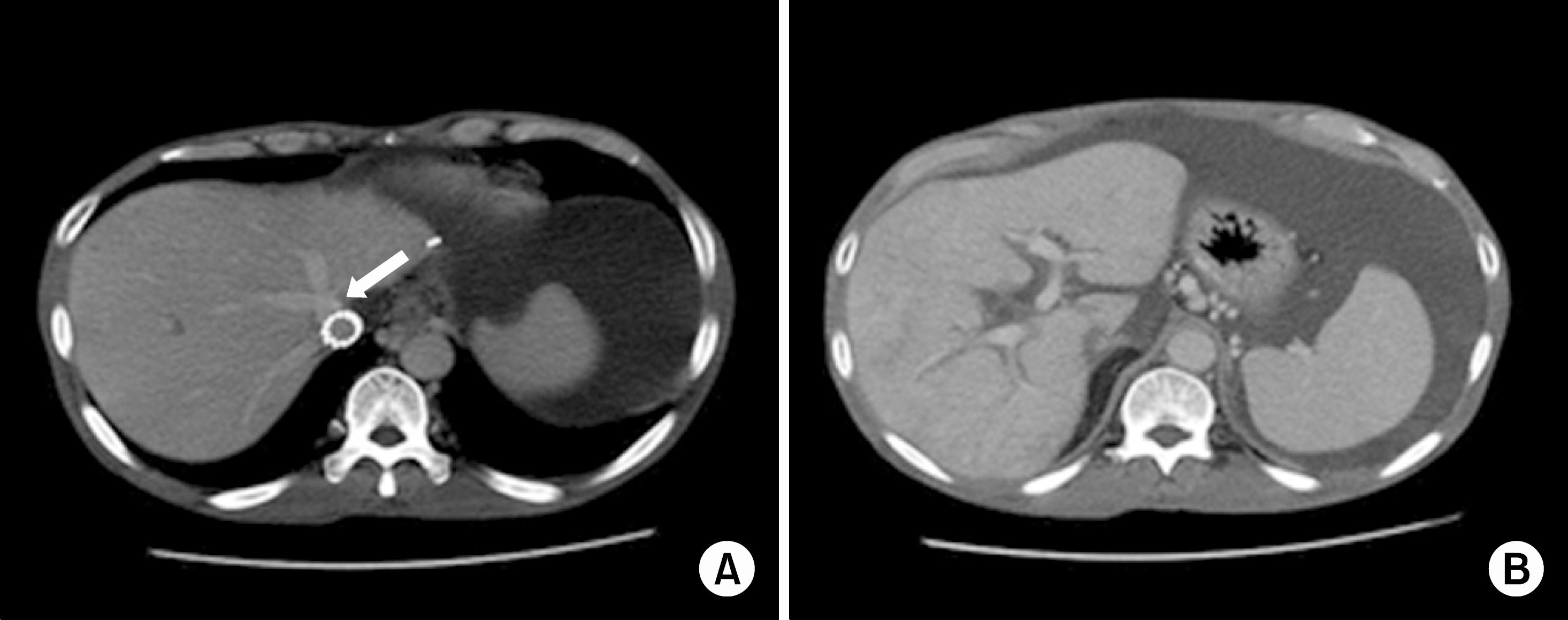
Fig. 3.
This contrast-enhanced abdominal computed tomography (CT) scan shows a collapsed inferior vena cava (IVC) chamber on the donor side, a large amount of ascites fluid and liver graft congestion. (A) In comparison with the previous CT scan, the donor side IVC chamber had collapsed from extrinsic compression of the Palmaz stent (arrow). (B) A mottled pattern of contrast enhancement was observed in the portal venous phase.


 PDF
PDF ePub
ePub Citation
 Print
Print


 XML Download