Abstract
The limited donor organ supply is a main problem for transplant surgeons in Korea, and forces them to use organs from extended sources. In one such case, we reused a transplanted kidney allograft in August 2012. This was the first successful case involving the reuse of a transplanted kidney allograft in Korea. The kidney donor was a 44-year-old man brain-dead due to spontaneous subdural hemorrhage. He received a kidney transplant from his sister in 2006. The second recipient was a 59-year-old man who had been receiving hemodialysis for 11 years. There were full human leukocyte antigen (HLA) matches between the first donor and the first recipient, and two HLA mismatches between the first donor and the second recipient. Fortunately, we were able to perform a crossmatch test between the first donor and the second recipient as well as the first recipient and the second recipient (with the first donor’ s agreement). We used the left iliac artery for perfusion instead of the aorta during organ procurement. The cold ischemic time was 4 hours and the initial kidney function was excellent. The patient has been doing well, without any significant complications or rejections, for 3 weeks. His last serum creatinine level was 0.91 mg/dL. Our case shows that the reuse of kidney allografts could be a possible solution for the shortage of donor kidneys. However, this method requires careful consideration and an agreement among participants before its performance.
REFERENCES
1). Lucarelli G, Bettocchi C, Battaglia M, Impedovo SV, Vavallo A, Grandaliano G, et al. Extended criteria donor kidney transplantation: comparative outcome analysis between single versus double kidney transplantation at 5 years. Transplant Proc. 2010; 42:1104–7.

2). Perico N, Ruggenenti P, Scalamogna M, Remuzzi G. Tackling the shortage of donor kidneys: how to use the best that we have. Am J Nephrol. 2003; 23:245–59.

3). Rentsch M, Meyer J, Andrassy J, Fischer-Fröhlich CL, Rust C, Mueller S, et al. Late reuse of liver allografts from brain-dead graft recipients: the Munich experience and a review of the literature. Liver Transpl. 2010; 16:701–4.

4). Graetz K, Cunningham D, Rigg K, Shehata M. Expansion S of the organ donor pool by the reuse of a previously transplanted kidney: is this ethically and scientifically val-id? Transplant Proc. 2002; 34:3102–3.
5). Celik A, Saglam F, Cavdar C, Sifil A, Gungor O, Bora S, et al. Successful reuse of a transplanted kidney: 3-year follow-up. Am J Kidney Dis. 2007; 50:143–5.

Fig. 1.
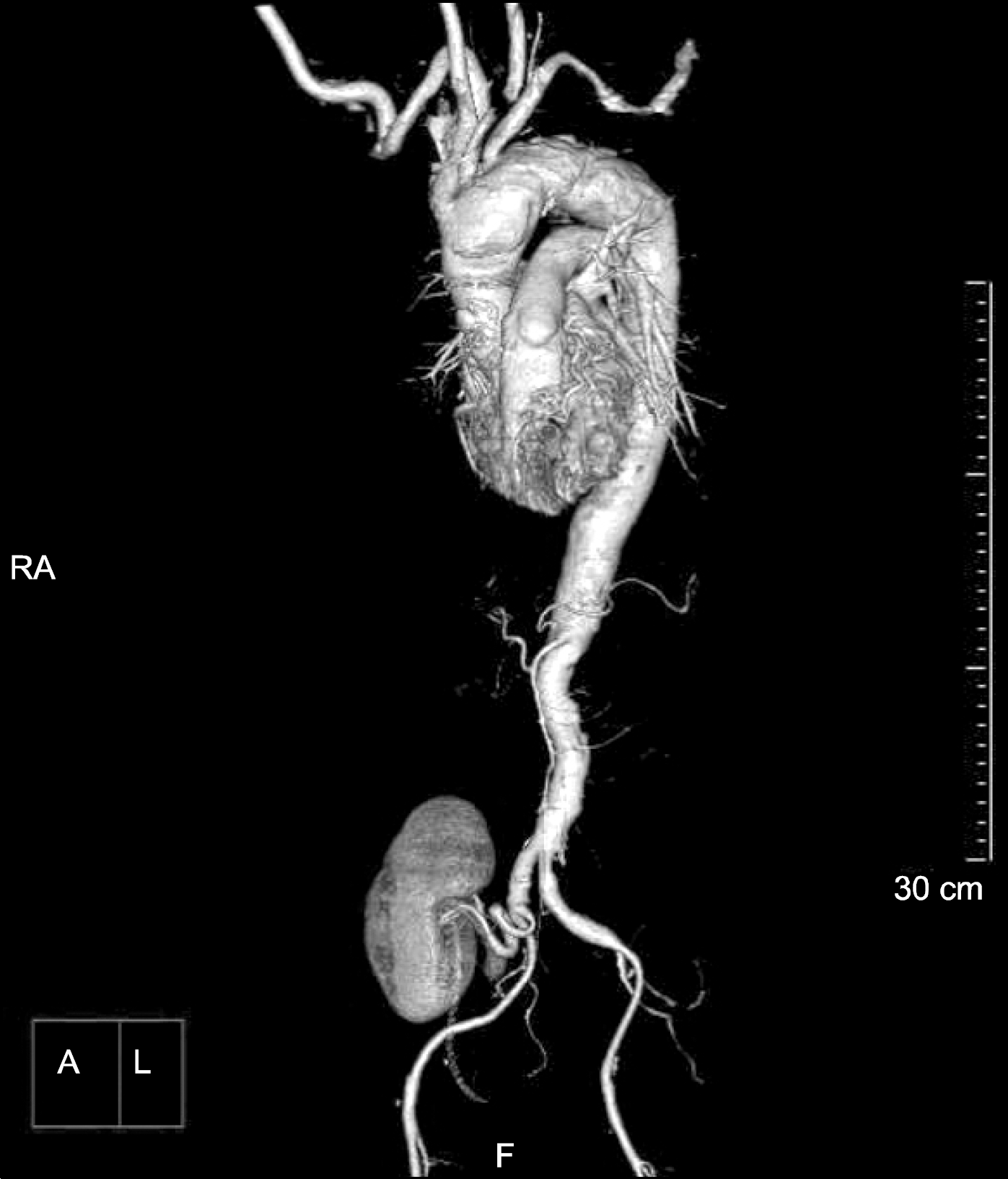
Catheter insertion for perfusate infusion. We inserted the catheter via left iliac artery and clamped the supraceliac aorta and right iliac artery at distal to kidney transplantation. Portal catheter was inserted via inferior mesenteric vein. Dot-line is the arterial shapes.
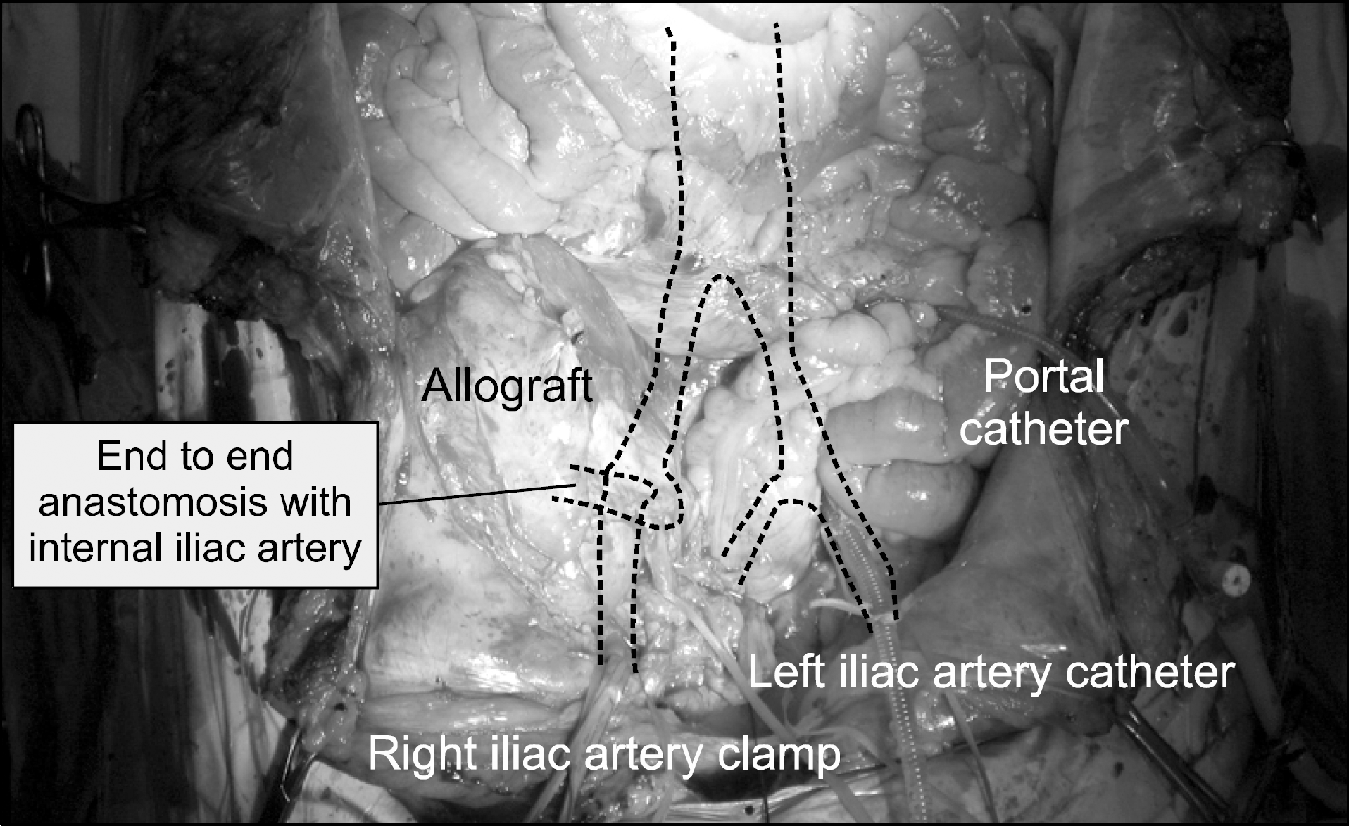
Fig. 2.
Harvested kidney allograft. Kidney was harvested with donor’ s right iliac artery and vein. A black arrow indicates the renal vein surrounding by donor’s iliac vein.
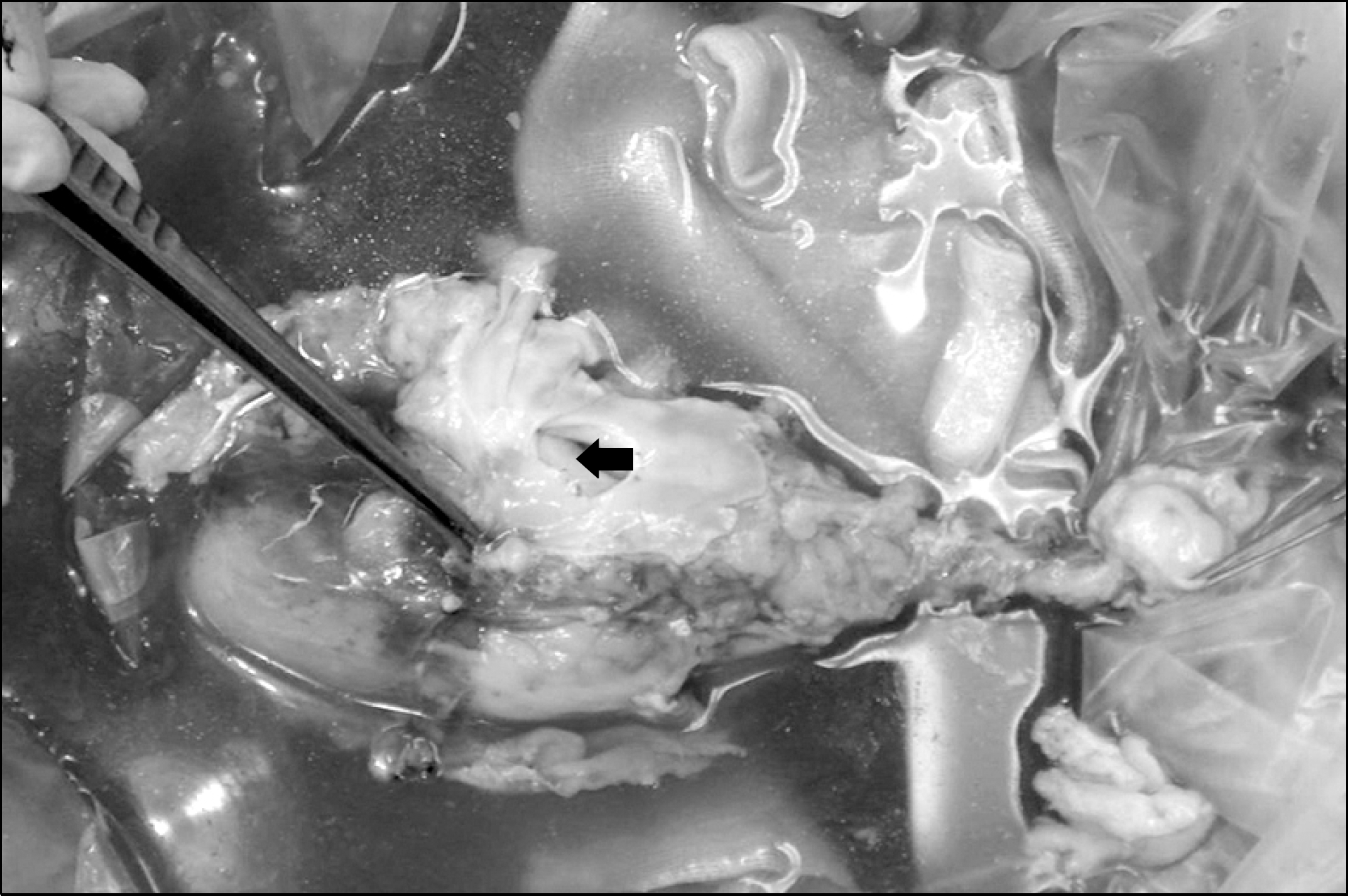
Fig. 3.
Operative findings of transplantation. Renal artery was reconstructed by end to end anastomosis between donor’ s iliac artery and recipient’s right internal iliac artery. Renal vein was reconstructed by end to side anastomosis between donor’s iliac vein patch and recipient’s right iliac vein. Ureteroneocystostomy was done with double J stent. A black arrow indicates the anastomosis of donor’ s renal artery and recipient’ s internal iliac arery. Abbreviations: IIA, internal iliac artery of recipient; EIA, external iliac artery of recipient; UT, ureter.
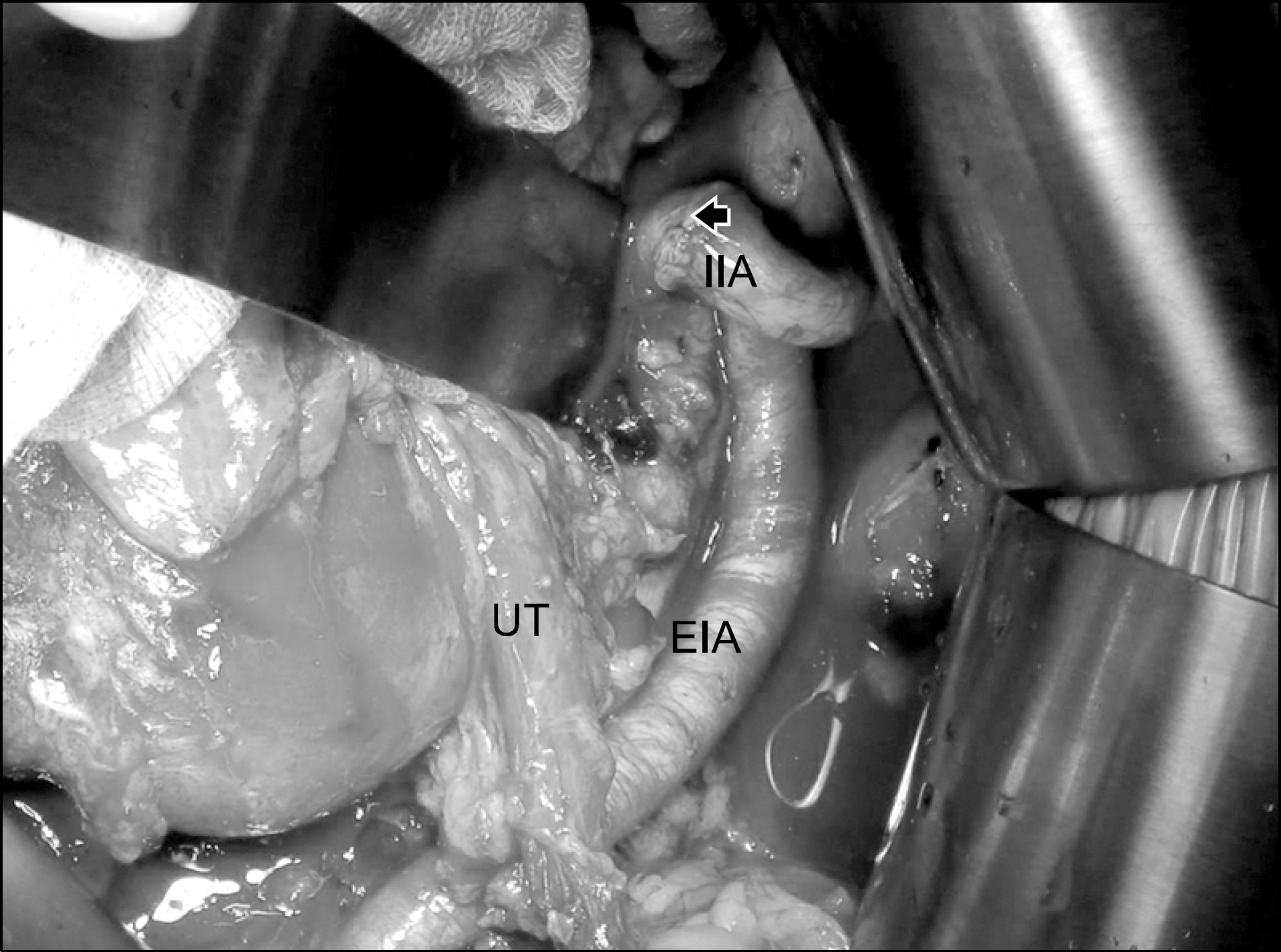
Fig. 5.
Microscopic findings of kidney biopsy. There is no abnor-mality except the hypertrophied glomerulus (PAS, ×200).
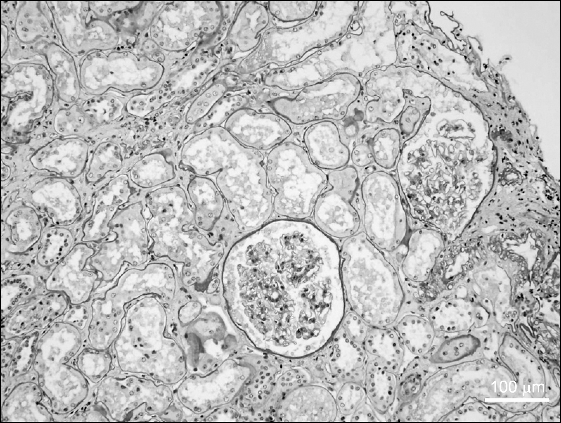
Table 1.
Brief clinical characteristics of first donor, first recipient, and second recipient


 PDF
PDF ePub
ePub Citation
 Print
Print


 XML Download