

 PDF
PDF ePub
ePub Citation
 Print
Print


Abstract
Although gallbladder (GB) perforation due to acalculous cholecystitis after kidney transplantation is rarely observed, it can be life threatening and result in cholecystectomy. Coronary artery aneurysm (CAA) is also rare and may require invasive therapy depending on its diameter. We report herein the case of a 69-year-old female who developed GB perforation due to acalculous cholecystitis after kidney transplantation and underwent cholecystectomy. The patient was later invasively treated when CCA was detected by coronary angiography.
REFERENCES
1). Lowell JA, Stratta RJ, Taylor RJ, Bynon JS, Larsen JL, Nelson NL. Cholelithiasis in pancreas and kidney transplant recipients with diabetes. Surgery. 1993; 114:858–63.
2). Hopkinson GB, Crowson MC, Barnes AD. Perforation of the acalculous gallbladder following renal transplantation. Transplant Proc. 1985; 17:2014–5.
3). Syed M, Lesch M. Coronary artery aneurysm: a review. Prog Cardiovasc Dis. 1997; 40:77–84.

4). Banerjee P, Houghton T, Walters M, Kaye GC. Giant right coronary artery aneurysm presenting as a media-stinal mass. Heart. 2004; 90:e50.
5). Becker CG, Dubin T, Glenn F. Induction of acute cholecystitis by activation of factor XII. J Exp Med. 1980; 151:81–90.
6). Barie PS, Eachempati SR. Acute acalculous cholecystitis. Gastroenterol Clin North Am. 2010; 39:343–57.
7). Lorber MI, Van Buren CT, Flechner SM, Williams C, Kahan BD. Hepatobiliary and pancreatic complications of cyclosporine therapy in 466 renal transplant recipients. Transplantation. 1987; 43:35–40.
8). Satran A, Bart BA, Henry CR, Murad MB, Talukdar S, Satran D, et al. Increased prevalence of coronary artery aneurysms among cocaine users. Circulation. 2005; 111:2424–9.
9). Wan S, LeClerc JL, Vachiery JL, Vincent JL. Cardiac tamponade due to spontaneous rupture of right coronary artery aneurysm. Ann Thorac Surg. 1996; 62:575–6.
10). Chia HM, Tan KH, Jackson G. Non-atherosclerotic cor-onary artery aneurysms: two case reports. Heart. 1997; 78:613–6.
Fig. 1.
Abdomen and pelvic com-puted tomography findings of the patient. (A) Cross section image revealing the irregular mucosa of gallbladder (GB) with suspicious wall defects and distended GB without definite radiopaque stone in GB. (B) Coronal section image showing a small amount of ascites in the perihepatic space and bowel ileus.
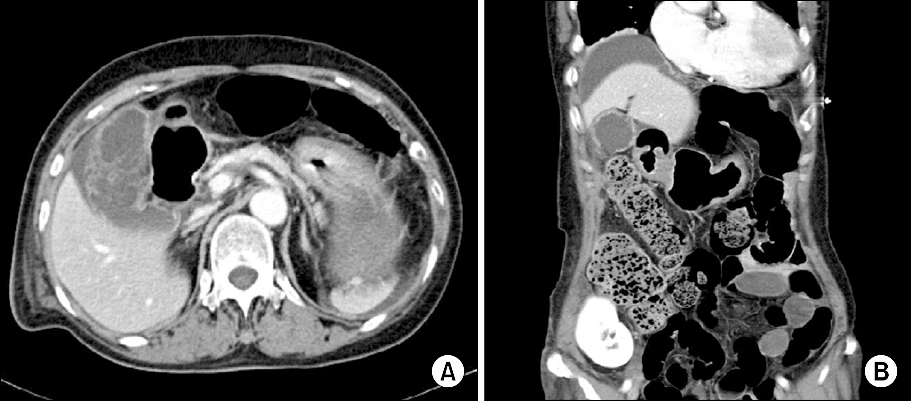
Fig. 2.
This gallbladder (GB) specimen consists of a previously opened GB measuring 10 cm in length and 4.5 cm in diameter. The serosa is dull gray tan and the mucosa is diffusely necrotic in appearance. There is no tumorous appearance and no gall-stones, GB sludge nor GB polyps in this specimen.
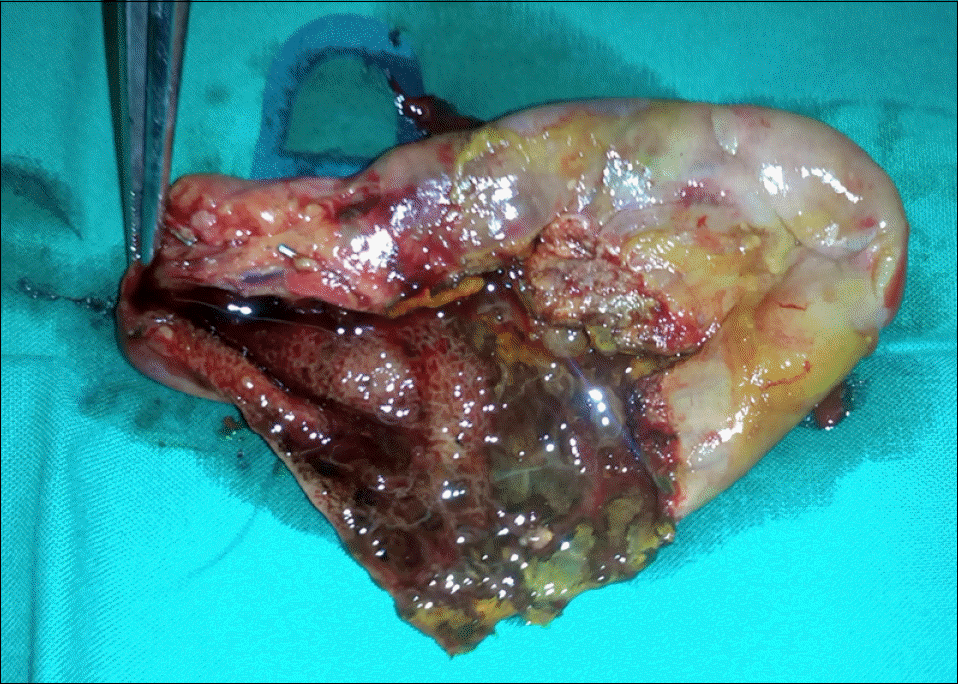
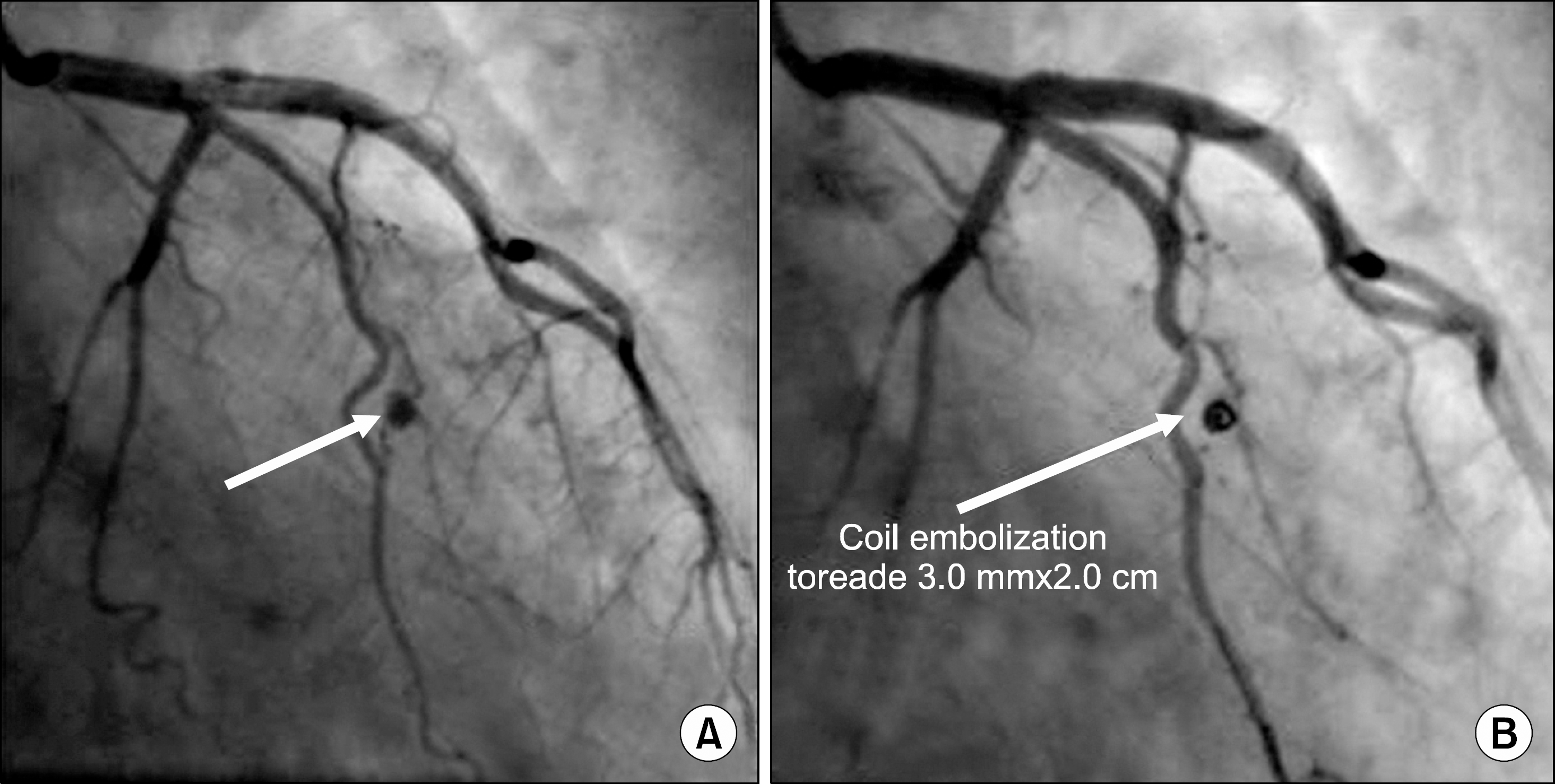
Fig. 3.
Coronary angiographic findings of the patient. (A) The short arrow indicates saccular aneurysmal change in the distal part of the obtuse marginal branch of the left circumflex artery. (B) The long arrow indicates the 3.0 mm×2.0 cm-sized tornado coil located in the aneurysmal sac. After this em-bolization, no more contrast agent filling the aneurysmal sac was observed and distal flow below aneurysmal sac disappeared.
 XML Download