Abstract
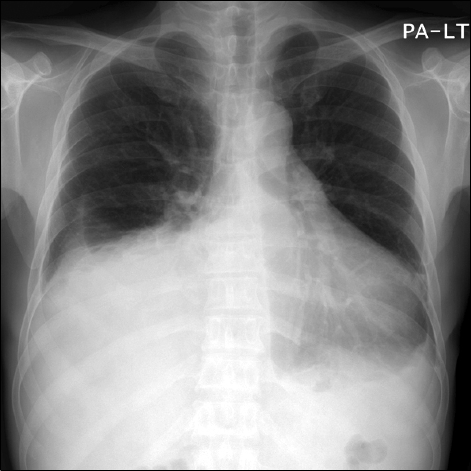
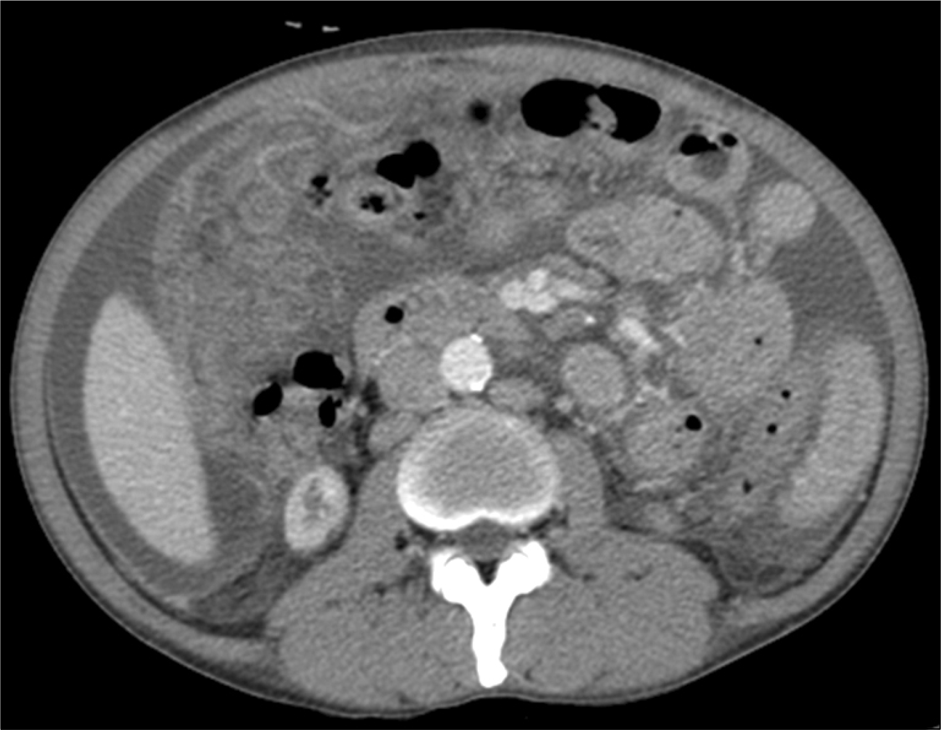
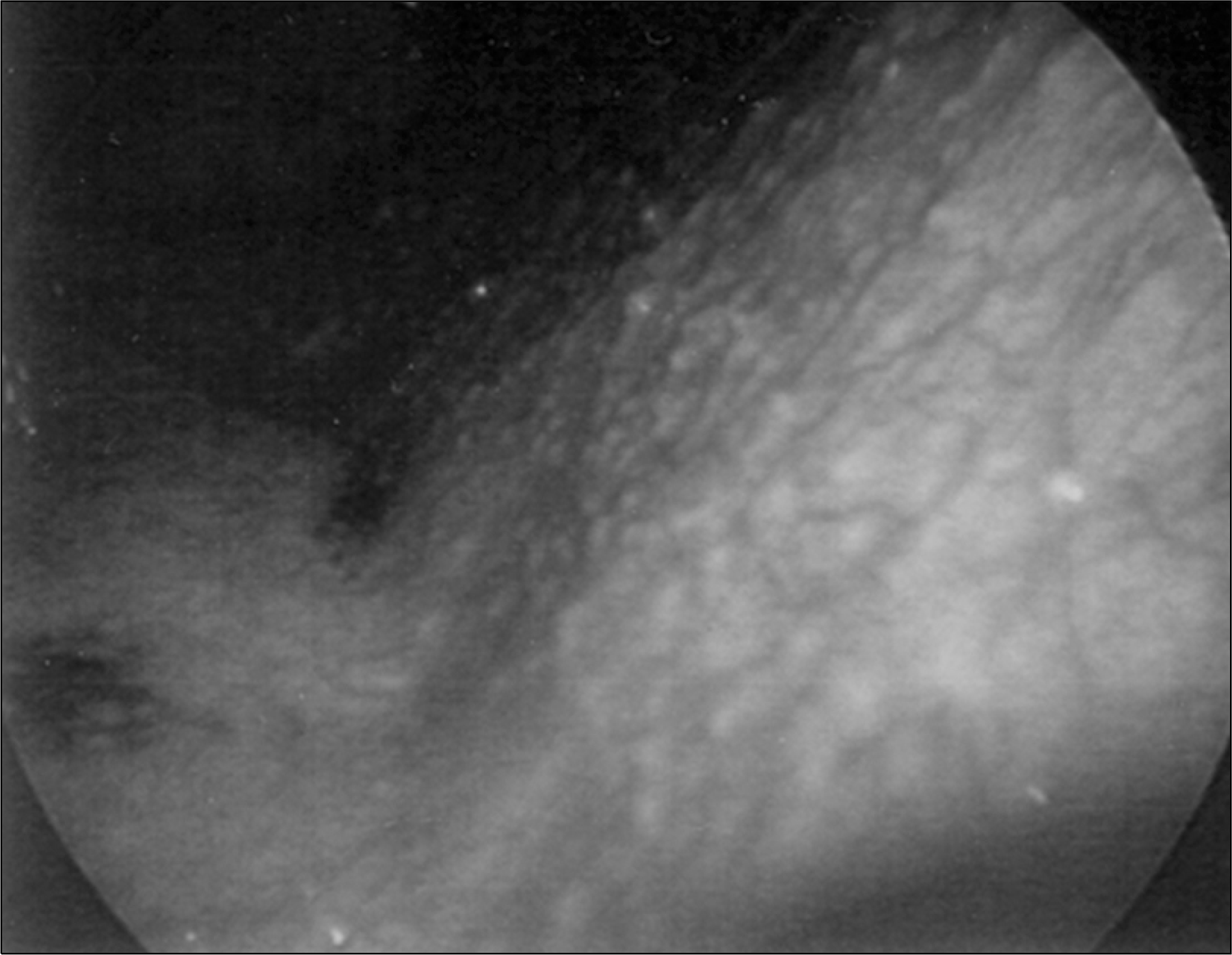
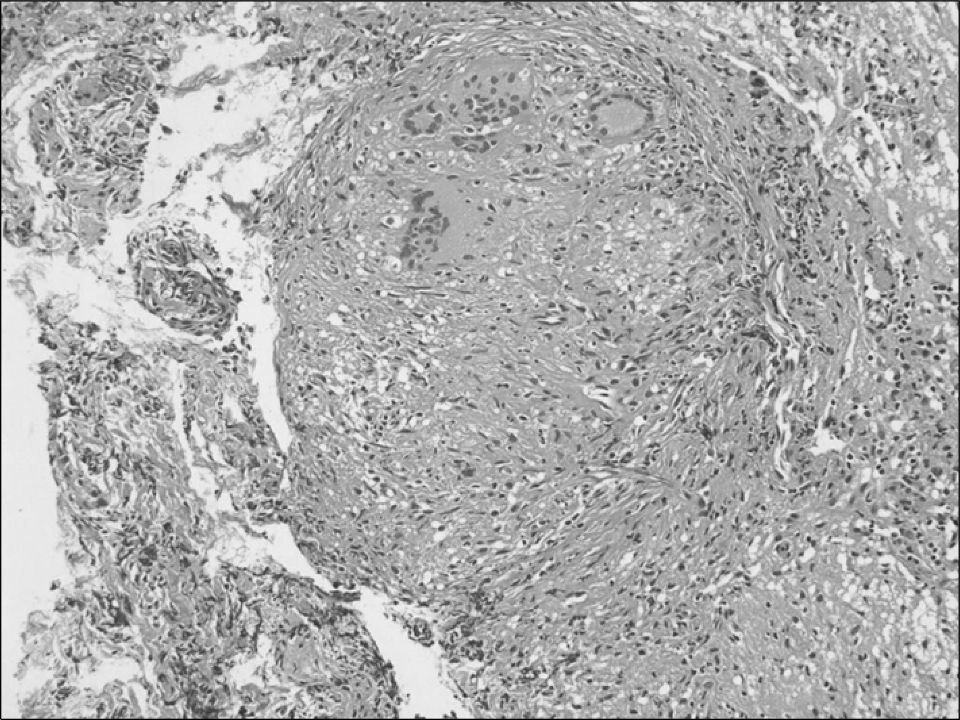
Tuberculosis is an opportunistic infection that causes significant morbidity and mortality in recipients of renal transplants. Although tuberculous peritonitis is easily diagnosed by paracentesis, it is difficult to diagnosis in the absence of ascites. Laparotomy and laparoscopic biopsies are needed for the diagnosis of tuberculous peritonitis. According to recent reports, the latter has a better outcome because of fewer associated complications. A case of tuberculous peritonitis in a renal transplant patient is reported that was diagnosed by laparoscopic peritoneal biopsy.
References
1). Fishman JA. Infection in solid organ transplant recipients. N Engl J Med. 2007; 357:2601–14.
2). Park JS, Kim MS, Lee JH, Chang J, Kim SK, Jeon KO, et al. Mycobacterial infection after kidney transplantation: single center experience. J Korean Soc Transplant. 2001; 15:39–46. (박준성, 김명수, 이종훈, 장준, 김세규, 전경옥 등. 신장 이식 후 발생한 결핵에 관한 고찰. 대한이식학회지 2001;15: 39–46.).
3). Park SB, Suk J, Joo I, Kim HC, Cho WH, Park CH. Tuberculosis complicating kidney transplantation. J Korean Soc Transplant. 1995; 9:95–102. (박성배, 석준, 주일, 김현철, 조원현, 박철회. 신이식 후 발생한 결핵에 대한 고찰. 대한이식학회지 1995;9: 95–102.).
4). Kim MS, Byun CG, Lee KY, Kim SI, Kim SK, Chang J, et al. Mycobacterial infections after renal transplantation. J Korean Soc Transplant. 1995; 9:103–9. (김명수, 변창규, 이강영, 김순일, 김세규, 장준 등. 신장 이식 후 발생한 결핵. 대한이식학회지 1995;9: 103–9.).
5). The Korean National Tuberculosis Association (KNTA) [Internet]. Seoul: KNTA;2008. Available from:. http://www.knta.or.kr.
6). EBPG Expert Group on Renal Transplantation. European best practice guidelines for renal transplantation. Section IV: Long-term management of the transplant recipient. IV. 7.2. Late infections. Tuberculosis. Nephrol Dial Transplant. 2002; 17(S4):39–43.
7). Sakhuja V, Jha V, Varma PP, Joshi K, Chugh KS. The high incidence of tuberculosis among renal transplant recipients in India. Transplantation. 1996; 61:211–5.

8). Jorgensen JO, Lalak NJ, Hunt DR. Is laparoscopy associated with a lower rate of postoperative adhesions than laparotomy? A comparative study in the rabbit. Aust N Z J Surg. 1995; 65:342–4.

9). Schippers E, Tittel A, Ottinger A, Schumpelick V. Laparoscopy versus laparotomy: comparison of adhesion-formation after bowel resection in a canine model. Dig Surg. 1998; 15:145–7.
10). Apaydin B, Paksoy M, Bilir M, Zengin K, Saribeyoglu K, Taskin M. Value of diagnostic laparoscopy in tuberculous peritonitis. Eur J Surg. 1999; 165:158–63.
11). al Quorain AA, Satti MB, al Gindan YM, al Ghassab GA, al Freihi HM. Tuberculous peritonitis: the value of laparoscopy. Hepatogastroenterology. 1991; 38(S1):37–40.
12). Cassidy MJ, van Zyl-Smit R, Pascoe MD, Swanepoel CR, Jacobson JE. Effect of rifampicin on cyclosporin A blood levels in a renal transplant recipient. Nephron. 1985; 41:207–8.

13). Offermann G, Keller F, Molzahn M. Low cyclosporin A blood levels and acute graft rejection in a renal transplant recipient during rifampin treatment. Am J Nephrol. 1985; 5:385–7.

14). Vachharajani TJ, Oza UG, Phadke AG, Kirpalani AL. Tuberculosis in renal transplant recipients: rifampicin sparing treatment protocol. Int Urol Nephrol 2002–3;34:. 551–3.

15). Lee SJ, Park JA, Chang YK, Choi BS, Yoon SA, Yang CW, et al. The treatment of post transplant tuberculosis: rifampin sparing regimen. J Korean Soc Transplant. 2009; 23:22–7. (이상주, 박진아, 장윤경, 최범순, 윤선애, 양철우 등. 신이식 후 발생한 결핵의 치료: Rifampin 제외 복합요법. 대한이식학회지 2009;23: 22–7.).
16). Kidney Disease. Improving Global Outcomes (KDIGO) Transplant Work Group. KDIGO clinical practice guideline for the care of kidney transplant recipients. Am J Transplant. 2009; 9(S3):1–155.


 PDF
PDF ePub
ePub Citation
 Print
Print


 XML Download