Abstract
Salmonella enterica serovar Enteritidis is one of the major food borne pathogens. Utilizing lytic bacteriophages against this pathogen can be a new and effective approach for the prevention of food-contamination and food-borne infection. In this study, we isolated and characterized a Salmonella Enteritidis specific lytic bacteriophage (phage SE2). The bacteriolytic activity of planktonic and biofilmed cells against an antibiotic resistant strain of Salmonella Enteritidis was also evaluated. Phage SE2 revealed an efficient bacteriolytic effect with biofilm dispersing ability and could maintain its virulence even at extreme pH and temperature. It can be a potential biotherapeutic agent against Salmonella Enteritidis.
REFERENCES
1). Rodrigue DC, Tauxe RV, Rowe B. International increase in Salmonella enteritidis: A new pandemic? Epidemiol infect. 1990; 105:21–7.
2). Guard-Petter J. The chicken, the egg and Salmonella Enteritidis. Environ Microbiol. 2001; 3:421–30.
3). Aarestrup FM, Wiuff C, Mølbak K, Threlfall EJ. Is it time to change fluoroquinolone breakpoints for Salmonella spp.? Antimicrob Agents Chemother. 2003; 47:827–9.
4). Kang ZW, Jung JH, Kim SH, Lee BK, Lee DY, Kim YJ, et al. Genotypic and Phenotypic Diversity of Salmonella Enteritidis Isolated from Chickens and Humans in Korea. J Vet Med Sci. 2009; 71:1433–8.
5). Lindgren MM, Kotilainen P, Huovinen P, Hurme S, Lukinmaa S, Webber MA, et al. Reduced fluoroquinolone susceptibility in Salmonella enterica isolates from travelers, Finland. Emerg Infect Dis. 2009; 15:809–12.
6). Tamang MD, Nam HM, Kim TS, Jang GC, Jung SC, Lim SK. Emergence of extended-spectrum β-Lactamase (CTX-M-15 and CTX-M-14)-producing nontyphoid Salmonella with reduced susceptibility to ciprofloxacin among food animals and humans in Korea. J Clin Microbiol. 2011; 49:2671–5.
7). Kusumaningrum HD, Riboldi G, Hazeleger WC, Beumer RR. Survival of foodborne pathogens on stainless steel surfaces and cross-contamination to foods. Int J Food Microbiol. 2003; 85:227–36.

8). Chorianopoulos NG, Giaouris ED, Kourkoutas Y, Nychas GJ. Inhibition of the early stage of Salmonella enterica serovar Enteritidis biofilm development on stainless steel by cell-free supernatant of a Hafnia alvei culture. Appl Environ Microbiol. 2010; 76:2018–22.
9). Solano C, García B, Valle J, Berasain C, Ghigo JM, Gamazo C, et al. Genetic analysis of Salmonella Enteritidis biofilm formation: critical role of cellulose. Mol Microbiol. 2002; 43:793–808.
10). Soares VM, Pereira JG, Viana C, Izidoro TB, Bersot Ldos S, Pinto JP. Transfer of Salmonella Enteritidis to four types of surfaces after cleaning procedures and cross-contamination to tomatoes. Food Microbiol. 2012; 30:453–6.
11). Van Houdt R, Michiels CW. Biofilm formation and the food industry, a focus on the bacterial outer surface. J Appl Microbiol. 2010; 109:1117–31.

12). Borysowski J, Weber-Dabrowska B, Górski A. Bacteriophage endolysins as a novel class of antibacterial agents. Exp Biol Med. 2006; 231:366–77.

13). Goodridge LD, Bisha B. Phage-based biocontrol strategies to reduce foodborne pathogens in foods. Bacteriophage. 2011; 1:130–7.

14). Mahony J, McAuliffe O, Ross RP, Van Sinderen D. Bacteriophages as biocontrol agents of food pathogens. Curr Opin Biotechnol. 2011; 22:157–63.

15). Andreatti Filho RL, Higgins JP, Higgins SE, Gaona G, Wolfenden AD, Tellez G, et al. Ability of bacteriophages isolated from different sources to reduce Salmonella enterica serovar Enteritidis in vitro and in vivo. Poult Sci. 2007; 86:1904–9.
16). Atterbury RJ, Van Bergen MA, Ortiz F, Lovell MA, Harris JA, De Boer A, et al. Bacteriophage therapy to reduce Salmonella colonization of broiler chickens. Appl Environ Microbiol. 2007; 73:4543–9.
17). Goode D, Allen VM, Barrow PA. Reduction of experimental Salmonella and Campylobacter contamination of chicken skin by application of lytic bacteriophages. Appl Environ Microbiol. 2003; 69:5032–6.
18). Guenther S, Huwyler D, Richard S, Loessner MJ. Virulent bacteriophage for efficient biocontrol of Listeria monocytogenes in ready-to-eat foods. Appl Environ Microbiol. 2009; 75:93–100.
19). Fu W, Forster T, Mayer O, Curtin JJ, Lehman SM, Donlan RM. Bacteriophage cocktail for the prevention of biofilm formation by Pseudomonas aeruginosa on catheters in an in vitro model system. Antimicrob Agents Chemother. 2010; 54:397–404.
20). Rahman M, Kim S, Kim SM, Seol SY, Kim J. Characterization of induced Staphylococcus aureus bacteriophage SAP-26 and its anti-biofilm activity with rifampicin. Biofouling. 2011; 27:1087–93.
21). Siringan P, Connerton PL, Payne RJ, Connerton IF. Bacteriophage-mediated dispersal of Campylobacter jejuni biofilms. Appl Environ Microbiol. 2011; 77:3320–6.
22). Lu TK, Collins JJ. Dispersing biofilms with engineered enzymatic bacteriophage. Proc Natl Acad Sci U S A. 2007; 104:11197–202.

23). Tiwari BR, Kim S, Kim J. Complete genomic sequence of Salmonella enterica serovar Enteritidis phage SE2. J Virol. 2012; 86:7712.
24). Merabishvili M, Pirnay JP, Verbeken G, Chanishvili N, Tediashvili M, Lashkhi N, et al. Quality-controlled small-scale production of a well-defined bacteriophage cocktail for use in human clinical trials. PLoS one. 2009; 4:e4944.

25). Allison DG, Ruiz B, SanJose C, Jaspe A, Gilbert P. Extracellular products as mediators of the formation and detachment of Pseudomonas fluorescens biofilms. FEMS Microbiol Lett. 1998; 167:179–84.
26). Giaouris E, Chorianopoulos N, Nychas GJ. Effect of temperature, pH, and water activity of biofilm formation by Salmonella enterica Enteritidis PT4 on stainless steel surfaces as indicated by the bead vortexing method and conductance measurments. J Food Prot. 2005; 68:2149–54.
27). Carey-Smith GV, Billington C, Cornelius AJ, Hudson JA, Heinemann JA. Isolation and characterization of bacteriophages infecting Salmonella spp. FEMS Microbiol Lett. 2006; 258:182–6.
28). O'Flynn G, Coffey A, Fitzgerald GF, Ross RP. The newly isolated lytic bacteriophages st104a and st104b are highly virulent against Salmonella enterica. J Appl Microbiol. 2006; 101:251–9.
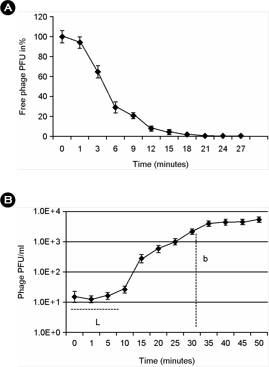
Figure 1.
Adsorption rate and burst size of Phage SE2. (A) Adsorption rate of Phage SE2. (B) Burst size of Phage SE2. L, Latent time (10 minutes); b, average burst size (155 PFU/host cells at 35 minutes).

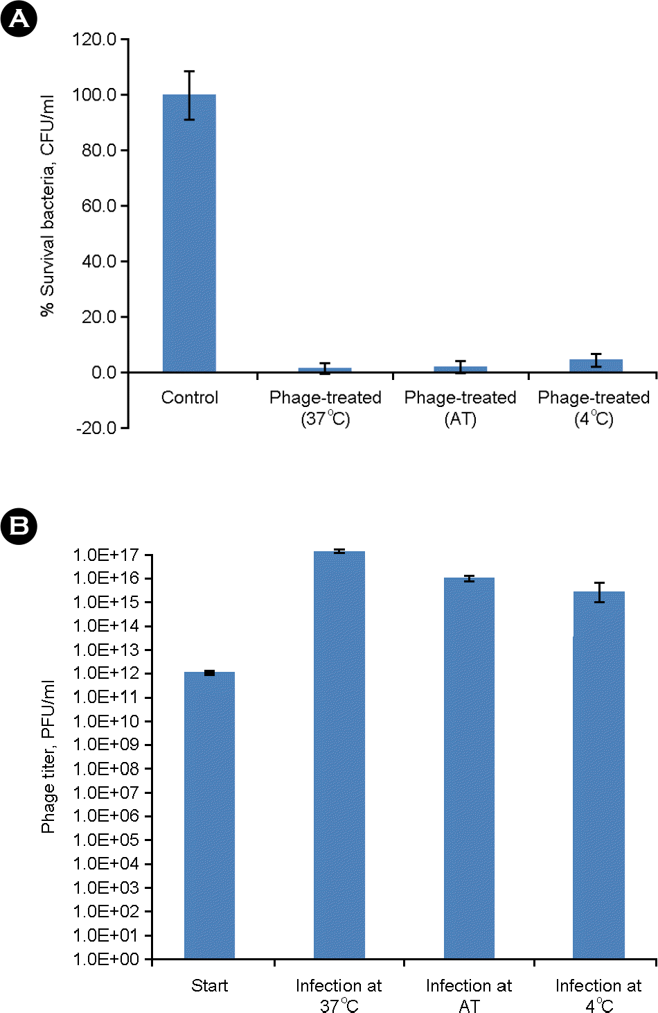
Figure 2.
Ability of phage SE2 to kill bacteria and produce progenies at different temperature. (A) Ability of phage SE2 to kill bacteria at 37°C, ambient temperature and 4°C. (B) Ability of phage to produce progenies during infection at 37°C, ambient temperature (AT) and 4°C.

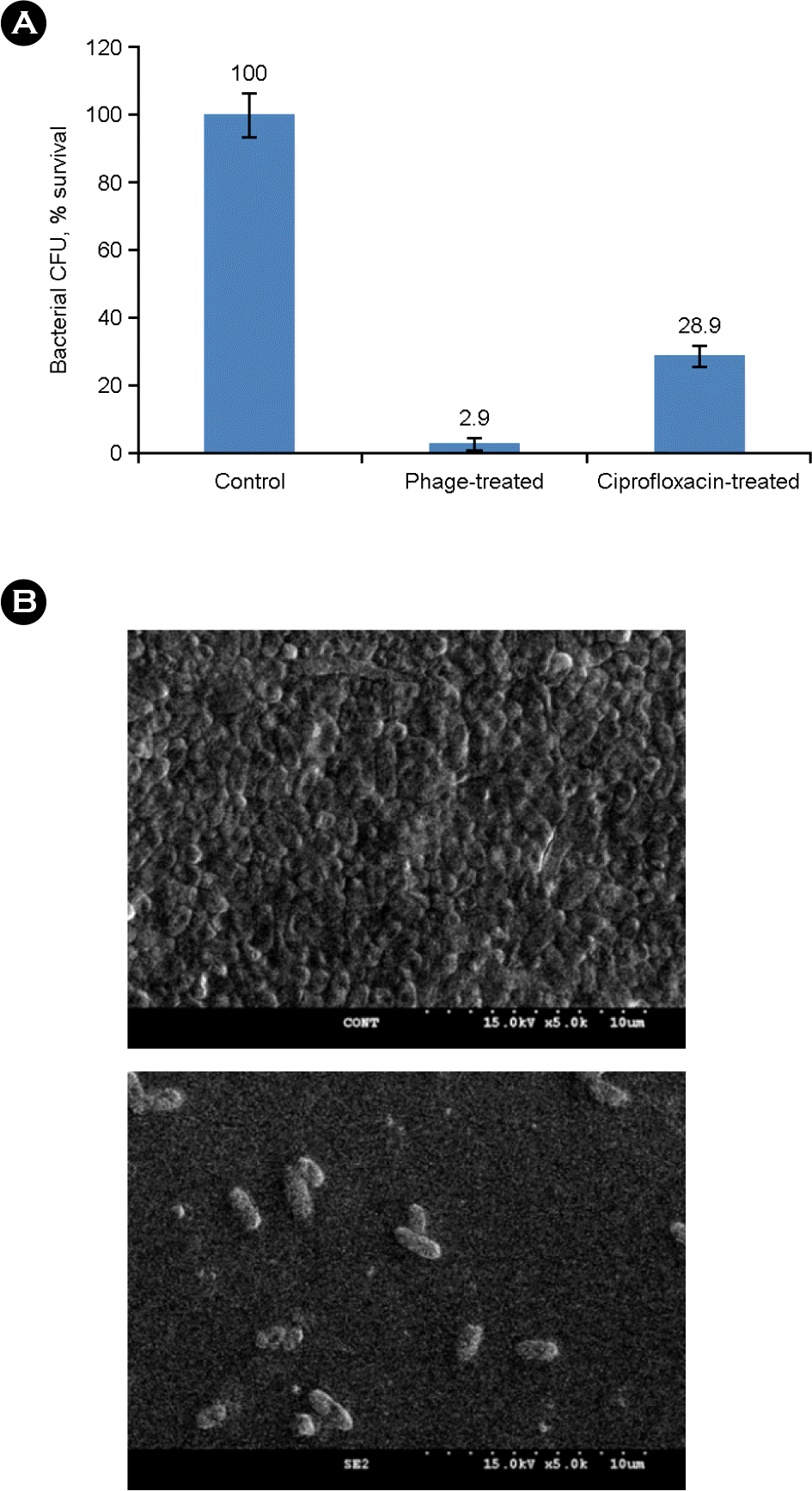
Figure 3.
Bacteriolytic activity of the phage SE2 to biofilmed cells. (A) Biofilmed cells of Salmonella Enteritidis JB-201 were treated with tryptic soy broth (control), phage SE2 (1011 PFU/ml) or ciprofloxacin (0.5 μg/ml) for 4 hours at 37°C. Mean bacterial CFU was calculated from the triplicates. (B) Field emission scanning electron microscopic (FE-SEM) images showing biofilm dispersion activity of phage SE2. FE-SEM images showed that almost all the biofilmed cells were lysed with the dispersion of biofilm's matrix by phage SE2, very few cells, cell debris and ghost like bacteria can be seen on phage treated biofilms images (lower) compared that of phage untreated control (upper). At least three independent experiments were performed.

Table 1.
Host range of phage SE2 against Salmonella enterica serovars Enteritidis and Gallinarum.
| Salmonella Serovars | Year of isolation | Plaques1 | Salmonella Serovars | Year of isolation | Plaques1 |
|---|---|---|---|---|---|
| S. Enteritidis 012 | 2000 | ++ | S. Gallinarum I | ? | ++ |
| S. Enteritidis 013 | 2000 | ++ | S. Gallinarum II | ? | ++ |
| S. Enteritidis 014 | 2000 | ++ | S. Gallinarum 08vs-38 | 2008 | ++ |
| S. Enteritidis 015 | 2002 | ++ | S. Gallinarum 08vs-178 | 2008 | ++ |
| S. Enteritidis 018 | 2002 | ++ | S. Gallinarum 08vs-3 | 2008 | ++ |
| S. Enteritidis 09 | 2005 | ++ | S. Gallinarum 08vs-4 | 2008 | ++ |
| S. Enteritidis 010 | 2005 | ++ | S. Gallinarum 08vs-5 | 2008 | ++ |
| S. Enteritidis 011 | 2005 | ++ | S. Gallinarum 08vs-6 | 2008 | ++ |
| S. Enteritidis 015 | 2006 | ++ | S. Gallinarum 08vs-8 | 2008 | ++ |
| S. Enteritidis 016 | 2006 | ++ | S. Gallinarum 08vs-10 | 2008 | ++ |
| S. Enteritidis 017 | 2006 | ++ | S. Gallinarum 08vs-18 | 2008 | ++ |
| S. Enteritidis 01 | 2009 | ++ | S. Gallinarum 08vs-19 | 2008 | ++ |
| S. Enteritidis 09 | 2009 | ++ | S. Gallinarum 08vs-20 | 2008 | ++ |
| S. Enteritidis PT-4 | ? | ++ | S. Gallinarum 08vs-21 | 2008 | ++ |
| S. Enteritidis Jb-10-201 | 2010 | ++ | S. Gallinarum 08vs-23 | 2008 | ++ |
| S. Enteritidis 5-17-6 | 2010 | ++ | S. Gallinarum 08vs-25 | 2008 | ++ |
| S. Enteritidis 5-6-5 | 2010 | + | S. Gallinarum 08vs-27 | 2008 | ++ |
| S. Enteritidis 5-6-2 | 2010 | + | S. Gallinarum 08vs-33 | 2008 | ++ |
| S. Gallinarum 08vs-34 | 2008 | ++ | |||
| S. Gallinarum 08vs-36 | 2008 | + | |||
| S. Gallinarum 08vs-38 | 2008 | + | |||
| S. Gallinarum 08vs-42 | 2008 | + | |||
| S. Gallinarum 08vs-43 | 2008 | ++ | |||
| S. Gallinarum 08vs-45 | 2008 | ++ | |||
| S. Gallinarum 08vs-51 | 2008 | ++ | |||
| S. Gallinarum 08vs-4 | 2008 | ++ | |||
| S. Gallinarum 08vs-52 | 2008 | ++ | |||
| S. Gallinarum 08vs-53 | 2008 | ++ | |||
| S. Gallinarum 08vs-56 | 2008 | ++ | |||
| S. Gallinarum 08vs-57 | 2008 | ++ | |||
| S. Gallinarum 08vs-165 | 2008 | ++ | |||
| S. Gallinarum 08vs-168 | 2008 | ++ | |||
| S. Gallinarum 08vs-173 | 2008 | ++ | |||
| S. Gallinarum 08vs-178 | 2008 | ++ | |||
| S. Gallinarum 08vs-188 | 2008 | ++ | |||
| S. Gallinarum 08vs-190 | 2008 | ++ | |||
| S. Gallinarum 08vs-191 | 2008 | ++ | |||
| S. Gallinarum 08vs-200 | 2008 | ++ | |||
| S. Gallinarum 08vs-202 | 2008 | ++ | |||
| S. Gallinarum 08vs-236 | 2008 | ++ | |||
| S. Gallinarum 08vs-240 | 2008 | ++ | |||
| S. Gallinarum 08vs-251 | 2008 | ++ | |||
| S. Gallinarum 09vs-6 | 2009 | + | |||
| S. Gallinarum 09vs-7 | 2009 | + | |||
| S. Gallinarum 09vs-8 | 2009 | + | |||
| S. Gallinarum 09vs-9 | 2009 | ++ |


 PDF
PDF ePub
ePub Citation
 Print
Print


 XML Download