

 PDF
PDF ePub
ePub Citation
 Print
Print


INTRODUCTION
The replication of adeno-associated virus (AAV), which is a small non-enveloped single stranded DNA virus, usually requires co-infection with helper virus (1). Among the 12 AAV serotype, AAV-2, 3, and 5 have been detected in human clinical samples (2, 3). They belong to the genus Dependovirus, family Parvoviridae (4).
Although AAV infection is yet thought to be nonpathogenic in human, there has been possibility of its association with early miscarriage and other reproductive problems during pregnancy (5~7). AAV DNA has been detected in uterus, early abortus, and female genital tissues (6, 8, 9). Its detection rate (12~77%) depends on the experimental methods and the sampling conditions. Reports on the detection rate of AAV in cervical secretion and prevalence of serum antibodies to AAV indicated that AAV infections were significantly more frequent in pregnant women than in non-pregnant controls (10, 11). AAV infection occurred frequently in male reproductive tract and viral DNA of AAV was mainly detected in sperm fractions of the semen. These findings indicate the possibility of AAV being sexually transmitted. In the previous studies, AAV was frequently detected in the semen from men with infertility (12, 13), which suggests that AAV might be a possible causative agent of male infertility. The presence of AAV in early abortus also indicates that AAV may be one of the causes of abortion (5, 7, 9). Additionally, HPV, which is a helper virus of AAV, is involved in male factor infertility and can reduce the motility of sperm (14, 15).
To assess the relationship between the presence of these viruses and male factor infertility and repeated miscarriages of their partners, the detection rates of AAV and HPV were evaluated in the semen.
MATERIALS AND METHODS
Patients
From May 2007 to February 2008, the informed consent was obtained from 99 male who visited the Hamchoon Women's Clinic for sperm donation, semen analysis or in vitro fertilization and embryo transfer (IVF-ET) programs. This study was approved by Institutional Review Board in both Seoul National University Hospital and Hamchoon Women's Clinic. Semen samples were collected by masturbation following three- or four-days' sexual abstinence. Thirty-six male were excluded from study; in 4 cases, there were no remaining samples or DNA after nucleic acid extraction procedure; in 32 cases, reproductivity could not be evaluated because they did not revisit the clinic or did not report whether pregnancy was maintained successfully or not.
The semen samples were examined by routine analysis according to WHO guidelines. Abnormal semen was evaluated according to sperm concentration (less than 20 × 106/ml) or sperm motility (less than 50%). Abnormal results were shown in 15 men and they were allocated to the male factor (MF) infertility group. Thirty-four men, whose wives had suffered from more than two miscarriages, were designated as the repeated miscarriages (RM) group. We defined these two groups as an impaired reproductivity group. Fourteen men were allocated to control group; they had normal results in semen analysis and their conception partner had ongoing pregnancy to mid-trimester without any treatment for miscarriages.
DNA extraction
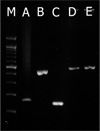
DNA extraction was performed using Real Genomics™ Genomic DNA Extraction Kit (Real Biotech Corporation, Taiwan) according to the manufacturer's protocol. Achondroplasia gene amplification was used as a control of the extraction and DNA integrity (Fig. 1A).
AAV DNA detection
All samples were examined for the presence of AAV by the nested PCR using AUS1/Pan3 primers (AUS1: 5'-ACA CCA TCT GGC TGT TTG GG-3', Pan3: AAA AAG TCT TTG ACT TCC TGC TT-3') for primary PCR and IN1/IN2 primers (IN1: GAG GCC ATA GCC CAC ACT GT-3', IN2: GAG AAT GGC TTT GGC CGA CT-3') for nested PCR (16). Two µL of extracted DNA of each sample was initially mixed with an AccuPower™ PCR PreMix (Bioneer, Korea) which contained 1.5 mM MgCl2, 250 µM of each dNTP, 10 mM Tris-HCl pH 9.0, 30 mM KCl, and 1.0 unit Taq polymerase. To this mixture, 20 pmole of each primer was added and adjusted to a final volume of 20 µl with deionized water. The same reaction formula was applied for nested PCR using 2 µl of tenfold-diluted primary PCR product as template. The primary PCR was carried out as follows: initial denaturation at 94℃ for 10 min, followed by 25 cycles for 30 sec at 94℃, 20 sec at 58℃, 10 sec at 72℃ and with a final extension at 72℃ for 7 min. The nested PCR reactions were carried out with an initial denaturation step at 94℃ for 10 min, 25 cycles of 30 sec at 94℃, 15 sec at 61℃ and 10 sec at 72℃ and with a final extension at 72℃ for 7 min. A volume of 5 µl products was subjected to electrophoresis on 2% agarose gel and visualized under UV illuminator with ethidium bromide staining. The primary and nested PCR resulted in 427 and 150 bp products, respectively (Fig. 1B and 1C). AAV2 strain (ATCC VR680) was used as a positive control. DNA and RNA free water was included in all PCR procedures as a negative control.
HPV DNA detection
HPV DNA detection was based on PCR amplification of the L1 region using the L1A/L1B primers (L1A: 5'-GCA CAG GGA CAT AAT AAT GG-3', L1B: 5'-CGT CCA AGA GGA TAC TGA TC-3') (17). The reaction mixture was same as that of AAV detection. Initial denaturation step was at 94℃ for 10 min, followed by 35 cycles for 30 sec at 94℃, 30 sec at 60℃, and 15 sec at 72℃, and with a final extension at 72℃ for 7 min. PCR product was a 452 bp fragment.
Statistical analysis
PASW Statistics 17.0 program was used to analyze the data. Differences in the means of the continuous interval were analyzed using one-way ANOVA. Frequencies of virus DNA detection were analyzed using χ2-test; odds ratio and 95% confidence interval were calculated where appropriate. In all analyses, the value of p < 0.05 was considered significant.
RESULTS
To access the relationship between the detection of AAV and HPV in the semen and male infertility and recurrent miscarriages, semen analysis and amplification of specific sequences of both viruses were done.
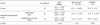
Table 1 demonstrates differences in the sperm count and the sperm motility of three tested groups: the control, MF infertility group, and RM group. Both the sperm count (25.9 ± 27.52 × 10⁶/ml) and the sperm motility (33.7 ± 13.95%) in MF infertility group were significantly lower than in control (62.1 ± 24.24 × 10⁶/ml and 67.5 ± 9.15%, respectively) and RM group (72.4 ± 23.75 × 10⁶/ml and 67.9 ± 9.62%, respectively).
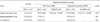
Cellular DNA was detected in all semen samples (achondroplasia sequence in chromosome 4p). AAV DNA in the semen was found in 50% (17/34) of RM and 60% (9/15) of MF infertility group, which was statistically significantly higher than control group (14%, 2/14) (Table 2). Odds ratio of the MF infertility group in AAV detection compared with the control group was 9.0 (95% CI 1.46~55.48) and that compared with the RM group was 6.0 (95% CI 1.16~30.96).
HVP DNA was detected in the semen. Table 2 shows that the detection rates of HPV in RM (15%, 5/34) and MF infertility group (7%, 1/15) were not statistically different from that of control group (21%, 3/14). The co-infections of AAV with HPV were 14, 12 and 0% in the control, RM and MF infertility group, respectively.
DISCUSSION
AAV has been investigated with the purpose of carrying out gene therapy and does not seem to be pathogenic in human. However, AAV has been found to infect the genital system of both man and woman and might be associated with early miscarriages and other prenatal problems (5, 7, 9, 12, 18).
Since normal delivery can be expected if the pregnancy is maintained through the second trimester, men whose spousal pregnancies were ongoing to mid-trimester without any treatment of repeated miscarriage were used as the control group.
AAV detection rate of MF infertility group was significantly higher than control group. MF infertility group was subdivided by their semen characteristics and compared for AAV detection rate with control group. AAV positive rates in oligozoospermia, asthenozoospermia, and oligoasthenozoospermia subgroups were 67%, 43% and 80%, respectively. Differences of AAV positive rate among these subgroups and control group were statistically significant (p < 0.05), but odd ratio (24.0, 95% CI 1.69~340.99) was statistically significant only in oligoasthenozoospermia subgroup when compared with control group. AAV DNA was detected in 6 of 8 patients with abnormal sperm count (OR 18.0, 95% CI 2.01~161.04), and in 7 of 12 patients with abnormal motility (OR 8.4, 95% CI 1.27~55.39). Higher detection rate of AAV in the semen with abnormal sperm parameters implies that there is a relationship between AAV infection and male factor infertility. Although the effect of AAV on the male factor infertility has not been clearly defined, AAV DNA could be detected in the sperm and the testis of azoospermia patients (13). This report suggests that AAV may exert the harmful effect on the sperm maturation.
Based on the report that semen containing lots of large sperms could cause repeated miscarriages because of the production of chromosomally abnormal embryos (19), sperm morphology test were fulfilled among patients. AAV DNA was present in 50% of RM group, which was significantly higher than that in the control group. The report showed that AAV infection was associated with adverse reproductive outcome and that AAV DNA was found in 73% of early abortus. It suggests that there could be a relationship between repeated miscarriages and AAV infection, although AAV DNA has not been surveyed in repeated miscarriages cases (7, 9). The miscarriages might have resulted from AAV infected sperms transmitting the virus to the egg. The ability of AAV to dysregulate the cell cycle might have a deleterious effect on embryo development. AAV infection induced the cessation of cleavage at 2-cell stage and injecting AAV DNA to the fertilized mouse embryos led to the developmental blockage at morula stage. Also the fetal death and early abortion were observed in AAV-infected pregnant mice (20).
Other study demonstrated that seropositivity of IgM antibody against AAV virus in maternal serum in the first trimester was 5.6 times more prevalent in pre-eclampsia, intrauterine growth retardation, and stillbirth cases, and 7.6 times more prevalent in pre-term deliveries than in control group (18). The higher detection of IgG antibody against AAV in the serum and AAV DNA in the cervical samples of the pregnant women than in non pregnant women might result from the reactivation of AAV persisting in woman's reproductive organ. This reactivation might be due to hormonal influences during pregnancy (10, 11). In general, patients with repeated miscarriages may be pregnant for much longer period of time and more frequently than normal pregnant women with regard to the total period of pregnancy. Therefore they have more chance to transmit AAV to their conception partners. This is one of the other possible explanations for higher detection rate of AAV DNA in the semen from RM group than that from the control group. This detection possibility, however, has limitation because only the semen was tested in our study. Therefore further research is warranted to look into this possibility in women in the future.
The detection rate of AAV DNA in our study was higher than the previous reports (12, 13). The differences in the prevalence of AAV infection due to racial or geographical disparity might have had influence on the result. Detection rate of AAV DNA in healthy women was 50% in Arkansas, but that in Jamaica was 0% even though same PCR protocol and primers were used (21, 22).
A recent report by Schlehofer et al showed no significant association of the presence of AAV DNA in semen samples or endocervical swabs with clinically relevant infertility factors (23). Their results were different from ours, but profiles of recruited patients and kinds of tested samples were also different from each other. Their patients were subfertile and especially over 80 percentage of patients were male factor infertility with reduced sperm motility. But the positive rate of AAV DNA in semen was lower than this study and their previous studies (12, 13). They did not include normal males, and did not explain the reasons of lower detection rates. Some additional future studies with sufficient number of well-characterized patients and normal persons are needed.
HPV, one of AAV helper viruses, is associated with male infertility. Sperm motility was decreased by the incubation with HPV DNA E6-E7 region for 24 h (15). However, other investigators reported that the detection of HPV DNA in semen was not related with the sperm quality, i.e. sperm counts and motility (24). The result of the detection of HPV DNA in the semen did not show significant correlation between the male infertility and HPV infection in our study. In several other reports, the prevalence of HPV DNA ranged from 4.5 to 36.5% in the semen of asymptomatic men (12, 13, 24, 25, 26). The finding that the prevalence of HPV DNA in the men (14.3%, 9/63) was lower than that of meta-analysis result (23.9%) in Korean women with normal cytology (27) is in line with the result from previous reports (28, 29).
The present findings suggest that AAV could be related to repeated miscarriages and male infertility. It will be worth the effort to learn the mechanism on how AAV affects to spermatogenesis and to confirm whether AAV and helper virus decrease the success rate of IVF-ET. Additionally, the further research with women should be included to better understand the relationship between repeated miscarriages and AAV infection.
 XML Download