

 PDF
PDF ePub
ePub Citation
 Print
Print


INTRODUCTION
Helicobacter pylori infection causes chronic gastritis, gastroduodenal ulcers, and gastric cancer. Currently, the initial management of H. pylori infection includes prescribing a proton pump inhibitor and a combination of two or more antibiotics such as amoxicillin, clarithromycin, or metronidazole (1~4). However, antibiotic resistance is an important problem (5, 6); H. pylori strains are increasingly resistant to metronidazole, clarithromycin, and amoxicillin (7, 8).
H. pylori resistance rates can vary among groups of patients according to age, sex, disease, and place of residence. In some countries, there is an evolution of resistance that often reflects the previous or current national consumption of a given antibacterial agent (9, 10). Antibiotic resistance testing of regional H. pylori isolates might be helpful for predicting the outcomes of standard treatment (11, 12). The aim of this study was to investigate antibiotic resistance rates of H. pylori isolated in Jinju between 1985 and 1999 and to compare our findings with isolates from Cheongju collected between 1995 and 1999.
MATERIALS AND METHODS
Patients
Strains were collected from the adult patients undergoing upper gastrointestinal endoscopy to evaluate abdominal pain or gastrointestinal bleeding at Gyeongsang National University (GNU) Hospital in 1985-1989, 1990-1994, and 1995-1999 and at Chungbuk National University in 1995-1999.
Biopsy specimens and H. pylori culture
Fresh antral and body biopsy specimens that were freshly obtained (used within 1 h) or kept frozen at the biobank (>1 year) were transported to the microbiology laboratory and cultured on Mueller-Hinton agar (MHA, Difco Co., Detroit, MI, USA) plates containing 10% bovine sera, vancomycin (10 µg/ml), nalidixic acid (25 µg/ml) and amphotericin B (5 µg/ml). The plates were incubated at 37℃ under microaerophilic conditions under a 10% CO2 atmosphere at 100% humidity for 3~5 days. All frozen biopsy specimens were provided by Gyeongsang National University Hospital, a member of the National Biobank of Korea, after the Institutional Review Board reviewed the research protocols (GNUHIRB-2009-007).
The organisms were identified as H. pylori by colony morphology; Gram staining; and positive urease, catalase, and peroxidase tests. Once cultured, the H. pylori were stored and distributed by the H. pylori Korean Type Culture Collection at Gyeongsang National University School of Medicine.
Susceptibility tests
The minimal inhibitory concentration (MIC) values of the H. pylori isolates to erythromycin (Sigma Chemical Co., St. Louis, MO, USA), clarithromycin (Sigma Chemical Co.), azithromycin (Pfizer Central Research, Groton, CT, USA), amoxicillin (Sigma Chemical Co.), tetracycline (Sigma Chemical Co.), metronidazole (Sigma Chemical Co.), furazolidone (Sigma Chemical Co.), levofloxacin (Sigma Chemical Co.), ciprofloxacin (Sigma Chemical Co.), moxifloxacin (Sigma Chemical Co.), and rifabutin (Yuyu Pharma Inc., Seoul, Korea) were examined using the serial two-fold agar dilution method as described in the Clinical and Laboratory Standards Institute guidelines (13). Briefly, bacteria were subcultured on MHA supplemented with 10% defibrinated bovine sera for 48 h. The bacterial suspension was adjusted to 1 × 10⁷ colony-forming units and was inoculated directly onto each antibiotic-containing agar dilution plate. MICs were determined after 72 h of incubation. The resistance breakpoints for clarithromycin, azithromycin, and erythromycin were all set as >1.0 µg/ml and those for amoxicillin, tetracycline, metronidazole, and furazolidone were defined as ≥0.5 µg/ml, >2.0 µg/ml, >8.0 µg/ml, and >1.0 µg/ml, respectively. The breakpoints for levoflaxacin, ciproflaxin, moxifloxacin, and rifabutin were provisionally defined as >1.0 µg/ml. Multidrug resistance was defined as resistance to >2 of the following antibiotics: macrolide, amoxicillin, tetracycline, metronidazole, furazolidone, fluoroquinolone, and rifabutin.
Statistical analysis
Data were analyzed using SPSS version 12.0 software (SPSS Inc., Chicago, IL, USA). The Wilcoxon rank-sum test and the Kruskal-Wallis tests were used for two-group and multiple-group comparisons, respectively. Chi-squared tests were used to compare sex, diagnosis, MIC distributions, and antibiotic resistance frequencies. Statistical significance was set at p < 0.05.
RESULTS
Changing pattern of MIC and antibiotic resistance in Jinju
Strains were collected from 170 patients (111 men [65.3%] and 59 women [34.7%]) with a median age of 28.8 years (range 16.0~71.0). Patients were diagnosed with gastritis (n = 155), gastric ulcer (n = 10), or duodenal ulcer (n = 5) (Table 1). There were no differences in distribution of sex or age or underlying disease among the three collection periods.
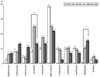
Resistance to moxifloxacin was significantly increased from 1985-1989 (0%) to 1995-1999 (14.9%) (p < 0.0001). The amoxicillin resistant rate was 7.5% in 1985-1989, 24.3% in 1990-1994, and 12.8% in 1995-1999. Resistance to amoxicillin in the 1990-1994 was significantly different from the other time points (p = 0.033) (Fig. 1).
There were no significant changes in other antibiotic resistances among H. pylori isolated from 1985-1999 in Jinju. Resistances to clarithromycin, azithromycin, and erythromycin were 1.9%, 7.5%, and 3.8% in 1985-1989; 5.7%, 7.1%, and 12.8% in 1990-1994; and 3.8%, 2.9%, and 10.6% in 1995-1999. Resistance to tetracycline showed a similar pattern to amoxicillin (9.4%, 17.1%, and 12.8% in 1985-1989, 1990-1994, and 1995-1999, respectively). Resistance to metronidazole decreased from 37.7% to 21.3% (p = 0.251) (Fig. 1). Resistance to furazolidone declined from 9.4% in 1985-1989 to 2.1% in 1995-1999. Rifabutin resistance remained low throughout (<5.0% in both 1985-1989 and 1995-1999).
The percentage of isolates that were resistant to >1 antibiotic was 56.6% in 1985-1989, 55.7% in 1990-1994, and 44.7% in 1995-1999 (p = 0.409). Multidrug resistance rate was 17.0% in 1985-1989, 21.4% in 1990-1994, and 21.3% in 1995-1999 (p = 0.803) (Table 2).
Comparing antibiotic resistance in Jinju and in Cheongju
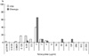
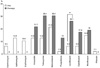
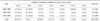
Strains were collected from 23 patients (16 men [69.6%] and 7 women [30.4%]) in Cheongju during 1995-1999, with a median age of 42.0 years (range 16.0~62.0). Patients were diagnosed with gastritis (n = 10), nodular gastritis (n = 1), gastric ulcer (n = 3), duodenal ulcer (n = 8), or gastric cancer (n = 1) (Table 1). Antibiotic resistance was compared with 47 H. pylori strains collected in Jinju between 1995 and 1999. MIC distributions of tetracycline (Fig. 2) and levofloxacin (Fig. 3) in H. pylori isolated from Jinju were lower than in those from Cheongju (p < 0.05).
The levofloxacin resistance rate was higher in Cheongju (26.1%) than in Jinju (6.4%, p = 0.023) (Fig. 4). No macrolide resistance was observed in H. pylori strains isolated from Cheongju. There were no statistical differences of macrolide resistance between Jinju and Cheongju. Resistances to amoxicillin, tetracycline, metronidazole, furazolidone, ciprofloxacin, and moxifloxacine were higher in Cheongju than in Jinju, but these differences were not statistically significant (Fig. 4). Rifabutin resistance was not observed in Cheongju.
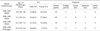
Out of 69 strains, 36 (52.2%) were resistant to one or more antibiotics, and the antibiotic resistance rate was statistically different between Jinju (43.5%) and Cheongju (69.6%, p = 0.041). The prevalence of strains with resistance to ≥2 antibiotics was 23.9% in Jinju and 34.8% in Cheongju (Table 3), and these values were not significantly different (p = 0.341).
In Jinju, triple resistance to macrolide, amoxicillin and metronidazole was found in two strains among 11 strains with resistance to ≥2 antibiotics. Three of 5 strains resistant to marolide were also resistant to amoxicillin or metronidazole. In Cheongju, five of the 6 strains resistant to amoxicillin were also resistant to tetracycline. Four to the 6 strains resistant to amoxicillin were also resistant to quinolone. Dual resistance to amoxicillin and metronidazole was found in two strains among 9 strains with resistance to ≥2 antibiotics.
DISCUSSION
The present study showed no remarkable increment of antimicrobial resistant rates of H. pylori strains isolated over 15-year period in Jinju. The distribution of MICs of amoxicillin, metronidazole, and tetracycline also showed no significant changes during the same periods. Resistance to moxifloxacin increased with time and the resistance to amoxicillin changed (increasing trend from 1985-1989 to 1990-1994 and then decreasing trend from 1990-1994 to 1995-1999). These results might be related with that moxifloxacin was introduced since 1990s and replaced the amoxicillin. In Seoul from 1987 to 2003, the MICs of amoxicillin, clarithromycin, metronidazole, tetracycline, azithromycin, and ciprofloxacin for H. pylori strains increased and rates to clarithromycin increased from 2.8% to 13.8% (14). Seoul is capital huge and crowded city having 10 million in Korea. Jinju is small city having 350, 000, located in the west Gyeongnam Province. Population movement is little in Jinju. Little changes of MICs and resistance of antibiotics were related to less movement of citizen and less change of doctors living in Jinu.
Comparing the antibiotic MICs and resistances of Cheongju and Jinju, those were different during same period. Different resistance rates to metronidazole, levofloxacin, and moxifloxacin reported between 2 institutions located in Seoul and Gyeonggi province and institutional difference of antibiotic resistance of H. pylori resulted in difference of eradication rate of H. pylori according to institution (15). Cheongju is bigger than Jinju and capital of Chungbuk Province, near to Seoul and Gyeonggi Province. The resistances of amoxicillin, tetracycline, metronidazole, and levofloxacin in Cheongju were more similar to Seoul than in Jinju. The difference of antibiotic resistance between Cheongju and Jinju might be resulted from different prescription of antibiotics in resident doctors.
International Guidelines have recommended a 7-day triple therapy consisting of clarithromycin, and either metronidazole or amoxicillin, with a proton pump inhibitor or ranitidine bismuth citrate as a first-line treatment for curing H. pylori infection (16~18). Clarithromycin is the drug of choice of chemotherapy for H. pylori infection. Macrolide resistance is important for management of H. pylori infection. Rates of primary resistance to clarithromycin increased from 2.8% in 1994 to 13.8% in 2003 (19). Resistance to clarithromycin increased in H. pylori isolated from patients in Jinju but increasing rate of clarithromycin resistance was lower than other reports in Korea (14, 20, 21). The range of macrolide resistance was 6.4~12.8% in Jinju, but no resistance to macrolides was observed in Cheoungju. Amoxicillin resistance was 21.7% in Cheongju and it is higher than in Jinju. This result suggested that clarithromycin-amoxicillin regimen is more effective than metronidazole-amoxicillin regimen in Cheongju. Previous reports in Korea, resistance to metronidazole was high, 50% (14). In Jinju, resistance rate to metronidazole decreased but there was no support with statistics. This results suggested that metronidazole and amoxicillin based chemotherapy is still effective regimen in Jinju, but the standard 7-day clarithromycin containing regimen is not valid as the empirical first-line eradication therapy in Jinju. Levofloxacin resistance is higher in Cheongju compared with that in Jinju. The high rate of resistance to levofloxacin may reflect that this antibiotic has already been extensively used in Cheongju.
In recent Korean study, H. pylori eradication rates of rifabutin- or levofloxacin-based triple therapy could not achieve enough eradication rate (22). The antibiotic resistances in Cheongju suggested that levofloxacin-based triple therapy would be low eradication rate as like the result. However, resistance to rifabutin was very low in both cities and no increment for 15-year period in Jinju. Further study is needed to assess the efficacy of rifabutin-based triple therapy for a rescue therapy in H. pylori infection.
There are some limitations to the present study including the small and different numbers of H. pylori isolates from Jinju and Cheongju, the different age distributions from Jinju and Cheonju, and no comparison with recently isolated H. pylori. The lately isolated H. pylori were obtained in 1999 and it is too old to provide valuable information for the current standard treatment. However, this result might be helpful to investigate the changing pattern of antimicrobial resistance in H. pylori isolated in Jinju and to compare the antimicrobial resistance in recently isolated H. pylori isolated in Jinju.
In conclusion, the MIC distributions of antimicrobial agents against H. pylori and antimicrobial resistant rates were different among strains depending on the regions of isolation. Pretreatment microbial susceptibility testing is highly recommended for success in eradication of H. pylori infection. In addition, changing pattern of antibiotic sensitivity should be identified according to region.


 XML Download