

 PDF
PDF ePub
ePub Citation
 Print
Print


INTRODUCTION
Vibrio vulnificus is a gram-negative halophilic bacterium that causes life-threatening septicemia and necrotizing wound infections mainly in patients with liver cirrhosis, hemochromatosis, or β-thalassemia. Several established and potential virulence factors have reported to play important roles in the pathogenesis of V. vulnificus infections, such as, capsular polysaccharides, lipopolysaccharides, iron-assimilation systems, flagella, pili, RTX toxin, and exotoxins such as cytolysins and proteases. These factors are under the control of global regulators, such as cyclic AMP (cAMP) and cAMP receptor protein (Crp) complex, and ferric uptake regulator (Fur) (1).
Elevated serum iron levels are well-known predisposing host factors. In mice, iron treatment reduces the intraperitoneal 50% lethal dose (LD50) from 106 cells to only 1 cell (2), and increases the mortality rate to 100% (3). V. vulnificus grows better in the blood of patients with hemochromatosis, when transferrin saturation by iron is increased, or if hematin is added (4). Although iron is an essential element for the survival and proliferation of most bacteria, some bacteria called ferrophilic or iron-sensitive bacteria have an impaired ability to acquire iron, and thus, cause diseases primarily in iron-overloaded hosts (5). In particular, V. vulnificus is a ferrophilic bacterium that requires higher levels of readily available iron for growth initiation than other pathogens (6).
To establish infection successfully, bacteria must be able to acquire iron from their hosts, and thus, most bacteria have evolved specific iron uptake systems. The ability to acquire iron is a well-established virulence factor in the pathogenesis of V. vulnificus infections, and V. vulnificus possesses multiple iron-uptake systems (1). Of these, the HupA-mediated iron-uptake system is not essential for survival but probably plays an important role in iron acquisition in vivo (7), because most iron within the human body is present as hemoglobin within red blood cells, and V. vulnificus produces several cytotoxins, such as, cytolysin (called VvhA) (8) and RTX toxin (9), which can destroy red blood cells, and thus, cause the release of intracellular iron in the form of hemoglobin, hemin, ferritin, and hemosiderin. In fact, a mutation in hupA has been reported to increase the intraperitoneal LD50 in mice (10).
Fur regulates hupA expression at the transcription level in response to iron availability (11, 12), and recently, Crp, a global regulator primarily responsible for catabolite repression (13), was found to act as a transcriptional activator of hupA expression (10). However, details of the roles played by Crp as an activator and Fur as a repressor of hupA expression have not been observed. It is known that the expressions of many genes are regulated by a combination of global regulators and specific local regulators (14). Therefore, this study was conducted to detail the coordinate roles of Crp as a global regulator and Fur as a local regulator in the regulation of hupA expression at various iron and glucose concentrations.
MATERIALS AND METHODS
Bacterial strains, plasmids, primers, media, and reagents
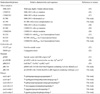
The bacterial strains, plasmids, and primers used in this study are listed in Tables 1 (15~24). Heart Infusion (HI; BD, Franklin Lakes, NJ, USA) agar or broth containing an additional 2.0% of NaCl, and Thiosulfate-Citrate-Bile Salt-Sucrose (TCBS, BD) agar were used to cultivate V. vulnificus strains. Luria-Bertani medium was used to cultivate Escherichia coli strains. Unless otherwise stated, all other reagents were purchased from Sigma-Aldrich (St. Louis, MO, USA).
Limitation and supplementation of iron or glucose
Two types of iron-limited media were used, that is, HI broth containing 200 µM α,α'-dipyridyl as an iron chelator was used for the adaption of V. vulnificus strains to iron limited conditions, and HI broth deferrated with 8-hydroxyquinoline as previously described (25) was used for main experiments. The concentration of residual iron in deferrated HI broth was less than 1 µg/dl. To observe the effect of iron on growth and gene expression, various concentrations (0~30 µM) of ferric chloride (FC) were added to deferrated HI broths. In addition, deferrated HI broth was used as a glucose-poor medium because it contains only a low, though undefined, level of glucose. To observe the effect of glucose, various concentrations of glucose were added to deferrated HI broths containing 100 mM N-trismethyl-2-aminoethanesulfonic acid (TES) as a buffering agent (TES-DF-HI), as previously described (18).
Culture conditions and measurements of bacterial growth and β-galactosidase activity
V. vulnificus strains were preconditioned in HI broth containing 200 µM dipyridyl at 37℃ overnight to adapt to iron-limited conditions. Preconditioned strains were then inoculated into test broths at a bacterial density of 5 × 10⁶ cells/ml and cultured with vigorous shaking at 37℃ for 12 h. Culture aliquots were withdrawn at appropriate times to determine bacterial growth and gene transcription levels. Bacterial growth levels were determined by measuring the optical densities of culture aliquots at 600 nm (OD600), and gene transcription levels were determined by measuring β-galactosidase activities on a per cell basis in culture aliquots, as previously described (26).
In frame deletion mutation and in trans complementation of genes
A crp-deleted CMM710 strain and a fur-deleted CMM-2303 strain were constructed as described previously (16, 17).
RC386 containing a deletion of cyaA was constructed as follows. Two pairs of PCR primers cyaA-up-1/cyaA-up-2 and cyaA-down-1/cyaA-down-2 were used to amplify the 5' and 3' ends of cyaA, respectively. The resulting PCR products were used as templates for second PCR amplification using the PCR primers, cyaA-up-1 with an XbaI overhang and cyaA-down-2 with a SmaI overhang. The XbaI-SmaI fragment containing deleted cyaA was cloned into pDM4, and the resulting plasmid (pRC316) was transformed into E. coli SY327 λpir and SM10 λpir, and subsequently transferred to M06-24/O by conjugation. To restore wild-type cyaA in RC386, a 3.16-kb BamHI-EcoRI fragment encompassing wild-type cyaA was amplified using the PCR primers, cyaA-comp-1 with a BamHI overhang and cyaA-comp-2 with an EcoRI overhang. The PCR product so obtained was subcloned into pLAFR3II, and the resulting plasmid pRC318 was transferred into RC386 by triparental mating using pRK2013.
Construction of lacZ-fused hupA transcription reporters
The lacZ-fused hupA transcription reporter RC120 strain was constructed by introducing the merodiploid PhupA::lacZ construct into CMM2101 with wild-type crp and fur as described previously (21). Using the same methodology, the merodiploid PhupA::lacZ construct was introduced into the crp-deleted RC100 strain and the fur-deleted CMM-2304 strain. Finally, the crp-deleted PhupA::lacZ transcription reporter strain and the fur-deleted PhupA::lacZ transcription reporter strain were named RC122 and RC124, respectively.
Preparation of His-tagged HupA protein and polyclonal anti-HupA antibody, and Western blotting
Recombinant HupA protein and rabbit polyclonal anti-HupA antibody were prepared and Western blotting for HupA was conducted as described previously (21). In brief, V. vulnificus strains were preconditioned and cultured as described above. Bacterial pellets containing approximately 1 × 10⁸ cells were then boiled for 10 min to obtain cell lysates, which were electrophoresed for Western blot analysis.
RESULTS
Effects of glucose on hupA expression in the presence or absence of Fur
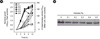
The effects of glucose on hupA transcription in the presence or absence of Fur were examined by culturing the two PhupA::lacZ transcriptional reporter strains, RC120 with wild-type fur and RC124 with mutated fur, in TES-DF-HI broths containing 5 or 25 µM FC at various glucose concentrations (0 to 0.5%). The growths of the two strains were slightly increased in the presence of glucose, but not in proportion to glucose concentrations (Figs. 1 and 2). However, in RC120, glucose dose-dependently repressed hupA transcription and completely repressed it at more than 0.4% (p < 0.05 by One Way ANOVA) (Fig. 1A). At 25 µM FC, hupA transcription in RC120 was completely repressed, and thus, the effects of glucose on hupA transcription could not be observed (data not shown). The effect of glucose on HupA production was also observed under the same conditions. At 5 µM FC (Fig. 1B), glucose dose-dependently repressed HupA production in M06-24/O and completely repressed it at 0.5%. At 25 µM FC, the effect of glucose on HupA production could not be observed in M06-24/O.
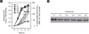
In RC124, hupA transcription appeared to be dose-dependently repressed by glucose (p < 0.05 by One Way ANOVA), but was maintained at considerable levels (approximately 40 Miller units) even in the presence of glucose at > 0.4% (Fig. 2A). Similar results were observed at 25 µM FC (data not shown). In contrast, HupA production in CMM2303 (Fig. 2B) was slightly reduced in the presence of glucose, but was maintained dose-independently at considerable levels in the presence of 0.5% glucose. Similar results were also observed at 25 µM FC (data not shown). A slight discrepancy was observed between hupA transcription and HupA protein levels. This discrepancy was probably due to the presence of TES as a pH-buffering agent and the likely low pH resulting from glucose catabolism, which could have synergistically inhibited the measurement of β-galactosidase activity. During a pilot experiment, we found that measured β-galactosidase activities were lower in media containing TES than in media not containing TES (data not shown).
Effects of cAMP on HupA expression
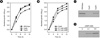
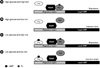
To find the connection between the effects of glucose and Crp on hupA expression, the effects of cAMP on V. vulnificus growth and HupA expression were determined by culturing the three strains, M06-24/O containing wild-type cyaA, RC386 containing mutated cyaA, and RC390 containing in trans complemented cyaA in deferrated HI broths containing 5 µM FC, and then comparing HupA production (Fig. 3A and 3C). The growth and HupA production of M06-24/O were inhibited by mutating cyaA, and these effects were prevented by complementing wild-type cyaA. In addition, bacterial growth and HupA production in RC386 were dose-dependently increased when exogenous cAMP was added to deferrated HI broths containing 5 µM FC (Fig. 3B and 3D).
Effects of iron on hupA expression in the presence or absence of Crp or Fur
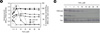
To observe the effects of iron on V. vulnificus growth and hupA transcription in the presence or absence of Crp or Fur, the three PhupA::lacZ transcriptional reporter strains, RC20 containing wild-type crp and fur, RC122 containing mutated crp, and RC124 containing mutated fur, were cultured in deferrated HI broths containing various concentrations of FC (0~30 µM) for 12 h (Fig. 4A). FC dose-dependently stimulated the growths of the three strains under severely iron-deficient media (< 5 µM FC), but not under moderately iron-deficient media (5 to 15 µM FC) and iron-sufficient media (> 15 µM FC). In RC120, FC dose-dependently increased hupA transcription under severely iron-deficient media, but dose-dependently repressed it under moderately iron-deficient media (p < 0.05 by One Way ANOVA) and completely repressed it under iron-sufficient media. The level of hupA transcription reached a peak at around 5 µM FC. In RC122, FC dose-dependently repressed hupA transcription even under severely iron-deficient media (p < 0.05 by One Way ANOVA) and completely repressed hupA transcription under moderately iron-deficient media. In RC124, FC dose-dependently increased hupA transcription under severely iron-deficient media (p < 0.05 by One Way ANOVA), but did not repress it under moderately iron-deficient or iron-sufficient media (p > 0.05 by One Way ANOVA). Moreover, under severely iron-deficient media, hupA transcription levels in RC122 were lower than in RC120 and RC124, and those in RC124 were lower than in RC120 but higher than in RC122 (p < 0.05 by Student's t-test).
The effects of iron on HupA production in the presence or absence of Crp or Fur were also determined by culturing M06-24/O with wild-type crp and fur, CMM710 with mutated crp, and CMM2303 with mutated fur, under the same conditions (Fig. 4B). In M06-24/O, FC appeared to have no significant effect on HupA production under severely iron-deficient media, but dose-dependently repressed it under moderately iron-deficient conditions and almost completely repressed it under iron-sufficient media. However, a slight discrepancy was observed between hupA transcription and HupA protein levels under severely iron-deficient conditions, suggesting the involvement of post-transcriptional mechanisms. In CMM710, FC dose-dependently repressed HupA production even under severely iron-deficient conditions and completely repressed it under moderately iron-deficient conditions. In CMM2303, FC appeared to have no significant effect on HupA production at any concentration examined. Moreover, HupA production levels under iron-deficient media were lower in CMM710 than in M06-24/O, and higher in CMM2303 than in CMM710.
DISCUSSION
Iron is essential for activating many catabolite enzymes, especially those involved in the electron transport system, and eventually, for efficient energy production and bacterial growth, although excessive iron is rather toxic to bacteria. This study shows that iron dose-dependently stimulates V. vulnificus growth under severely iron-deficient conditions, but not under moderately iron-deficient or iron-sufficient conditions. This iron-dependent biphasic growth suggests that iron availability acts as a double-edged sword in V. vulnificus.
Bacteria acquire energy for growth via catabolism, and Crp is a global regulator responsible for catabolite repression. Furthermore, glucose is the preferred energy source in most bacteria including V. vulnificus (13). The presence of glucose represses the expressions of a large number of genes associated with catabolism, and conversely, the absence of glucose stimulates the expressions of these genes via Crp. Accordingly, bacterial growth under glucose-deficient contions is certainly Crp-dependent. This study shows that V. vulnificus growth is severely inhibited in the absence of Crp regardless of iron concentration, whereas, in a previous study, we found that growth inhibition in the absence of Crp was recovered by glucose supplementation (19). These findings indicate that Crp is essential for energy production and V. vulnificus growth, especially under glucose-deficient conditions.
We propose a model for coordinated regulation of hupA expression by Crp and Fur, as in Fig. 5. The present study confirms that Crp positively affects hupA expression. A previous study demonstrated that Crp regulates hupA expression by directly binding to the -186 to -166 positions from the hupA transcription start site (10). In the present study, hupA expression was observed despite the absence of Crp under severely iron-deficient conditions, and hupA expression levels were higher in the presence of Crp than in the absence of Crp. In addition, under the same conditions, iron increased hupA expression in the presence of Crp, but repressed it in the absence of Crp. These findings indicate that iron limitation or the absence of Fur-mediated repression is an essential signal for constitutional hupA expression, whereas Crp is not essential for constitutional hupA expression but is required for optimal hupA expression, and that the effect of Crp as an activator is greater than the effect of Fur as a repressor in severely iron-deficient conditions. Furthermore, the present study shows that glucose represses hupA expression, and that a mutation in cyaA encoding adenylate cyclase required for cAMP synthesis represses HupA production, and this repression is recovered by adding exogenous cAMP. These findings indicate that the glucose- or Crp-mediated regulation of hupA expression is mediated by cAMP. Accordingly, it is likely that glucose deprivation increases cAMP by activating adenylate cyclase, cAMP binds to and activates Crp, and cAMP-Crp complex regulates hupA expression by directly binding the Crp-inding site, as described above.
Other iron-uptake systems in V. vulnificus are also likely to be under the positive control of Crp. Our recent studies show that Crp positively regulates the expression of vuuA encoding vulnibactin receptor (16) and the expression of iutA encoding E. coli aerobactin receptor (27). Furthermore, it has been reported that Crp also positively regulates the expression of vvhBA encoding a cytolysin/hemolysin involved in the release of intracellular iron, including hemoglobin (28, 29). This Crp- or glucose-mediated regulation of iron-uptake systems implies that glucose deprivation is an important signal for the optimal induction of iron-uptake systems, and that Crp is involved in the coordination of iron and carbon metabolisms (14). Accordingly, it is likely that the acquisition of iron should be increased to stimulate catabolism and to produce energy efficiently, especially under glucose-deficient conditions, and that eventually, this metabolically coordinated acquisition and effective utilization of iron is crucial for successful establishment of infection.
In contrast, the absence of Fur can also affect bacterial growth by increasing iron uptake, especially under iron-deficient conditions, because iron is also essential for efficient energy production. However, in this study, the absence of Fur appeared to have no significant effect on V. vulnificus growth. This may be because deferrated HI broths containing < 5 µM FC used in this study represents iron-deficient conditions (iron is deficient but its availability is not limited). Conversely, in our previous study, the absence of Fur could stimulate V. vulnificus growth under iron-limited conditions (sufficient iron is present but its availability is severely limited), for example, in deferrated HI broths containing transferrin-bound iron as the sole iron source [17]. Accordingly, the effect of Fur on V. vulnificus growth is likely to be considerably condition-dependent.
It is known that hupA expression is negatively controlled by iron or Fur (11, 12). Similarly, in the present study, iron repressed hupA expression in the presence of Fur, but derepressed it in the absence of Fur under moderately iron-deficient and iron-sufficient conditions. Moreover, glucose completely repressed hupA expression in the presence of Fur but not in the absence of Fur. These results indicate that Fur or iron is essential for the repression of hupA expression regardless of the presence or absence of Crp or glucose availability. However, direct Fur binding to hupA promoter has not been demonstrated, although, based on sequence analysis, Fur is likely to bind to the -37 to -19 positions from the hupA transcription start site to prevent the binding of RNA polymerase (12). This Fur-mediated complete repression of hupA expression implies that iron limitation is an essential signal for the induction of hupA expression and that Fur acts as a specific or local regulator that controls hupA expression in response to iron levels (14). Similarly, the expressions of other V. vulnificus iron-uptake systems are also known to be under the negative control of Fur or iron (30~33). Furthermore, in a previous study, we found that Fur negatively regulated the expression of vvhBA encoding a cytolysin/hemolysin, which destroys a variety of cells, including red blood cells, and causes the release of intracellular iron, including hemoglobin (20).
In the present study, hupA expression levels under severely iron-deficient conditions were higher in the wild-type background than in the fur-mutated background, which suggests that fur mutation negatively affects hupA expression. Probably, other iron-uptake systems including the VuuA-mediated iron-uptake system would be de-repressed, iron uptake would be increased, and intracellular iron would be increased in the fur-mutated strain, and these would partially repress hupA expression versus the wild-type strain.
In conclusion, this study shows that hupA expression is under the coordinate control of Crp, which responds to glucose availability, and of Fur, which responds to iron availability, and that Crp is not essential for constitutional hupA expression but is required for optimal hupA expression, whereas Fur is essential for the prevention of hupA over-expression.
 XML Download