Abstract
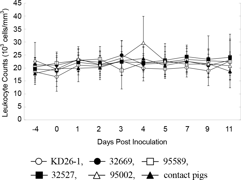
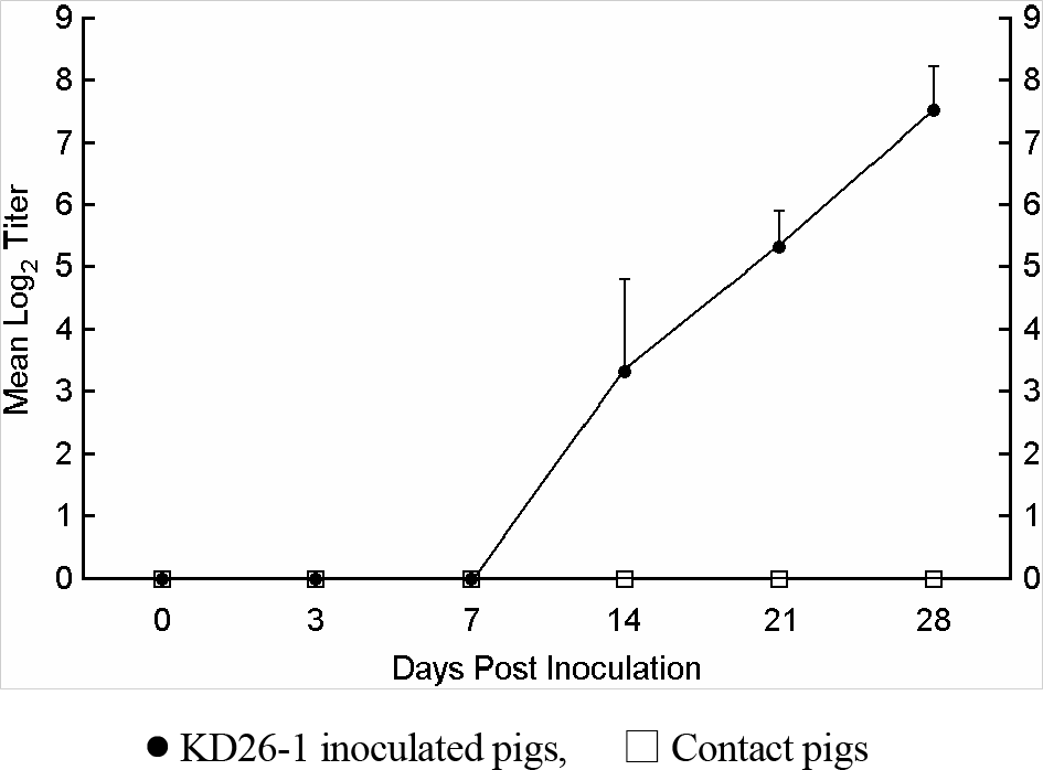
To select a less pathogenic bovine viral diarrhea virus (BVDV) strain for the construction of chimeric pestivirus harboring classical swine fever virus (CSFV) E2 gene, five Korean BVDV isolates (four type 1 isolates and a type 2 isolate) were evaluated for their pathological and biological properties with respect to porcine infection. Each of five groups of 100-day-old pigs was inoculated intranasally with one of the five BVDV isolates. No clinical sign or leukopenia was observed in any pig throughout the duration of the experiment, but viruses were detected in blood, nasal discharges and postmortem samples using RT-PCR. These results indicated that although the five BVD viruses could infect pigs, they did not cause clinically apparent symptoms. Because of its proper infection dynamics shown in this preliminary animal study and its fast growth rate and quick CPE in cell culture, one isolate (KD26-1) was chosen among the five isolates to test its virulence and immunogenic properties in 40-day-old piglets. Neither clinical sign nor pathological lesion was observed in 40-day-old piglets during the course of infection of isolate KD26-1. The first neutralizing antibodies were detectable 14 days post-inoculation (PI) and increased to 1:128~1:256 28 days PI. A BVDV specific gene was detectable by RT-PCR in tonsil, spleen, inguinal lymph node and brain until 14 days PI. According to this study, it can be concluded that isolate KD26-1 has little pathological effect in pigs and is a candidate for construction of chimeric pestivirus harboring CSFV E2 gene.
Go to : 
REFERENCES
1). Thiel HJ., Plagemann PGW., Moenning V. Pestiviruses. Fields BN, Knipe DM, Howley PM, editors. editors.Fields Virology. 3rd ed.New York: Raven Press;1996. p. 1059–73.
2). Carbrey EA., Stewart WC., Kresse JI., Snyder ML. Natural infection of pigs with bovine viral diarrhea virus and its differential diagnosis from hog cholera. J Am Vet Med Assoc. 1976. 169:1217–9.
3). Fernelius AL., Amtower WC., Lambert G., McClurkin AW., Matthews PJ. Bovine viral diarrhea virus in swine: characteristics of virus recovered from naturally and experimentally infected swine. Can J Comp Med. 1973. 37:13–20.
4). Paton DJ., Simpson V., Done SH. Infection of pigs and cattle with bovine viral diarrhoea virus on a farm in England. Vet Rec. 1992. 131:185–8.

5). Terpstra C., Wensvoort G. Natural infections of pigs with bovine viral diarrhoea virus associated with signs resembling swine fever. Res Vet Sci. 1988. 45:137–42.

6). Ridpath JF., Bolin SR., Dubovi EJ. Segregation of bovine viral diarrhea virus into genotypes. Virology. 1994. 205:66–74.

7). Kummerer BM., Tautz N., Becher P., Thiel H., Meyers G. The genetic basis for cytopathogenicity of pestiviruses. Vet Microbiol. 2000. 77:117–28.
8). Tautz N., Meyers G., Stark R., Dubovi EJ., Thiel HJ. Cytopathogenicity of a pestivirus correlates with a 27-nucleotide insertion. J Virol. 1996. 70:7851–8.

9). Vassilev VB., Donis RO. Bovine viral diarrhea virus induced apoptosis correlates with increased intracellular viral RNA accumulation. Virus Res. 2000. 69:95–107.

10). Graham DA., Calvert V., German A., McCullough SJ. Pestiviral infections in sheep and pigs in Northern Ireland. Vet Rec. 2001. 148:69–72.

11). Fernelius AL., Amtower WC., Malmquist WA., Lambert G., Matthews PJ. Bovine viral diarrhea virus in swine: neutralizing antibody in naturally and experimentally infected swine. Can J Comp Med. 1973. 37:96–102.
12). Loeffen WL., van Beuningen A., Quak S., Elbers AR. Seroprevalence and risk factors for the presence of ruminant pestiviruses in the Dutch swine population. Vet Microbiol. 2009. 136:240–5.

13). Wieringa-Jelsma T., Quak S., Loeffen WL. Limited BVDV transmission and full protection against CSFV transmission in pigs experimentally infected with BVDV type 1b. Vet Microbiol. 2006. 118:26–36.

14). Walz PH., Baker JC., Mullaney TP., Maes RK. Experimental inoculation of pregnant swine with type 1 bovine viral diarrhoea virus. J Vet Med B Infect Dis Vet Public Health. 2004. 51:191–3.

15). Kulcsar G., Soos P., Kucsera L., Glavits R., Palfi V. Pathogenicity of a bovine viral diarrhoea virus strain in pregnant sows: short communication. Acta Vet Hung. 2001. 49:117–20.
16). Dong XN., Chen YH. Marker vaccine strategies and candidate CSFV marker vaccines. Vaccine. 2007. 25:205–30.

17). Reimann I., Depner K., Trapp S., Beer M. An avirulent chimeric Pestivirus with altered cell tropism protects pigs against lethal infection with classical swine fever virus. Virology. 2004. 322:143–57.

18). van Gennip HG., van Rijn PA., Widjojoatmodjo MN., de Smit AJ., Moormann RJ. Chimeric classical swine fever viruses containing envelope protein E(RNS) or E2 of bovine viral diarrhoea virus protect pigs against challenge with CSFV and induce a distinguishable antibody response. Vaccine. 2000. 19:447–59.

19). Gilbert SA., Burton KM., Prins SE., Deregt D. Typing of bovine viral diarrhea viruses directly from blood of persistently infected cattle by multiplex PCR. J Clin Microbiol. 1999. 37:2020–3.

20). Anonymous. Bovine Viral Diarrhoea. Manual of Diagnostic Tests and Vaccines for Terrestrial Animals (mammals, birds and bees). 6th ed.Paris: OIE;2008. p. 698–711.
21). Cho IS. Studies on serological and genetical characteristics of bovine viral diarrhea viruses isolated in Korea [PhD thesis]. Seoul: Konkuk University. 2001.
Go to : 
 | Figure 1.Leukocyte counts (103 cells/mm3) during the course of intranasal infection in the first animal study. Mean leukocyte counts in pigs inoculated with isolates KD26-1, 32669, 95589, 32527 and 95002 and those of contact pigs are given. Standard deviations are shown as error bars. |
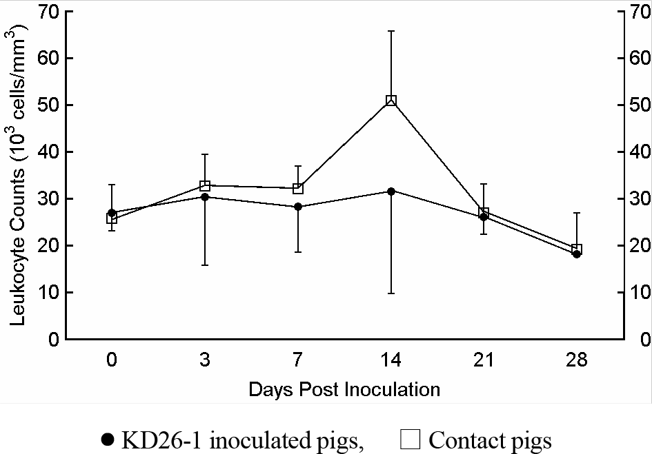
 | Figure 2.Leukocyte counts (103/mm3) of pigs after intramuscular inoculation with KD26-1 (107.5 TCID50/head). Mean leukocyte counts in pigs inoculated with isolates KD26-1 and those of contact pigs are given. Standard deviations are shown as error bars. |
 | Figure 3.Virus neutralization titers of sera from pigs after intramuscular inoculation with KD26-1 (107.5 TCID50/head). Mean virus neutralization titers in pigs inoculated with isolates KD26-1 and those of contact pigs are given. Standard deviations are shown as error bars. |
Table 1.
The properties of five BVDV isolates used in animal experiments
| BVD isolate | Origin | CPE (dpia) | Genotypeb |
|---|---|---|---|
| KD26-1 | Diarrhea | 3~4 | 1 |
| 32669 | Persistent infection | 4~5 | 1 |
| 95002 | Respiratory infection | 6~7 | 2 |
| 95589 | Diarrhea | 4~5 | 1 |
| 32527 | Diarrhea | 4~5 | 1 |
Table 2.
RT-PCR results for detection of viral antigen from nasal swabs of five pig groups during the course of intranasal infection
| Group | Inoculated virus | Days post inoculation | |||||||||
|---|---|---|---|---|---|---|---|---|---|---|---|
| -4 | 0 | 1 | 2 | 3 | 4 | 5 | 7 | 9 | 11 | ||
| I | KD26-1 | 0/6a | 0/6 | 0/6 | 2/6 | 0/6 | 1/6 | 0/6 | 2/6 | 0/4 | 0/2 |
| Contact | 0/2 | 0/2 | 0/2 | 0/2 | 0/2 | 0/2 | 1/2 | 1/2 | 0/1 | 1/1 | |
| II | 32669 | 0/6 | 0/6 | 1/6 | 1/6 | 0/6 | 0/6 | 0/6 | 0/6 | 0/4 | 0/2 |
| Contact | 0/2 | 0/2 | 0/2 | 0/2 | 0/2 | 0/2 | 0/2 | 0/2 | 0/1 | 0/1 | |
| III | 95002 | 0/6 | 0/6 | 2/6 | 0/6 | 0/6 | 1/6 | 1/6 | 2/6 | 0/4 | 0/2 |
| Contact | 0/2 | 0/2 | 0/2 | 0/2 | 0/2 | 0/2 | 0/2 | 0/2 | 0/1 | 0/1 | |
| IV | 95589 | 0/6 | 0/6 | 0/6 | 0/6 | 1/6 | 0/6 | 1/6 | 1/6 | 0/4 | 0/2 |
| Contact | 0/2 | 0/2 | 0/2 | 1/2 | 1/2 | 0/2 | 1/2 | 0/2 | 0/1 | 0/1 | |
| V | 32527 | 0/6 | 0/6 | 0/6 | 0/6 | 2/6 | 1/6 | 1/6 | 0/6 | 0/4 | 0/2 |
| Contact | 0/2 | 0/2 | 0/2 | 0/2 | 0/2 | 0/2 | 1/2 | 1/2 | 1/1 | 0/1 | |
Table 3.
RT-PCR results for detection of viral antigen from blood samples of five pig groups during the course of intranasal infection
| Group | Inoculated virus | Days post inoculation | |||||||||
|---|---|---|---|---|---|---|---|---|---|---|---|
| -4 | 0 | 1 | 2 | 3 | 4 | 5 | 7 | 9 | 11 | ||
| I | KD26-1 | 0/6a | 0/6 | 0/6 | 0/6 | 1/6 | 1/6 | 1/6 | 0/6 | 0/4 | 0/2 |
| Contact | 0/2 | 0/2 | 0/2 | 0/2 | 0/2 | 0/2 | 1/2 | 0/2 | 0/1 | 0/1 | |
| II | 32669 | 0/6 | 0/6 | 0/6 | 0/6 | 0/6 | 1/6 | 1/6 | 1/6 | 0/4 | 0/2 |
| Contact | 0/2 | 0/2 | 0/2 | 0/2 | 0/2 | 0/2 | 0/2 | 0/2 | 0/1 | 0/1 | |
| III | 95002 | 0/6 | 0/6 | 0/6 | 0/6 | 0/6 | 0/6 | 1/6 | 0/6 | 0/4 | 0/2 |
| Contact | 0/2 | 0/2 | 0/2 | 0/2 | 0/2 | 1/2 | 0/2 | 0/2 | 0/1 | 0/1 | |
| IV | 95589 | 0/6 | 0/6 | 0/6 | 0/6 | 0/6 | 2/6 | 2/6 | 0/6 | 0/4 | 0/2 |
| Contact | 0/2 | 0/2 | 0/2 | 0/2 | 0/2 | 0/2 | 0/2 | 0/2 | 0/1 | 0/1 | |
| V | 32527 | 0/6 | 0/6 | 0/6 | 0/6 | 0/6 | 0/6 | 0/6 | 0/6 | 0/4 | 0/2 |
| Contact | 0/2 | 0/2 | 0/2 | 0/2 | 0/2 | 0/2 | 0/2 | 1/2 | 0/1 | 0/1 | |
Table 4.
RT-PCR detection of viral antigen from tissues samples of five pig groups at 7, 9, and 11 days PI
| Group | Inoculated virus | 7 dpia | 9 dpi | 11 dpi |
|---|---|---|---|---|
| I | KD26-1 | -b | Tc, Bd, Le | T, B, L |
| Contact | – | NPf | – | |
| II | 32669 | T, 2Bg, L | L | L |
| Contact | T | NP | – | |
| III | 95002 | – | – | – |
| Contact | – | NP | – | |
| IV | 95589 | – | T | – |
| Contact | – | NP | – | |
| V | 32527 | – | – | T |
| Contact | – | NP | – |
Table 5.
Summary of viral antigen positive pigs (%) in thirty 100-day-old pigs inoculated intranasally with BVDV type 1 and 2
| Virus type | Antigen positive samples | No. of pigs tested | ||
|---|---|---|---|---|
| Nasal swabs | Blood | Tissues | ||
| Type 1 | 10 (42) | 9 (38) | 8 (33) | 24 (100) |
| Type 2 | 4 (67) | 1 (17) | 0 (0) | 6 (100) |
Table 6.
RT-PCR results for detection of viral antigen from organs of euthanized pigs after intramuscular inoculation with KD26-1 (107.5 TCID50/head)
| Organ | KD26-1 inoculated pigs | Contact pigs | ||||||||
|---|---|---|---|---|---|---|---|---|---|---|
| 3 da | 7 d | 14 d | 21 d | 28 d | 3 d | 7 d | 14 d | 21 d | 28 d | |
| Tonsil | 1/2b | 1/2 | 1/2 | 0/2 | 0/2 | 0/1 | 0/1 | 0/1 | 0/1 | 0/1 |
| Lung | 0/2 | 0/2 | 0/2 | 0/2 | 0/2 | 0/1 | 0/1 | 0/1 | 0/1 | 0/1 |
| Heart | 0/2 | 0/2 | 0/2 | 0/2 | 0/2 | 0/1 | 0/1 | 0/1 | 0/1 | 0/1 |
| Spleen | 0/2 | 1/2 | 0/2 | 0/2 | 0/2 | 0/1 | 0/1 | 0/1 | 0/1 | 0/1 |
| Liver | 0/2 | 0/2 | 0/2 | 0/2 | 0/2 | 0/1 | 0/1 | 0/1 | 0/1 | 0/1 |
| Kidney | 0/2 | 0/2 | 0/2 | 0/2 | 0/2 | 0/1 | 0/1 | 0/1 | 0/1 | 0/1 |
| Mesenteric LN | 0/2 | 0/2 | 0/2 | 0/2 | 0/2 | 0/1 | 0/1 | 0/1 | 0/1 | 0/1 |
| Ileum | 0/2 | 0/2 | 0/2 | 0/2 | 0/2 | 0/1 | 0/1 | 0/1 | 0/1 | 0/1 |
| Caecum | 0/2 | 0/2 | 0/2 | 0/2 | 0/2 | 0/1 | 0/1 | 0/1 | 0/1 | 0/1 |
| Inguinal LN | 0/2 | 0/2 | 1/2 | 0/2 | 0/2 | 0/1 | 0/1 | 0/1 | 0/1 | 0/1 |
| Brain | 1/2 | 0/2 | 0/2 | 0/2 | 0/2 | 0/1 | 0/1 | 0/1 | 0/1 | 0/1 |


 PDF
PDF ePub
ePub Citation
 Print
Print


 XML Download