Abstract
The infections by human papillomaviruses (HPVs) are clearly associated with the subsequent development of cervical cancer. In this study, HPV genotype distribution and prevalence were detected in Korean women from January to December 2008 using PCR-DNA sequencing. A total of 2,562 cervical samples from Korean women having routine Pap smear cytology screening were used. HPV DNA was extracted from cervical swab samples and amplified by PCR in L1 region of HPV. HPV DNA was detected in 23.2% and 65.5% from the groups of normal and abnormal Pap cytology, respectively. The prevalence of high-risk types of HPV had the highest frequency in the <30 year-olds' group (50.6%). The prevalence of HPV in normal, ASCUS, LSIL and HSIL groups was 23.2%, 58.1%, 96.3% and 97.0%, respectively. Moreover, the frequencies of the high-risk types of HPV were 16.2% in the normal Pap cytology, 44.7% in the ASCUS, 76.1% in the LSIL and 94.1% in the HSIL groups. The prevalence of the high-risk types of HPV increased in proportion to the severity of the cytological classification. In the HSIL group, HPV type 16 was the most frequently found at 32.4%, followed by types 58, 53 and 33 at 17.6%, 14.7% and 11.8%, respectively. HPV type 82 was found in 5.6% of the HSIL group and was not detected in the normal Pap cytology group. The frequency of high-risk type of HPV 82 is firstly reported in Korean women. This finding could be an informative basis for the development of future HPV vaccination strategies in Korean women.
REFERENCES
1). Bosch FX., Manos MM., Munoz N., Sherman M., Jansen AM., Peto J., Schiffman MH., Moreno V., Kurman R., Shah KV. Prevalence of human papillomavirus in cervical cancer: a worldwide perspective. International biological study on cervical cancer (IBSCC) Study Group. J Natl Cancer Inst. 1995. 87:796–802.
2). zur Hausen H. Papillomaviruses causing cancer: evasion from host-cell control in early events in carcinogenesis. J Natl Cancer Inst. 2000. 92:690–8.
3). Lazo PA. Papillomavirus integration: prognostic marker in cervical cancer? Am J Obstet Gynecol. 1997. 176:1121–2.

5). Lorincz AT., Reid R., Jenson AB., Greenberg MD., Lancaster W., Kurman RJ. Human papillomavirus infection of the cervix: relative risk associations of 15 common anogenital types. Obstet Gynecol. 1992. 79:328–37.
6). Munoz N., Bosch FX., de Sanjose S., Herrero R., Castellsague X., Shah KV., Snijders PJ., Meijer CJ. Epidemiologic classification of human papillomavirus types associated with cervical cancer. N Engl J Med. 2003. 348:518–27.

7). Walboomers JM., Jacobs MV., Manos MM., Bosch FX., Kummer JA., Shah KV., Snijders PJ., Peto J., Meijer CJ., Munoz N. Human papillomavirus is a necessary cause of invasive cervical cancer worldwide. J Pathol. 1999. 189:12–9.

8). Bollmann R., Mehes G., Torka R., Speich N., Schmitt C., Bollmann M. Determination of features indicating progression in atypical squamous cells with undetermined significance: human papillomavirus typing and DNA ploidy analysis from liquid-based cytologic samples. Cancer. 2003. 99:113–7.
9). Klug SJ., Hukelmann M., Hollwitz B., Duzenli N., Schopp B., Petry KU., Iftner T. Prevalence of human papilloma-virus types in women screened by cytology in Germany. J Med Virol. 2007. 79:616–25.

10). Weintraub J. Experience with new technologies within the context of Swiss practice and conclusions. Ann Pathol. 1999. 19:S96–8.
11). Jacobs MV., de Roda Husman AM., van den Brule AJ., Snijders PJ., Meijer CJ., Walboomers JM. Group-specific differentiation between high- and low-risk human papillomavirus genotypes by general primer-mediated PCR and two cocktails of oligonucleotide probes. J Clin Microbiol. 1995. 33:901–5.

12). Ylitalo N., Bergstrom T., Gyllensten U. Detection of genital human papillomavirus by single-tube nested PCR and type-specific oligonucleotide hybridization. J Clin Microbiol. 1995. 33:1822–8.

13). Coutlee F., Gravitt P., Kornegay J., Hankins C., Richardson H., Lapointe N., Voyer H., Franco E. Use of PGMY primers in L1 consensus PCR improves detection of human papillomavirus DNA in genital samples. J Clin Microbiol. 2002. 40:902–7.

14). van den Brule AJ., Pol R., Fransen-Daalmeijer N., Schouls LM., Meijer CJ., Snijders PJ. GP5+/6+ PCR followed by reverse line blot analysis enables rapid and high-throughput identification of human papillomavirus genotypes. J Clin Microbiol. 2002. 40:779–87.

15). Choi BS., Kim O., Park MS., Kim KS., Jeong JK., Lee JS. Genital human papillomavirus genotyping by HPV oligonucleotide microarray in Korean commercial sex workers. J Med Virol. 2003. 71:440–5.

16). Choi YD., Jung WW., Nam JH., Choi HS., Park CS. Detection of HPV genotypes in cervical lesions by the HPV DNA Chip and sequencing. Gynecol Oncol. 2005. 98:369–75.

17). Klaassen CH., Prinsen CF., de Valk HA., Horrevorts AM., Jeunink MA., Thunnissen FB. DNA microarray format for detection and subtyping of human papillomavirus. J Clin Microbiol. 2004. 42:2152–60.

18). Oh TJ., Kim CJ., Woo SK., Kim TS., Jeong DJ., Kim MS., Lee S., Cho HS., An S. Development and clinical evaluation of a highly sensitive DNA microarray for detection and genotyping of human papillomaviruses. J Clin Microbiol. 2004. 42:3272–80.

19). Lee KO., Seong HS., Chung SJ., Jung NY., Lee HJ., Kim KT. Genotype frequency of human papillomavirus determined by PCR and DNA sequencing in Korean women. Korean J Clinical Lab Sci. 2006. 38:99–104.
20). Speich N., Schmitt C., Bollmann R., Bollmann M. Human papillomavirus (HPV) study of 2916 cytological samples by PCR and DNA sequencing: genotype spectrum of patients from the west German area. J Med Microbiol. 2004. 53:125–8.

21). Cope JU., Hildesheim A., Schiffman MH., Manos MM., Lorincz AT., Burk RD., Glass AG., Greer C., Buckland J., Helgesen K., Scott DR., Sherman ME., Kurman RJ., Liaw KL. Comparison of the hybrid capture tube test and PCR for detection of human papillomavirus DNA in cervical specimens. J Clin Microbiol. 1997. 35:2262–5.

22). Coste-Burel M., Besse B., Moreau A., Imbert BM., Mensier A., Sagot P., Lopes P., Billaudel S. Detection of human papillomavirus in squamous intraepithelial lesions by consensus and type-specific polymerase chain reaction. Eur J Obstet Gynecol Reprod Biol. 1993. 52:193–200.

23). Hwang HS., Park M., Lee SY., Kwon KH., Pang MG. Distribution and prevalence of human papillomavirus genotypes in routine Pap smear of 2,470 Korean women determined by DNA Chip. Cancer Epidemiol Biomarkers Prev. 2004. 13:2153–6.
24). Cuzick J. Human papillomavirus testing for primary cervical cancer screening. JAMA. 2000. 283:108–9.

25). de Roda Husman AM., Walboomers JM., Van den Brule AJ., Meijer CJ., Snijders PJ. The use of general primers GP5 and GP6 elongated at their 3′ ends with adjacent highly conserved sequences improves human papilloma-virus detection by PCR. J Gen Virol. 1995. 76:1057–62.

26). Altschul SF., Madden TL., Schaffer AA., Zhang J., Zhang Z., Miller W., Lipman DJ. Gapped BLAST and PSI-BLAST: a new generation of protein database search programs. Nucleic Acids Res. 1997. 25:3389–402.

27). Agarossi A., Ferrazzi E., Parazzini F., Perno CF., Ghisoni L. Prevalance and type distribution of high-risk human papillomavirus infection in women undergoing voluntary cervical cancer screening in Italy. J Med Virol. 2009. 81:529–35.
28). Kim KH., Yoon MS., Na YJ., Park CS., Oh MR., Moon WC. Development and evaluation of a highly sensitive human papillomavirus genotyping DNA chip. Gynecol Oncol. 2006. 100:38–43.

29). Kim CJ., Jeong JK., Park M., Park TS., Park TC., Namkoong SE., Park JS. HPV oligonucleotide microarray-based detection of HPV genotypes in cervical neoplastic lesions. Gynecol Oncol. 2003. 89:210–7.

30). Kahng J., Lee HJ. Clinical efficacy of HPV DNA chip test in the era of HPV vaccination. Korean J Lab Med. 2008. 28:70–8.
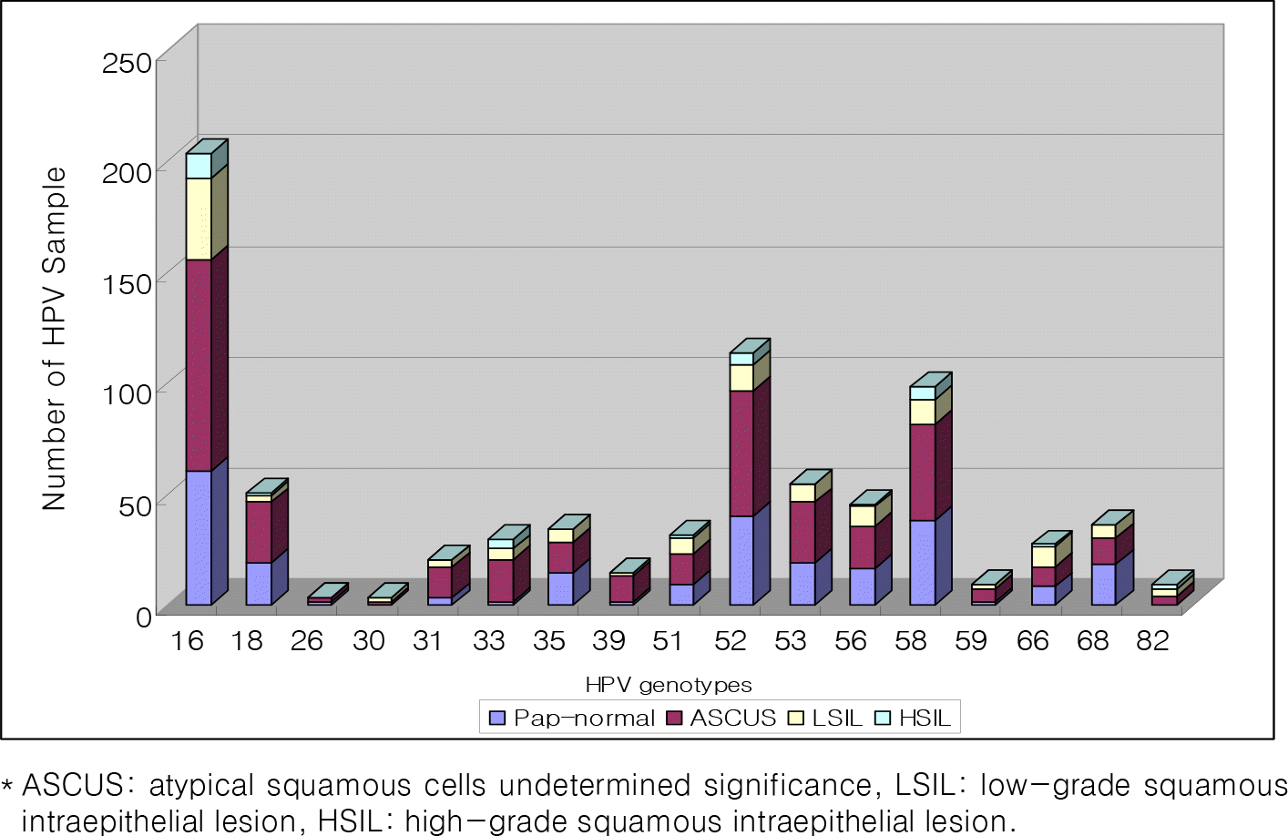
Figure 1.
Distribution of carcinogenic HPV types in cytological normal women and abnormal women, including ASCUS, LSIL and HSIL, respectively.
∗ ASCUS: atypical squamous cells undetermined significance, LSIL: low–grade squamous intraepithelial lesion, HSIL: high–grade squamous intraepithelial lesion.

Table 1.
Age distribution and prevalence of high- or low-risk HPVs in Koreans
| >30 years | 31~40 years | 41~50 years | >50 years | |
|---|---|---|---|---|
| High-risk types HPVa | 37.8% (220/582) | 31.3% (271/867) | 29.8% (207/699) | 23.0% (85/369) |
| Low-risk types HPVb | 10.3% (60/582) | 5.4% (47/867) | 5.6% (39/699) | 7.9% (29/369) |
| Uncharacterized | 2.6% (15/582) | 2.4% (21/867) | 2.0% (14/699) | 3.0% (11/369) |
| Total % of HPV | 50.6% (295/582) | 39.1% (339/867) | 37.4% (261/699) | 34.7% (125/369) |
Table 2.
Type-specific HPV prevalence determined by PCR-sequencing in normal and abnormal cytology groups
| Pap-normal | Pap-abnormal | ASCUS | LSIL | HSIL | Total | P value | |||||||
|---|---|---|---|---|---|---|---|---|---|---|---|---|---|
| n | % | n | % | n | % | n | % | N | % | n | % | ||
| Total No | 1529 | 59.7 | 1033 | 40.3 | 836 | 32.6 | 163 | 6.4 | 34 | 1.3 | 2562 | 100 | <0.05 |
| Any type | 355 | 23.2 | 676 | 65.5 | 486 | 58.1 | 157 | 96.3 | 33 | 97.0 | 1031 | 40.2 | |
| High risk types of HPV (Carcinogenic types) | |||||||||||||
| 16a | 60 | 3.9 | 143 | 13.8 | 95 | 11.4 | 37 | 22.7 | 11 | 32.4 | 203 | 7.9 | <0.05 |
| 18 | 19 | 1.2 | 31 | 3.0 | 27 | 3.2 | 3 | 1.8 | 1 | 2.9 | 50 | 2.0 | cNS |
| 26 | 1 | 0.1 | 2 | 0.2 | 2 | 0.2 | 0 | 0.0 | 0 | 0.0 | 3 | 0.1 | NS |
| 30 | 0 | 0.0 | 3 | 0.3 | 1 | 0.1 | 2 | 1.2 | 0 | 0.0 | 3 | 0.1 | NS |
| 31 | 3 | 0.2 | 17 | 1.6 | 14 | 1.7 | 3 | 1.8 | 0 | 0.0 | 20 | 0.8 | NS |
| 33a | 1 | 0.1 | 28 | 2.7 | 19 | 2.3 | 5 | 3.1 | 4 | 11.8 | 29 | 1.1 | <0.05 |
| 35 | 14 | 0.9 | 20 | 1.9 | 14 | 1.7 | 6 | 3.7 | 0 | 0.0 | 34 | 1.3 | NS |
| 39 | 1 | 0.1 | 13 | 1.3 | 12 | 1.4 | 1 | 0.6 | 0 | 0.0 | 14 | 0.5 | NS |
| 51 | 9 | 0.6 | 22 | 2.1 | 14 | 1.7 | 7 | 4.3 | 1 | 2.9 | 31 | 1.2 | NS |
| 52a | 40 | 2.6 | 73 | 7.1 | 56 | 6.7 | 12 | 7.4 | 5 | 14.7 | 113 | 4.4 | <0.05 |
| 53 | 19 | 1.2 | 35 | 3.4 | 27 | 3.2 | 8 | 4.9 | 0 | 0.0 | 54 | 2.1 | NS |
| 56 | 16 | 1.0 | 29 | 2.8 | 19 | 2.3 | 9 | 5.5 | 1 | 2.9 | 45 | 1.8 | NS |
| 58a | 38 | 2.5 | 60 | 5.8 | 43 | 5.1 | 11 | 6.7 | 6 | 17.6 | 98 | 3.8 | <0.05 |
| 59 | 1 | 0.1 | 8 | 0.8 | 6 | 0.7 | 2 | 1.2 | 0 | 0.0 | 9 | 0.4 | NS |
| 66 | 8 | 0.5 | 19 | 1.8 | 9 | 1.1 | 9 | 5.5 | 1 | 2.9 | 27 | 1.1 | NS |
| 68 | 18 | 1.2 | 18 | 1.7 | 12 | 1.4 | 6 | 3.7 | 0 | 0.0 | 36 | 1.4 | NS |
| 82a | 0 | 0.0 | 9 | 0.9 | 4 | 0.5 | 3 | 1.8 | 2 | 5.9 | 9 | 0.4 | <0.05 |
| subtotal | 248 | 16.2 | 530 | 51.3 | 374 | 44.7 | 124 | 76.1 | 32 | 94.1 | 778 | 30.4 | b<0.0001 |
| Low risk types of HPV | |||||||||||||
| 6 | 10 | 0.7 | 7 | 0.7 | 2 | 0.2 | 5 | 3.1 | 0 | 0.0 | 17 | 0.7 | NS |
| 11 | 1 | 0.1 | 4 | 0.4 | 1 | 0.1 | 3 | 1.8 | 0 | 0.0 | 5 | 0.2 | NS |
| 32 | 2 | 0.1 | 1 | 0.1 | 1 | 0.1 | 0 | 0.0 | 0 | 0.0 | 3 | 0.1 | NS |
| 34 | 2 | 0.1 | 4 | 0.4 | 4 | 0.5 | 0 | 0.0 | 0 | 0.0 | 6 | 0.2 | NS |
| 40 | 2 | 0.1 | 2 | 0.2 | 2 | 0.2 | 0 | 0.0 | 0 | 0.0 | 4 | 0.2 | NS |
| 42 | 0 | 0.0 | 4 | 0.4 | 2 | 0.2 | 2 | 1.2 | 0 | 0.0 | 4 | 0.2 | NS |
| 43 | 0 | 0.0 | 9 | 0.9 | 8 | 1.0 | 1 | 0.6 | 0 | 0.0 | 9 | 0.4 | NS |
| 54 | 14 | 0.9 | 7 | 0.7 | 6 | 0.7 | 0 | 0.0 | 1 | 2.9 | 21 | 0.8 | NS |
| 61 | 12 | 0.8 | 16 | 1.5 | 12 | 1.4 | 4 | 2.5 | 0 | 0.0 | 28 | 1.1 | NS |
| 62 | 16 | 1.0 | 17 | 1.6 | 13 | 1.6 | 4 | 2.5 | 0 | 0.0 | 33 | 1.3 | NS |
| 70 | 21 | 1.4 | 19 | 1.8 | 15 | 1.8 | 4 | 2.5 | 0 | 0.0 | 40 | 1.6 | NS |
| Low risk types of HPV | |||||||||||||
| 81 | 3 | 0.2 | 8 | 0.8 | 6 | 0.7 | 2 | 1.2 | 0 | 0.0 | 11 | 0.4 | NS |
| 84 | 2 | 0.1 | 3 | 0.3 | 3 | 0.4 | 0 | 0.0 | 0 | 0.0 | 5 | 0.2 | NS |
| subtotal | 85 | 5.6 | 101 | 9.8 | 75 | 9.0 | 25 | 15.3 | 1 | 2.9 | 186 | 7.3 | NS |
| Uncharacterized | 22 | 1.4 | 45 | 4.4 | 37 | 4.4 | 8 | 4.9 | 0 | 0.0 | 67 | 2.6 | |
Note: ASCUS; atypical squamous cells undetermined significance, LSIL; low-grade squamous intraepithelial lesion, HSIL; high-grade squamous intraepithelial lesion.
a : P value was observed between normal cytology group and HSIL group. Five high-risk types of HPV (HPV-16, 33, 52, 58, 82) were statistically significant between two groups.


 PDF
PDF ePub
ePub Citation
 Print
Print


 XML Download