Abstract
During 3 years surveillance (January 2001 through December 2003) for acute gastroenteritis in human in Daejeon region, 432 out of 4,869 stool samples were selected as rotavirus-positive specimens by means of antigen-capture enzyme-linked immunosorbent assay (ELISA). The P (VP4) and G (VP7) genotypes for 432 stool samples were investigated by reverse transcription polymerase chain reaction (RT-PCR) and nested multiplex PCR. The most prevalent P subtype was P[8] (44.9%), followed by P[4] (25.7%) and P[6] (17.1%). No cases for P[10] and P[9] subtypes were found through the study. In G subtyping, G1 (53.2%) was the most frequently found G type, followed by G2 (23.1%), G3 (9.5%), G4 (6.7%), and G9 (0.9%). The order of detection rates for G2, G3 and G4 was variable by years. The most common G- and P- type combination found in this study was G1P[8] (33.1%), followed by G2P[4] (20.4%), G1P[6] (10.0%), G3P[8] (7.2%) and G4P[6] (4.2%). The mixed types of G and P were observed most frequently in P[8] (1.4%) and G1 (3.2%), respectively. This is the first molecular epidemiological study for Group A rotavirus in Daejeon region. The results might be useful data for evaluating the epidemiological status of rotaviral diarrhea in the region.
Go to : 
References
1). 김은정, 서병태, 박석기, 이정자. 서울지역 설사환자에 서 분리한 A군 Rotavirus의 Multiplex PCR을 이용한 VP4, VP7 유전자형. 대한미생물학회지. 32:291–297. 2002.
2). Barnes GL. Etiology of acute gastroenteritis in hospitalized children in Melbourne, Australia, from April 1980 to March 1993. J Clin Microbiol. 36:133–138. 1998.
3). Beards GM, Dessel BU, Flewett TH. Temporal and geographical distributions of human rotavirus serotypes, 1983 to 1988. J Clin Microbiol. 27:2827–2833. 1989.

4). Bern C, Martines J, Zoysa I, Glass RI. The magnitude of the global problem of diarrheal disease: a ten-year up-date. Bull WHO. 70:705–714. 1992.
5). Bishop RF. Natural history of human rotavirus infections. pp. p. 131–167. In. Vial Infections of the Gastrointerstinal Tract. 2nd ed.Kapikian AZ, editor. (Ed).Marcel Dekker;New York: 1994.
6). Bishop RF, Davidson GP, Holmes IH, Ruck BJ. Detection of a new virus by electron microscopy of fecal extracts from children with acute gastroenteritis. Lancet. 1:149–151. 1973.
7). Bresee JS, Parashar UD, Gentsch JR, Glass RI. Rotavirus vaccines:review, Rationale and prospects. Vaccines Children Practice. 2:8–11. 1999.
8). Cha KJ, Song JO, Cho HC. Serotype and nucleotide analysis of human rotavirus isolates in Korea. J Korean Soc Virology. 29:75–85. 1999.
9). Chung JK, Song HJ, Kim SH, Seo JJ, Kee HY, Kim ES, Ha DR, Ryu PY, Lee JI. Epidemiological study of viral diarrhea in Gwangju area during 2000–2002. JBV. 36:195–203. 2006.

10). Cubitt WD, Steele AD, Iturriza M. Characterization of rota-virus from children treated at a London hospital during 1996: emergence of strain G9P2A[6] and G3P2A[6]. J Med Virol. 61:150–154. 2000.
11). Estes MK, Kapikian A. Rotavirus and their replication. pp. p. 1917–1974. In. Fields Virology Vol 2. 5th ed. Knipe D, editor. (Ed),. Lippincott Willians and Wilkins;Philadelphia: 2007.
12). Gentsch JR, Glass RI, Woods P. Identification of group A rotavirus gene 4 types by polymerase chain reaction. J Clin Microbiol. 30:1365–1373. 1992.

13). Gouvea V, Castro L, Timenetsky MD, Greenberg H, Santos N. Rotavirus serotype G5 associated with diarrhea in Brazilian children. J Clin Microbiol. 32:1408–1409. 1994.

14). Gouvea V, Glass RI, Woods P, Taniguchi K, Clark HF, Forrester B, Fang ZY. Polymerase chain reaction amplification and typing of rotavirus nucleic acid from stool specimens. J Clin Microbiol. 28:276–282. 1990.

15). Griffin DD, Kirkwood CD, Parashar UD, Woods PA, Bresee JS, Glass RI, Genttsch JR. Surveillance of rotavirus strains in the United States: identification of unusual strains. J Clin Microbiol. 38:2784–2787. 2000.

16). Gulati BR, Deepa R, Singh BK, Rao CD. Diversity in Indian equine rotaviruses; identification of genotype g10, p6[1] and g1 strains and a new VP7 genotype (g16) strain in specimens from diarrheic foals in India. J Clin Microbiol. 45:2354. 2007.
17). Iturriza-Gomara M, Green J, Brown DW, Ramsay M, Desselberger U, Gray JJ. Molecular epidemiology of human group A rotavirus infections in the United Kingdom between 1995 and 1998. J Clin Microbiol. 38:4394–4401. 2000.

18). Jain V, Das BK, Bhan MK, Glass RI, Gentsch JR. Great diversity of group A rotavirus strains and high prevalence of mixed rotavirus infections in India. J Clin Microbiol. 39:3524–3529. 2001.

19). Kang JO, Kilgore P, Kim JS, Nyambat B, Kim JG, Suh HS, Yoon YM, Jang SJ, Chang CH, Choi SW, Kim MN, Gentsch J, Bresee J, Glass R. Molecular epidemiological profile of rotavirus in South Korea, July 2002 through June 2003: Emergence of G4P[6] and G9P[8] strains. JID. 192(Suppl 12):S57–S63. 2005.

20). Khamrin P, Maneekarn N, Peerakome S, Chan-it W, Yagyu F, Okitsu S, Ushijima H. Novel porcine rotavirus of genotype P[27] shares new phylogenetic lineage with G2 porcine rotavirus strain. Virology. 361:243–252. 2007.

21). Kim JS, Kang JO, Cho SC, Jang YT, Min SA, Park TH, Nyambat B, Jo DS, Gentsch J, Bresee JS, Mast TC, Kilgore PE. Epidemiological profile of rotavirus infection in the Republic of Korea: results from prospective surveillance in the Jeongeub district, 1 July 2002 through 30 June 2004. JID. 192(Suppl 1):S49–S56. 2005.
22). Kirkwood CD, Palombo EA. Genetic characterization of the rotavirus nonstructural protein, NSP4. Virology. 236:258–265. 1997.

23). Lee JS, Kim HH. Clinical study of acute diarrhea in infants and children. J Korean Pediatr Soc. 20:30–46. 1977.
24). Martella V, Ciarlet M, Bányai K, Lorusso E, Arista S, Lavazza A, Pezzotti G, Decaro N, Cavalli A, Lucente MS, Corrente M, Elia G, Camero M, Tempesta M, Buonavoglia C. Identification of group A porcine rotavirus strains bearing a novel VP4 (P) Genotype in Italian swine herds. J Clin Microbiol. 45:577–580. 2007.

25). Min BS, Noh YJ, Shin JH, Baek SY, Kim JO, Min KI, Ryu SR, Kim BG, Kim DK, Lee SH, Min HK, Ahn BY, Park SN. Surveillance study (2000 to 2001) of G- and P-type human rotavirus circulating in South Korea. J Clin Microbiol. 42:4297–4299. 2004.
26). Murphy TV, Gargiullo PM, Massoudi MS, Nelson DB, Jumaan AO, Okoro CA, Zanardi LR, Setia S, Fair E., LeBaron CW, Wharton M, Livinggood JR. Susception among infants given an oral rotavirus vaccine. N Engl J Med. 344:564–572. 2001.
27). Nguyen TV, Van PL, Huy CL, Weintraub A. Diarrhea caused by rotavirus in children less than 5 years of age in Hanoi, Vietnam. J Clin Microbiol. 42:5745–5750. 2004.

28). Palombo EA, Masendyca PJ, Bugg HC, Bogdanovic S, Akran N, Barnes GL, Bishop RF. Emergence of serotype G9 human rotavirus in Australia. J Clin Microbiol. 38:1305–1306. 2000.
29). Park HK, Woo SV, Seoh JU, Chong YH, Seo JW. VP7 genotypes of diarrhea by reverse transcription-polymerase chain reaction. J Korean Soc Microbiol. 32:675–683. 1997.
30). Rácz ML, Kroeff SS, Munford V, Caruzo TAR, Durigon EL, Hayashi Y, Gouvea V, Palombo EA. Molecular characterization of porcine rotaviruses from the southern region of Brazil: characterization of an atypical genotype G[9] strain. J Clin Microbiol. 38:2443–2446. 2000.

31). Sato T, Suzuki H, Kitaoka S. Patterns of polypeptide synthesis in human rotavirus infected cell. Arch Virol. 90:29–40. 1986.
32). Seo JK, Sim JG. Overview of rotavirus infection in Korea. Pediat Internat. 42:406–410. 2000.
33). Steyer A, Poljsak-Prijatelj M, Barlic-Maganja D, Jamnikar U, Mijovski JZ, Marin J. Molecular characterization of a new porcine rotavirus P genotype found in an asymptomatic pig in Slovenia. Virology. 359:275–282. 2007.

34). Unicomb LE, Podder G, Gentsch JR, Woods PA, Hasan KZ, Faruque AS, Albert MJ, Glass RI. Evidence of high-frequency genomic reassortment group A strain Bangladesh emergence of type G9 in 1995. J Clin Microbial. 37:1885–1991. 1999.
35). Zhou Y, NaKayama M, Hasegawa A. Serotypes of humman rotaviruses in 7 regions of Japan from 1984 to 1997. Kansenshogaku Zasshi. 73:35–42. 1999.
Go to : 
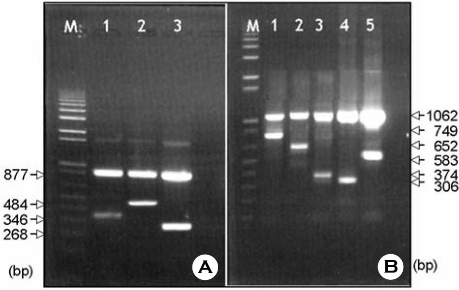
 | Figure 1.Patterns of nested multiplex PCR for P and G genotypes of Group A rotavirus detected from stool specimens.
(A) Lane M; 100 bp DNA ladder, Lanes 1; P[8], 2; P[4], 3: P[6].
(B) Lane M; 100 bp DNA ladder, Lanes 1; G1, 2; G2, 3; G3, 4; G9, 5; G4.
|
Table 1.
Distribution of P and G genotypes of rotavirus detected from stool samples of diarrheic patients in Daejeon region during 2001∼2003


 PDF
PDF ePub
ePub Citation
 Print
Print


 XML Download