Journal List > Immune Netw > v.9(2) > 1033822

Yang, Yuk, and Jo: The Role of Nitric Oxide in Mycobacterial Infections

Abstract

Although tuberculosis poses a significant health threat to the global population, it is a challenge to develop new and effective therapeutic strategies. Nitric oxide (NO) and inducible NO synthase (iNOS) are important in innate immune responses to various intracellular bacterial infections, including mycobacterial infections. It is generally recognized that reactive nitrogen intermediates play an effective role in host defense mechanisms against tuberculosis. In a murine model of tuberculosis, NO plays a crucial role in antimycobacterial activity; however, it is controversial whether NO is critically involved in host defense against Mycobacterium tuberculosis in humans. Here, we review the roles of NO in host defense against murine and human tuberculosis. We also discuss the specific roles of NO in the central nervous system and lung epithelial cells during mycobacterial infection. A greater understanding of these defense mechanisms in human tuberculosis will aid in the development of new strategies for the treatment of disease.

INTRODUCTION

Tuberculosis (TB) is a bacterial infectious disease caused by the obligate human pathogen Mycobacterium tuberculosis (MTB). TB remains an urgent global health problem, with a third of the global population latently infected and eight million new cases each year. Although only 5~10% of infected individuals develop active TB, the fatality rate is nearly two million people annually (1-3). Following exposure to MTB, a series of immune responses are triggered that ultimately define the course of the infection (4,5). The pathogenesis of infection is complicated; however, recent discoveries have attracted great attention due to their association with host-derived and microbial factors. Advances in free radical research have revealed that the production of reactive oxygen and nitrogen oxide species such as superoxide (O2-) and nitric oxide (NO) by innate immune cells is a relatively effective host defense mechanism against bacterial, viral, parasitic, and fungal infections (5,6).
The host cells that are protective against TB include macrophages, dendritic cells, T lymphocytes, and alveolar epithelial cells (2,3,7). Macrophages are believed to play a pivotal role in the immune response against mycobacteria through the production of cytokines such as tumor necrosis factor (TNF)-α and interleukin (IL)-1β. TNF-α and IL-1β, along with interferon (IFN)-γ, which is produced by T lymphocytes, can induce NO production in macrophages via the action of inducible forms of the enzyme NO synthase (iNOS) (8-10). NO and related reactive nitrogen intermediates (RNI)s can kill and/or inhibit intracellular pathogens such as mycobacteria (11-14). The actions of iNOS and the production of NO correlate well with antimycobacterial defense in murine models of TB infection (10,12,15). Although it has been demonstrated that iNOS expression is up-regulated in macrophages from human TB lesions (16), few reports have examined the antimycobacterial effects of cytokines and NO released by human macrophages (17,18). These data suggest that human macrophages possess a NO-independent antimicrobial mechanism, although a role for NO in human host defense cannot be excluded.
MTB infects the airways and stimulates alveolar macrophages, epithelial cells, and macrophages. As a result, NO is produced in response to the stimulation of cytokines and chemokines (19). By producing NO, alveolar epithelial cells can actively participate in alveolar inflammatory processes and defense mechanisms against MTB. In this review, we discuss the role of NO in defense mechanisms against MTB and the mechanisms regulating the production of NO in macrophages, including microglia and alveolar epithelial cells.

OVERVIEW OF NO PRODUCTION AND FUNCTION

NO is a gaseous free radical molecule with pleiotropic functions in pathophysiology that is synthesized by a two-step enzymatic reaction involving a monooxygenase (12,13). One molecule of L-arginine is oxidized at the terminal nitrogen in guanidine to produce Nω-OH-L-arginine as an intermediate. This intermediate is then further oxidized to form one molecule each of NO and L-citrulline (13,14). L-arginine (a conditionally essential amino acid) is obtained from exogenous (food) and endogenous sources, including whole-body protein degradation and, to a lesser extent, de novo synthesis from citrulline by renal arginosuccinate synthase (20,21). Two sequential reactions are catalyzed by NOSs, resulting in the constitutive expression of enzymes primarily in endothelial cells (eNOS) and neuronal cells (nNOS), and as an inducible isoform (iNOS). Constitutively produced NOSs contribute to several physiological processes including vasorelaxation and neurotransmission. In contrast, iNOS is expressed in various cells including macrophages, neutrophils, epithelial cells, and hepatocytes, and it produces excessive NO during infection, inflammation, and states of physiological stimulation (22-24).
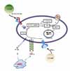
Th1 cytokines such as IFN-γ, IL-1β, and TNF-α stimulate the expression of macrophage iNOS, leading to NO production. In contrast, under the influence of Th2 cytokines such as IL-4, IL-10, and IL-13, arginine is depleted by arginases (8-10). NO is one of several RNIs with antimicrobial activity (18,25). The increase in RNIs is mediated through reactive nitrogen oxides (e.g., peroxynitrite (ONOO-)) generated by the reaction of NO with O2- (13,24) (Fig. 1). NO and RNIs can modify bacterial DNA, proteins, and lipids in both the microbe and host. NO can also deaminate and directly damage bacterial DNA by generating abasic sites and strand breaks (7). Other potential killing mechanisms by NO include interactions with accessory protein targets such as iron-sulfur groups, heme groups, thiols, aromatic or phenolic residues, tyrosyl radicals, and amines. These reactions result in enzymatic inactivation and/or other protein malfunctions (26).

THE ROLE OF NO IN HOST DEFENSE AGAINST MICROBIAL INFECTIONS

During infection with Mycobacterium, Salmonella, Streptococcus, Leishmania, or Bordetella, excessive NO is produced after the induction of iNOS. In many cases, excessive NO production results in innate resistance to bacterial infection. In a study of Bordetella pertussis infection in wild-type (WT) and iNOS-knockout (iNOS KO) mice, the iNOS KO mice displayed increased bacterial growth and susceptibility to infection as compared with the WT mice (27). In a study of murine salmonellosis (Salmonella typhimurium), the use of a NO inhibitor, Nω-monomethyl-L-arginine (L-NMMA), or iNOS KO mice led to similar antimicrobial effects (12,28). In these studies, the lack of NO production was associated with extensive damage, including increased bacterial growth, increased apoptosis, and the exacerbation of histopathological characteristics in mouse livers infected with Salmonella enterica serovar Typhimurium (12). Although NO shows antimicrobial activity against bacteria, fungi, and parasites, some studies suggest dual functions during viral infection. NO produced by macrophages and phagocytic cells can act as an effector molecule during innate host defense mechanisms. For example, NO shows antiviral activity in response to certain viruses such as coxsackievirus (29-31), Epstein-Barr virus (32), and herpes simplex virus (HSV)-1 (33-35). In contrast to the antibacterial activity observed with NO, this antiviral activity is associated with nonspecific damage to host cells and tissues, leading to an exacerbation of viral pathogenesis in many infections such as influenza (36), tick-born virus (37), sendai virus (38), HSV-1 (39,40), and cytomegalovirus (41,42). Therefore, despite the antiviral activity of NO, excessive NO production may facilitate viral pathogenesis. These dual functions of NO may lead to differential outcomes during viral infection.

THE ROLE OF NO IN MYCOBACTERIAL INFECTION: MURINE STUDIES

NO plays a key role in innate immunity and host defense against mycobacteria (1,2,7,43). For example, iNOS KO and immunodeficient mice infected with MTB are at a significantly higher risk of dissemination and mortality as compared with control mice (1,43). In addition, macrophages from mice with the Bcg/natural resistance associated macrophage protein-1 resistance phenotype show inhibition of MTB survival through NO production (3,44). Mycobacterial species exhibit variations in susceptibility to NO and its RNIs. For example, murine macrophages have been shown to inhibit the intracellular growth of M. leprae, M. bovis, and MTB H37Rv (7,11,17,45). When IFN-γ-treated rat alveolar macrophages were infected with M. avium, the growth of the bacterium was significantly inhibited by NO synthesized from L-arginine (46).
Contrasting data have been reported in murine and human macrophages infected with M. avium (6); neither competitive inhibition by L-NMMA nor depletion of L-arginine by arginase had any effect on M. avium growth in murine peritoneal macrophages or human monocyte-derived macrophages (6). In addition, no significant inhibitory effects of NO produced by rat macrophages were observed on the growth of M. intracellulare (45). In murine models of latent infection, both NO-dependent (iNOS- and IFN-γ-dependent antimycobacterial mechanisms) and -independent (CD4+ T cells required for preventing reactivation of the disease) mechanisms maintain latent TB (4,47); however, the applicability of these reports to humans is uncertain.

THE ROLE OF NO IN MYCOBACTERIAL INFECTION: HUMAN STUDIES

In contrast to the murine model of TB, there is controversy surrounding the role of NO in the killing and inhibition of MTB in humans (5). The early inhibition of mycobacterial growth by human alveolar macrophages has been shown to be NO-independent (48). Specifically, exogenous IFN-γ failed to produce mycobactericidal effects in human alveolar macrophages (48). Nevertheless, a growing body of evidence suggests that NO production by MTB-infected human monocytes/macrophages, macrophage-like cell lines, and epithelial cells induces mycobacteriostatic activity against MTB (16, 49-52). For example, alveolar macrophages from healthy control subjects infected with MTB produce NO, and this production is correlated with the intracellular inhibition of MTB growth (51). One study demonstrated increased NO production in TB patients as compared with healthy controls following the infection of peripheral blood mononuclear cells (PBMC)s with MTB (50). Other studies have also demonstrated that alveolar macrophages are able to kill mycobacteria and that these antimycobacterial activities are dependent on iNOS expression (49,53). These results suggest a significant role for NO in host defense against mycobacterial infection. Moreover, increased iNOS expression and pulmonary NO production have been reported in alveolar macrophages and PBMCs from TB patients as compared with healthy controls (8,10,16,54). In those studies, NO played a role in the enhancement of TNF-α and IL-1β secretion, which subsequently affected NO production via feedback loops (8). These data indicate an autoregulatory role for NO. It has also been shown that iNOS and nitrotyrosine (a tissue marker of NO metabolism) are expressed in macrophages within granulomatas and areas of TB pneumonitis (15,55). Human PBMCs and bronchial epithelial cells may produce NO when stimulated with MTB-produced NO (50). In addition, the avirulent strain H37Ra was shown to induce significantly higher levels of NO production as compared with the virulent strain H37Rv (50). Recent studies have shown that L-arginine depletion induces the down-regulation of CD3ζ, thereby impairing T cell signaling, whereas the addition of L-arginine leads to CD3ζ re-expression and the recovery of T cell proliferation (18,56). In addition, T cells from TB patients show reduced CD3ζ expression, which is correlated with arginase-induced L-arginine deficiency. These expression levels were normalized with successful TB treatment (21). Taken together, these data suggest that NO plays a contributory role in human host defense against MTB infection.

THE ROLE OF NO IN MYCOBACTERIAL INFECTION OF THE CENTRAL NERVOUS SYSTEM (CNS)

The roles of NO and iNOS in host defense against infection of the CNS by intracellular pathogens have been reported in previous studies of several intracellular pathogens (e.g., Toxoplasma gondii and Sindbis virus) (57,58). The role of microglial cells in neuropathogenesis following CNS infection has been a topic of growing research interest (59). It was previously reported that in contrast to astrocytes, iNOS was not expressed in human microglia following stimulation by IL-1β or IFN-γ (9,60). These findings suggest that NO and iNOS expression may be dependent on cell type and species. Indeed, previous reports demonstrated NO production in activated murine microglia, but not in human microglia (61). Recently, significant effort has been devoted to developing appropriate models of TB infection in the CNS (CNS-TB) using rabbits or mice. Intracerebral inoculation with MTB or M. bovis BCG resulted in mononuclear cell infiltration, microglial cell activation, and an increase in the number of bacterial cells within the CNS in a mouse model (62,63). In addition, inoculation with MTB or M. bovis BCG led to the up-regulation of IL-1β, TNF-α, IL-6, and IFN-γ within the CNS (63). Recently, Michael et al. (64) reported that iNOS KO mice infected intracerebrally with MTB developed clinical manifestations of CNS-TB, including high mortality rates and histopathological abnormalities resembling human tuberculous meningitis throughout the meninges. The above clinical manifestations were absent in WT mice. These studies underscore the importance of NO in defense against CNS-TB.

THE ROLE OF NO IN THE MYCOBACTERIAL INFECTION OF EPITHELIAL CELLS

Alveolar epithelial cells are able to actively participate in the pathogenesis of pulmonary inflammatory diseases by producing several cytokines and chemokines (65-67). Alveolar epithelial cells produce NO and various innate immune effectors including chemokines (IL-8), which regulate immune activation. In addition, normal T cells express and secrete RANTES in response to MTB infection (19,67). Strong NO production via iNOS also occurs in human lung epithelial cells (19,66); however, the amount of NO released in response to MTB is not mycobactericidal (65,66). Various cytokines (IFN-γ, TNF-α, and IL-1β; alone or in combination) and mycobacterial components stimulate MTB-infected epithelial cells, inducing NO production and mycobactericidal effects (65,66). These factors may contribute to innate immune control in epithelial cells against intracellular pathogens such as MTB.

CONCLUDING REMARKS

NO is a nonspecific, chemically reactive molecule that is important in host defense against a wide variety of microbial pathogens. However, it is becoming increasingly clear that specific killing mechanisms and cell types are not sufficient to kill mycobacteria in vivo. Although NO is not required for mycobactericidal activity in mouse models, the lack of a role for NO or its products (e.g., ONOO-) has not been definitively proven in humans. Nevertheless, a substantial body of evidence indicates a role for NO in human host defenses against MTB. Additional studies are necessary to define the role of NO in relevant human cells including alveolar macrophages, microglia, and epithelial cells. Additionally, it would be useful to generate conditions that mimic in vivo environments, such as the co-culture of relevant cells. Such studies, which will refine our understanding of the importance and specific role of NO in TB defense, may lead to innovative strategies for TB treatment.

Figures and Tables

Figure 1
Yang et al. Synthesis, regulation, and antimycobacterial function of NO in mycobacterial infection. Activated inducible nitric oxide synthase (iNOS) produces Nω-OH-L-arginine from L-arginine, and then Nω-OH-L-arginine is transduced to form NO and L-citruline. Synthesis of NO and reactive nitrogen oxides (RNI) are positively regulated by Th1 cytokines, whereas they are negatively regulated by Th2 cytokines. Produced NO and RNIs, which combined with NO and O2-, can directly kill intracellular MTB in the infected cells (including macrophages, epithelial and glial cells), although the action of NO is dependent on the species and specific cell types.
in-9-46-g001

ACKNOWLEDGEMENTS

This research was supported by the Korea Science & Engineering Foundation through the Infection Signaling Network Research Center (R13-2007-020-01000-0) at Chungnam National University. The authors declare that they have no competing financial interests.

Notes

The authors have no financial conflict of interest.

References

1. Adams LB, Dinauer MC, Morgenstern DE, Krahenbuhl JL. Comparison of the roles of reactive oxygen and nitrogen intermediates in the host response to Mycobacterium tuberculosis using transgenic mice. Tuber Lung Dis. 1997. 78:237–246.
crossref
2. Chan J, Xing Y, Magliozzo RS, Bloom BR. Killing of virulent Mycobacterium tuberculosis by reactive nitrogen intermediates produced by activated murine macrophages. J Exp Med. 1992. 175:1111–1122.
crossref
3. Rojas M, Barrera LF, Puzo G, Garcia LF. Differential induction of apoptosis by virulent Mycobacterium tuberculosis in resistant and susceptible murine macrophages: role of nitric oxide and mycobacterial products. J Immunol. 1997. 159:1352–1361.
4. Flynn JL, Scanga CA, Tanaka KE, Chan J. Effects of aminoguanidine on latent murine tuberculosis. J Immunol. 1998. 160:1796–1803.
5. Nathan C, Shiloh MU. Reactive oxygen and nitrogen intermediates in the relationship between mammalian hosts and microbial pathogens. Proc Natl Acad Sci U S A. 2000. 97:8841–8848.
crossref
6. Bermudez LE. Differential mechanisms of intracellular killing of Mycobacterium avium and Listeria monocytogenes by activated human and murine macrophages. The role of nitric oxide. Clin Exp Immunol. 1993. 91:277–281.
crossref
7. Chan J, Tanaka K, Carroll D, Flynn J, Bloom BR. Effects of nitric oxide synthase inhibitors on murine infection with Mycobacterium tuberculosis. Infect Immun. 1995. 63:736–740.
crossref
8. Kuo HP, Wang CH, Huang KS, Lin HC, Yu CT, Liu CY, Lu LC. Nitric oxide modulates interleukin-1beta and tumor necrosis factor-alpha synthesis by alveolar macrophages in pulmonary tuberculosis. Am J Respir Crit Care Med. 2000. 161:192–199.
crossref
9. Lee SC, Dickson DW, Liu W, Brosnan CF. Induction of nitric oxide synthase activity in human astrocytes by interleukin-1beta and interferon-gamma. J Neuroimmunol. 1993. 46:19–24.
crossref
10. Wang CH, Lin HC, Liu CY, Huang KH, Huang TT, Yu CT, Kuo HP. Upregulation of inducible nitric oxide synthase and cytokine secretion in peripheral blood monocytes from pulmonary tuberculosis patients. Int J Tuberc Lung Dis. 2001. 5:283–291.
11. Adams LB, Franzblau SG, Vavrin Z, Hibbs JB Jr, Krahenbuhl JL. L-arginine-dependent macrophage effector functions inhibit metabolic activity of Mycobacterium leprae. J Immunol. 1991. 147:1642–1646.
12. Alam MS, Akaike T, Okamoto S, Kubota T, Yoshitake J, Sawa T, Miyamoto Y, Tamura F, Maeda H. Role of nitric oxide in host defense in murine salmonellosis as a function of its antibacterial and antiapoptotic activities. Infect Immun. 2002. 70:3130–3142.
crossref
13. MacMicking J, Xie QW, Nathan C. Nitric oxide and macrophage function. Annu Rev Immunol. 1997. 15:323–350.
crossref
14. Mayer B, Hemmens B. Biosynthesis and action of nitric oxide in mammalian cells. Trends Biochem Sci. 1997. 22:477–481.
crossref
15. Choi HS, Rai PR, Chu HW, Cool C, Chan ED. Analysis of nitric oxide synthase and nitrotyrosine expression in human pulmonary tuberculosis. Am J Respir Crit Care Med. 2002. 166:178–186.
crossref
16. Nicholson S, Bonecini-Almeida Mda G, Lapa e Silva JR, Nathan C, Xie QW, Mumford R, Weidner JR, Calaycay J, Geng J, Boechat N, Linhares C, Rom W, Ho JL. Inducible nitric oxide synthase in pulmonary alveolar macrophages from patients with tuberculosis. J Exp Med. 1996. 183:2293–2302.
crossref
17. Denis M. In vivo modulation of atypical mycobacterial infection: adjuvant therapy increases resistance to Mycobacterium avium by enhancing macrophage effector functions. Cell Immunol. 1991. 134:42–53.
crossref
18. Rodriguez PC, Zea AH, DeSalvo J, Culotta KS, Zabaleta J, Quiceno DG, Ochoa JB, Ochoa AC. L-arginine consumption by macrophages modulates the expression of CD3zeta chain in T lymphocytes. J Immunol. 2003. 171:1232–1239.
crossref
19. Robbins RA, Barnes PJ, Springall DR, Warren JB, Kwon OJ, Buttery LD, Wilson AJ, Geller DA, Polak JM. Expression of inducible nitric oxide in human lung epithelial cells. Biochem Biophys Res Commun. 1994. 203:209–218.
crossref
20. Paton NI, Chua YK, Earnest A, Chee CB. Randomized controlled trial of nutritional supplementation in patients with newly diagnosed tuberculosis and wasting. Am J Clin Nutr. 2004. 80:460–465.
crossref
21. Wu G, Morris SM Jr. Arginine metabolism: nitric oxide and beyond. Biochem J. 1998. 336:1–17.
crossref
22. Moncada S, Palmer RM, Higgs EA. Nitric oxide: physiology, pathophysiology, and pharmacology. Pharmacol Rev. 1991. 43:109–142.
23. Nathan C, Xie QW. Regulation of biosynthesis of nitric oxide. J Biol Chem. 1994. 269:13725–13728.
crossref
24. Zaki MH, Akuta T, Akaike T. Nitric oxide-induced nitrative stress involved in microbial pathogenesis. J Pharmacol Sci. 2005. 98:117–129.
crossref
25. Ralph AP, Kelly PM, Anstey NM. L-arginine and vitamin D: novel adjunctive immunotherapies in tuberculosis. Trends Microbiol. 2008. 16:336–344.
crossref
26. Gow AJ, Thom SR, Ischiropoulos H. Nitric oxide and peroxynitrite-mediated pulmonary cell death. Am J Physiol. 1998. 274:L112–L118.
27. Canthaboo C, Xing D, Wei XQ, Corbel MJ. Investigation of role of nitric oxide in protection from Bordetella pertussis respiratory challenge. Infect Immun. 2002. 70:679–684.
crossref
28. Umezawa K, Akaike T, Fujii S, Suga M, Setoguchi K, Ozawa A, Maeda H. Induction of nitric oxide synthesis and xanthine oxidase and their roles in the antimicrobial mechanism against Salmonella typhimurium infection in mice. Infect Immun. 1997. 65:2932–2940.
crossref
29. Zaragoza C, Ocampo C, Saura M, Leppo M, Wei XQ, Quick R, Moncada S, Liew FY, Lowenstein CJ. The role of inducible nitric oxide synthase in the host response to coxsackievirus myocarditis. Proc Natl Acad Sci U S A. 1998. 95:2469–2474.
crossref
30. Zaragoza C, Ocampo CJ, Saura M, Bao C, Leppo M, Lafond-Walker A, Thiemann DR, Hruban R, Lowenstein CJ. Inducible nitric oxide synthase protection against coxsackievirus pancreatitis. J Immunol. 1999. 163:5497–5504.
31. Zaragoza C, Ocampo CJ, Saura M, McMillan A, Lowenstein CJ. Nitric oxide inhibition of coxsackievirus replication in vitro. J Clin Invest. 1997. 100:1760–1767.
crossref
32. Mannick JB, Asano K, Izumi K, Kieff E, Stamler JS. Nitric oxide produced by human B lymphocytes inhibits apoptosis and Epstein-Barr virus reactivation. Cell. 1994. 79:1137–1146.
crossref
33. Croen KD. Evidence for antiviral effect of nitric oxide. Inhibition of herpes simplex virus type 1 replication. J Clin Invest. 1993. 91:2446–2452.
crossref
34. Gamba G, Cavalieri H, Courreges MC, Massouh EJ, Benencia F. Early inhibition of nitric oxide production increases HSV-1 intranasal infection. J Med Virol. 2004. 73:313–322.
crossref
35. MacLean A, Wei XQ, Huang FP, Al-Alem UA, Chan WL, Liew FY. Mice lacking inducible nitric-oxide synthase are more susceptible to herpes simplex virus infection despite enhanced Th1 cell responses. J Gen Virol. 1998. 79:825–830.
crossref
36. Akaike T, Okamoto S, Sawa T, Yoshitake J, Tamura F, Ichimori K, Miyazaki K, Sasamoto K, Maeda H. 8-nitroguanosine formation in viral pneumonia and its implication for pathogenesis. Proc Natl Acad Sci U S A. 2003. 100:685–690.
crossref
37. Kreil TR, Eibl MM. Nitric oxide and viral infection: NO antiviral activity against a flavivirus in vitro, and evidence for contribution to pathogenesis in experimental infection in vivo. Virology. 1996. 219:304–306.
crossref
38. Yoshitake J, Akaike T, Akuta T, Tamura F, Ogura T, Esumi H, Maeda H. Nitric oxide as an endogenous mutagen for Sendai virus without antiviral activity. J Virol. 2004. 78:8709–8719.
crossref
39. Adler H, Beland JL, Del-Pan NC, Kobzik L, Brewer JP, Martin TR, Rimm IJ. Suppression of herpes simplex virus type 1 (HSV-1)-induced pneumonia in mice by inhibition of inducible nitric oxide synthase (iNOS, NOS2). J Exp Med. 1997. 185:1533–1540.
crossref
40. Fujii S, Akaike T, Maeda H. Role of nitric oxide in pathogenesis of herpes simplex virus encephalitis in rats. Virology. 1999. 256:203–212.
crossref
41. Bolovan-Fritts CA, Spector SA. Endothelial damage from cytomegalovirus-specific host immune response can be prevented by targeted disruption of fractalkine-CX3CR1 interaction. Blood. 2008. 111:175–182.
crossref
42. Zhang M, Xin H, Atherton SS. Murine cytomegalovirus (MCMV) spreads to and replicates in the retina after endotoxin-induced disruption of the blood-retinal barrier of immunosuppressed BALB/c mice. J Neurovirol. 2005. 11:365–375.
crossref
43. MacMicking JD, North RJ, LaCourse R, Mudgett JS, Shah SK, Nathan CF. Identification of nitric oxide synthase as a protective locus against tuberculosis. Proc Natl Acad Sci U S A. 1997. 94:5243–5248.
crossref
44. Arias M, Rojas M, Zabaleta J, Rodríguez JI, París SC, Barrera LF, García LF. Inhibition of virulent Mycobacterium tuberculosis by Bcg(r) and Bcg(s) macrophages correlates with nitric oxide production. J Infect Dis. 1997. 176:1552–1558.
crossref
45. Flesch IE, Hess JH, Kaufmann SH. NADPH diaphorase staining suggests a transient and localized contribution of nitric oxide to host defence against an intracellular pathogen in situ. Int Immunol. 1994. 6:1751–1757.
crossref
46. Doi T, Ando M, Akaike T, Suga M, Sato K, Maeda H. Resistance to nitric oxide in Mycobacterium avium complex and its implication in pathogenesis. Infect Immun. 1993. 61:1980–1989.
crossref
47. Scanga CA, Mohan VP, Yu K, Joseph H, Tanaka K, Chan J, Flynn JL. Depletion of CD4(+) T cells causes reactivation of murine persistent tuberculosis despite continued expression of interferon gamma and nitric oxide synthase 2. J Exp Med. 2000. 192:347–358.
crossref
48. Aston C, Rom WN, Talbot AT, Reibman J. Early inhibition of mycobacterial growth by human alveolar macrophages is not due to nitric oxide. Am J Respir Crit Care Med. 1998. 157:1943–1950.
crossref
49. Jagannath C, Actor JK, Hunter RL Jr. Induction of nitric oxide in human monocytes and monocyte cell lines by Mycobacterium tuberculosis. Nitric Oxide. 1998. 2:174–186.
crossref
50. Kwon OJ. The role of nitric oxide in the immune response of tuberculosis. J Korean Med Sci. 1997. 12:481–487.
crossref
51. Rich EA, Torres M, Sada E, Finegan CK, Hamilton BD, Toossi Z. Mycobacterium tuberculosis (MTB)-stimulated production of nitric oxide by human alveolar macrophages and relationship of nitric oxide production to growth inhibition of MTB. Tuber Lung Dis. 1997. 78:247–255.
crossref
52. Rockett KA, Brookes R, Udalova I, Vidal V, Hill AV, Kwiatkowski D. 1,25-Dihydroxyvitamin D3 induces nitric oxide synthase and suppresses growth of Mycobacterium tuberculosis in a human macrophage-like cell line. Infect Immun. 1998. 66:5314–5321.
crossref
53. Nozaki Y, Hasegawa Y, Ichiyama S, Nakashima I, Shimokata K. Mechanism of nitric oxide-dependent killing of Mycobacterium bovis BCG in human alveolar macrophages. Infect Immun. 1997. 65:3644–3647.
crossref
54. Wang CH, Liu CY, Lin HC, Yu CT, Chung KF, Kuo HP. Increased exhaled nitric oxide in active pulmonary tuberculosis due to inducible NO synthase upregulation in alveolar macrophages. Eur Respir J. 1998. 11:809–815.
crossref
55. Facchetti F, Vermi W, Fiorentini S, Chilosi M, Caruso A, Duse M, Notarangelo LD, Badolato R. Expression of inducible nitric oxide synthase in human granulomas and histiocytic reactions. Am J Pathol. 1999. 154:145–152.
crossref
56. Kropf P, Baud D, Marshall SE, Munder M, Mosley A, Fuentes JM, Bangham CR, Taylor GP, Herath S, Choi BS, Soler G, Teoh T, Modolell M, Muller I. Arginase activity mediates reversible T cell hyporesponsiveness in human pregnancy. Eur J Immunol. 2007. 37:935–945.
crossref
57. Gazzinelli RT, Eltoum I, Wynn TA, Sher A. Acute cerebral toxoplasmosis is induced by in vivo neutralization of TNF-alpha and correlates with the down-regulated expression of inducible nitric oxide synthase and other markers of macrophage activation. J Immunol. 1993. 151:3672–3681.
58. Tucker PC, Griffin DE, Choi S, Bui N, Wesselingh S. Inhibition of nitric oxide synthesis increases mortality in Sindbis virus encephalitis. J Virol. 1996. 70:3972–3977.
crossref
59. Rock RB, Gekker G, Hu S, Sheng WS, Cheeran M, Lokensgard JR, Peterson PK. Role of microglia in central nervous system infections. Clin Microbiol Rev. 2004. 17:942–964.
crossref
60. Rock RB, Hu S, Deshpande A, Munir S, May BJ, Baker CA, Peterson PK, Kapur V. Transcriptional response of human microglial cells to interferon-γ. Genes Immun. 2005. 6:712–719.
crossref
61. Peterson PK, Hu S, Anderson WR, Chao CC. Nitric oxide production and neurotoxicity mediated by activated microglia from human versus mouse brain. J Infect Dis. 1994. 170:457–460.
crossref
62. Mazzolla R, Puliti M, Barluzzi R, Neglia R, Bistoni F, Barbolini G, Blasi E. Differential microbial clearance and immunoresponse of Balb/c (Nramp1 susceptible) and DBA2 (Nramp1 resistant) mice intracerebrally infected with Mycobacterium bovis BCG (BCG). FEMS Immunol Med Microbiol. 2002. 32:149–158.
crossref
63. van Well GT, Wieland CW, Florquin S, Roord JJ, van der Poll T, van Furth AM. A new murine model to study the pathogenesis of tuberculous meningitis. J Infect Dis. 2007. 195:694–697.
crossref
64. Olin MR, Armien AG, Cheeran MC, Rock RB, Molitor TW, Peterson PK. Role of nitric oxide in defense of the central nervous system against Mycobacterium tuberculosis. J Infect Dis. 2008. 198:886–889.
crossref
65. Kwon OJ, Kim JH, Kim HC, Suh GY, Park JW, Chung MP, Kim H, Rhee CH. Nitric oxide expression in airway epithelial cells in response to tubercle bacilli stimulation. Respirology. 1998. 3:119–124.
crossref
66. Roy S, Sharma S, Sharma M, Aggarwal R, Bose M. Induction of nitric oxide release from the human alveolar epithelial cell line A549: an in vitro correlate of innate immune response to Mycobacterium tuberculosis. Immunology. 2004. 112:471–480.
crossref
67. Sharma M, Sharma S, Roy S, Varma S, Bose M. Pulmonary epithelial cells are a source of interferon-gamma in response to Mycobacterium tuberculosis infection. Immunol Cell Biol. 2007. 85:229–237.
crossref
TOOLS
Similar articles