Journal List > Immune Netw > v.9(6) > 1033813

Park, Nam, Seo, Seo, and Kim: Tiul1 and TGIF are Involved in Downregulation of TGFβ1-induced IgA Isotype Expression

Abstract

TGF-β1 is well known to induce Ig germ-line α (GLα) transcription and subsequent IgA isotype class switching recombination (CSR). Homeodomain protein TG-interacting factor (TGIF) and E3-ubiquitin ligases TGIF interacting ubiquitin ligase 1 (Tiul1) are implicated in the negative regulation of TGF-β signaling. In the present study, we investigated the roles of Tiul1 and TGIF in TGFβ1-induced IgA CSR. We found that over-expression of Tiul1 decreased TGFβ1-induced GLα promoter activity and strengthened the inhibitory effect of Smad7 on the promoter activity. Likewise, overexpression of TGIF also diminished GLα promoter activity and further strengthened the inhibitory effect of Tiul1, suggesting that Tiul1 and TGIF can down-regulate TGFβ1-induced GLα expression. In parallel, overexpression of Tiul1 decreased the expression of endogenous IgA CSR-predicitive transcripts (GLTα, PSTα, and CTα) and TGFβ1-induced IgA secretion, but not GLTγ3 and IgG3 secretion. Here, over-expressed TGIF further strengthened the inhibitory effect of Tiul1. These results suggest that Tiul1 and TGIF act as negatively regulators in TGFβ1-induced IgA isotype expression.

INTRODUCTION

TGF-β generates signals through TGF-β receptors (type I and type II serine/threonine kinase receptors) and receptor-regulated Smads (R-Smads) such as Smad2 and Smad3. Either Smad2 or Smad3 is phosphorylated and complexed with Smad4 (1-4). These Smad complexes translocate to the nucleus where they bind specific DNA sequences in target promoters, thereby acting as transcriptional activators for TGF-β-responsive genes. On the other hand, Smad6 and Smad7, termed inhibitory Smads (I-Smads), antagonize TGF-β signaling by inhibiting the phosphorylation of R-Smads (5-7). In addition, TGF-β signaling is regulated by ubiquitin-dependent degradation. First, HECT type E3-ubiquitin ligases such as Smad ubiquitination regulatory factor 1 (Smurf1) and Smurf2 can be recruited to the activated type I receptor (TβRI) by interacting with the Smad7, resulting in receptor ubiquitination and degradation, and reduced signaling (8-10). It has been reported that Smurfs can interact with R-Smad and Runx leading to degradation of these proteins (11-15). Secondly, RING finger type E3 ligase, Arkadia, interacts with Smad7 leading to degradation of Smad7 (16). Third, another HECT type E3 ligase, TG-interacting factor (TGIF) interacting ubiquitin ligase 1 (Tiul1) interacts constitutively with Smad7 and induces degradation of TβRI without affecting level of Smad7 expression. Tiul1 can interact with Smad2/Smad3 and TGIF upon activation of TGF-β1 signaling. The interaction of Tiul1 with TGIF allows this ubiquitin ligase to target Smad2 and Smad3 for degradation, leading to a diminution of TGF-β1 signaling (17). Furthermore, TGIF functions as a negative regulator in TGF-β signaling through recruiting HDACs to a Smad target promoter (18, 19) and inhibition of R-Smads phosphorylation (20).
We have previously shown that TGF-β1, acting mainly through Smad3/4 and Runx3, induces germ-line α (GLα transcription and subsequent class switching recombination (CSR) to IgA, but Smad7 inhibited this TGFβ-induced GLα transcription (21,22). Moreover, we found that Smurfs reinforces the inhibitory effect of Smad7 on TGFβ1-induced IgA CSR while Arkadia antagonizes the action of Smurf causing an increase of IgA CSR (23). In the present study, we provide evidence that Tiul1 and TGIF may be involved in downregulation of TGFβ1-induced IgA isotype expression.

MATERIALS AND METHODS

Cell lines and cell culture

The murine B cell lymphoma line, A20.3 was provided by Dr. J. Stavnezer (University of Massachusetts Medical School, Worcester, MA, USA). CH12F3-2A was provided by Dr. T. Honjo (Kyoto University, Japan). Cells were cultured at 37℃ in a humidified atmosphere containing 5% CO2 in RPMI-1640 medium (Sigma) supplemented with 10% FBS, 50 µM 2-ME, 5 mM HEPES, penicillin (100 U/ml)/streptomycin (100 µg/ml).

Gene expression and reporter constructs

Genes encoding Smad7 (7) subcloned into Flag-pcDNA3 (6) were provided by Dr. M. Kawabata (The Cancer Institute, Tokyo, Japan). For Tiul1 expression constructs, the open reading frame was amplified from the γ-ZAP11 clone by PCR and subcloned into p3xFlag-CMV-10 expression vector (Sigma). A similar approach was used to clone TGIF fragments in pcDNA3-HA (17).

Transfection and luciferase assays

Transfection were performed by electroporation with a Gene Pulser II (Bio-Rad, USA) as described (21). Reporter plasmids were cotransfected with expression plasmids and pCMVβgal (Stratagene), and luciferase and β-gal assays were performed as described (21).

RT-PCR

RNA preparation, reverse transcription, and PCR were performed as described previously (21). Primers for PCR were synthesized by Bioneer Corp. (Seoul, Korea). The primers for GLTα were: forward primer, 5'-CTACC ATAGG GAAGA TAGCC T-3', and reverse primer, 5'-TAATC GTGAA TCAGG CAG-3' (product size, 267 bp); PSTα were: forward primer, 5'-CTCTG GCCCT GCTTA TTGTT G-3', and reverse primer, 5'-GAGCT GGTGG GAGTG TCAGT G-3' (product size, 267 bp); CTα were: forward primer, 5'-CTACC ATAGG GAAGA TAGCC T-3', and reverse primer, 5'-TCTGA ACCTT CAAGG ATGCT CTTG-3' (product size, 365 bp); GLTγ3 were: forward primer, 5'-CAAGT GGATC TGAAC ACA-3', and reverse primer, 5'-GGCTC CATAG TTCCA TT-3' (product size, 349 bp); β-actin were: forward primer, 5'-CATGT TTGAG ACCTT CAACA CCCC-3', and reverse primer, 5'-GCCAT CTCCT GCTCG AAGTC TAG-3' (product size, 320 bp). All reagents for RT-PCR were purchased from Promega Corp. PCR reactions for β-actin were performed in parallel in order to normalize cDNA concentrations within each set of samples. Aliquots of the PCR products were resolved by electrophoresis on 2% agarose gels.

Isotype-specific ELISA

ELISAs were performed as described previously (21). The reaction products were measured at 405 nm with an ELISA reader (VERSAMAX reader, Molecular Devices, Sunnyvale, CA).

RESULTS AND DISCUSSION

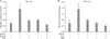
Tiul1 and TGIF inhibit TGFβ1-induced GLα promoter activity

Although Tiul1 down-regulates TGF-β signaling by inducing degradation of the activated type I receptor and R-Smads (17), it is not known if Tiul1 is involved in TGFβ1-induced IgA CSR. Therein, we investigated the effect of Tiul1 on TGFβ1-induced GLα transcription in A20.3 B lymphoma cell lines, using a GLα promoter reporter. As shown in Fig. 1A, over-expression of Tiul1 decreased promoter activity by twofold. In addition, it strengthened the inhibitory effect of Smad7 on the promoter activity (Fig. 1A). TGIF down-regulates TGF-β signaling through recruiting HDACs to a Smad target promoter (18,19) and inhibiting R-Smads phosphorylation (20). Further, TGIF interacts with Tiul1 in the nucleus leading to the degradation of R-Smads (17). We tested the effects TGIF along with Tiul1 on TGF-β induced GLα promoter activity. As shown in Fig. 1B, overexpression of TGIF decreased the TGF-β induced GLα promoter activity. Moreover, TGIF strengthened the inhibitory effect of Tiul1 on the promoter activity. Taken together, these results suggest that Tiul1 not only interacts with Smad7 but also with TGIF, both of which lead to the downregulation of GLα gene expression.

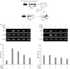
Effect of Smad7 and Tiul1 on the expression of endogenous IgA transcripts and IgA secretion

Thus far, we observed that Tiul1 acts as the negative regulator in TGFβ1-induced GLα promoter activity. To gain evidence that this phenomenon is physiologically relevant, we asked if Tiul1 actually inhibits the expression of transcripts associated with IgA CSR. As shown in the diagram in Fig. 2A, once CSR to IgA occurs, the GLµ promoter, which becomes associated with the Cα gene and continues to be active, generates transcripts termed α post-switch transcripts (PSTα) (24,25). Furthermore, the DNA sequences between Sµ and Sα are looped out of the chromosome as switch circles during CSR, and another type of transcript, termed a circle transcript (CT), in this case consisting of the Iα exon spliced to the Cµ exon (CTα), is transcribed from the switch circle owing to the presence of the active Iα promoter (26). Thus, expression of PSTα and CTα as well as GLTα can be used as indicatives of active IgA CSR. As in the case of the GLα promoter reporter, overexpression of Smad7 decreased TGFβ1-induced GLTα expression (Fig. 2B). In this, Tiul1 again strengthened the inhibitory effect of Smad7 on the GLα transcription, but not GLTγ3. Similarly, Smad7 and Tiul1 in combination downregulated the expression of PSTα and CTα. Finally, we examined the effects of Smad7 and Tiul1 on IgA secretion. As shown in Fig. 2C, over-expression of either Smad7 or Tiul1 alone decreased TGFβ1-induced IgA secretion, and the combination markedly diminished IgA secretion. Not addressed specifically, these results implicate that Tiul1 degrades TβR1 through interacting with Smad7 as shown before (17), resulting in reduction of TGFβ1-induced IgA production.

Effect of Tiul1 and TGIF on TGFβ1-induced IgA expression

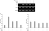
Since we observed that Tiul1 in cooperation with Smad7 downregulate TGF-β induced IgA expression, we examined if Tiul1 together with TGIF can also regulate TGFβ1-induced IgA expression. As shown in Fig. 3A, either overexpression of Tiul1 or TGIF decreased the TGFβ1-induced GLTα transcription. Overexpression of both molecules more dramatically decreased the expression of GLTα but not GLTγ3. In fact, this was the case for the TGFβ1-induced IgA secretion (Fig. 3B). These results indicate the possibility that Tiul1, in cooperation with TGIF, can inhibit TGFβ1-induced IgA production.

Concluding remarks

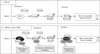
In the present study, we have shown that Tiul1 and TGIF can down-regulate TGFβ1-induced IgA CSR. Possible mechanisms underlying this phenomenon are illustrated in Fig. 4. In this model, Tiul1 downregulates TGFβ1-induced IgA CSR through degradation of activated TβR-I. Secondly, TGIF inhibits TGFβ1-induced IgA CSR by the inhibition of R-Smads. Third, Tiul1 along with TGIF decreased TGFβ1-induced IgA CSR via degradation of R-Smads such as Smad2 and Smad3. Since we observed in the present study that Tiul1 can act a negative regulator in association with Smad7 and TGIF toward TGFβ1-induced IgA CSR, it would be important to elucidate the dynamics of interrelation among Smad7, Tiul1, and TGIF along with Smurfs (23) in the context of TGFβ1-induced IgA expression in the future.

Figures and Tables

Figure 1
Effects of Smad7, Tiul1, and TGIF on TGFβ1-induced GLα promoter activity. (A) A20.3 B lymphoma cells were transfected with expression plasmids (10 µg) for Smad7, Tiul1 and GLα-Luc reporter (15 µg). TGF-β1 (1 ng/ml) was added and luciferase activity measured 16 h later. Transfection efficiency was normalized to β-gal activities. (B) A20.3 B lymphoma cells were transfected with expression plasmids (10 µg) for Tiul1, TGIF and GLα-Luc reporter (15 µg). TGF-β1 (1 ng/ml) was added and luciferase activity measured 16 h later. Transfection efficiency was normalized to β-gal activities. Data represent average luciferase activity from three independent transfections with SEMs (bars).
in-9-248-g001
Figure 2
Effects of Smad7 and Tiul1 on the levels of Ig GLTs and Ig secretion by mouse B lymphoma cells. (A) Diagram of DNA recombination occurring during switching to IgA. Rectangles and ovals represent exons and S regions, respectively. RNA transcripts are indicated beneath the DNA diagrams. (B) CH12F3-2A B lymphoma cells were transfected with expression plasmid for Smad7, Tiul1 or pcDNA3 (30 µg of each). They were then cultured with LPS (12.5 µg/ml) and TGF-β1 (1 ng/ml), and after 24 h, total RNA isolated and measured levels of endogenous GLTα, GLTγ3, PSTα, and CTα transcripts by RT-PCR. (C) After 3 days of culture, supernatant were collected and secretion of IgA and IgG3 was determined by isotype-specific ELISA. Data are means of triplicate samples±SEM.
in-9-248-g002
Figure 3
Effects of Tiul1 and TGIF on TGFβ1-induced GLTα transcription and IgA secretion in mouse B cells. CH12F3-2A B lymphoma cells were transfected with expression plasmid for Tiul1, TGIF or pcDNA3 (30 µg of each). They were then cultured with LPS (12.5 µg/ml) and TGF-β1 (1 ng/ml), and after 24 h, total RNA isolated and measured levels of endogenous GLTα and GLTγ3 transcripts by RT-PCR (Panel A). After 3 days of culture, supernatant were collected and secretion of IgA and IgG3 was determined by ELISA (Panel B). Data are means of triplicate samples±SEM.
in-9-248-g003
Figure 4
Proposed mechanisms by which Tiul1 and TGIF downregulate TGFα1-induced IgA isotype expression. (I) Upon stimulation by TGFβ1, Smad3 becomes phosphorylated by the activated TGF-β1 receptors and forms complexes with Smad4. These Smads complexes translocate to the nucleus where they bind SBEs in the GLα promoter, thereby activating transcription. (II) In the cytoplasm, Smad7 can inhibit TGF-β1 signaling by deactivating Smad3 phosphorylation. Tiul1 interacts with Smad7 leading to ultimate degradation of the activated type I receptor. On the other hand, TGIF prevents the ligand-dependent phosphorylation of Smad3. In addition, Tiul1 interacts with TGIF in the nucleus resulting in degradation of R-Smads such as Smad2 and Smad3.
in-9-248-g004

ACKNOWLEDGEMENTS

This research was supported by Basic Science Research Program through the National Research Foundation of Korea (NRF) funded by the Ministry of Education, Science and Technology (2009-0066852) to PH Kim and by the second stage of the Brain Korea 21 program. It was carried out in the facilities of the Vascular System Research Center and Institute of Bioscience & Biotechnology of Kangwon National University.

Notes

The authors have no financial conflict of interest.

References

1. Lagna G, Hata A, Hemmati-Brivanlou A, Massagué J. Partnership between DPC4 and SMAD proteins in TGF-beta signalling pathways. Nature. 1996. 383:832–836.
crossref
2. Wu RY, Zhang Y, Feng XH, Derynck R. Heteromeric and homomeric interactions correlate with signaling activity and functional cooperativity of Smad3 and Smad4/DPC4. Mol Cell Biol. 1997. 17:2521–2528.
crossref
3. Zhang Y, Musci T, Derynck R. The tumor suppressor Smad4/DPC 4 as a central mediator of Smad function. Curr Biol. 1997. 7:270–276.
crossref
4. Nakao A, Imamura T, Souchelnytskyi S, Kawabata M, Ishisaki A, Oeda E, Tamaki K, Hanai J, Heldin CH, Miyazono K, ten Dijke P. TGF-beta receptor-mediated signalling through Smad2, Smad3 and Smad4. EMBO J. 1997. 16:5353–5362.
crossref
5. Hayashi H, Abdollah S, Qiu Y, Cai J, Xu YY, Grinnell BW, Richardson MA Jr, Topper JN, Gimbrone MA Jr, Wrana JL, Falb D. The MAD-related protein Smad7 associates with the TGFbeta receptor and functions as an antagonist of TGFbeta signaling. Cell. 1997. 89:1165–1173.
crossref
6. Imamura T, Takase M, Nishihara A, Oeda E, Hanai J, Kawabata M, Miyazono K. Smad6 inhibits signalling by the TGF-beta superfamily. Nature. 1997. 389:622–626.
7. Nakao A, Afrakhte M, Morén A, Nakayama T, Christian JL, Heuchel R, Itoh S, Kawabata M, Heldin NE, Heldin CH, ten Dijke P. Identification of Smad7, a TGFbeta-inducible antagonist of TGF-beta signalling. Nature. 1997. 389:631–635.
crossref
8. Ebisawa T, Fukuchi M, Murakami G, Chiba T, Tanaka K, Imamura T, Miyazono K. Smurf1 interacts with transforming growth factor-beta type I receptor through Smad7 and induces receptor degradation. J Biol Chem. 2001. 276:12477–12480.
crossref
9. Kavsak P, Rasmussen RK, Causing CG, Bonni S, Zhu H, Thomsen GH, Wrana JL. Smad7 binds to Smurf2 to form an E3 ubiquitin ligase that targets the TGF beta receptor for degradation. Mol Cell. 2000. 6:1365–1375.
crossref
10. Kim KI, Baek SH. SUMOylation code in cancer development and metastasis. Mol Cells. 2006. 22:247–253.
11. Zhu H, Kavsak P, Abdollah S, Wrana JL, Thomsen GH. A SMAD ubiquitin ligase targets the BMP pathway and affects embryonic pattern formation. Nature. 1999. 400:687–693.
crossref
12. Zhao M, Qiao M, Oyajobi BO, Mundy GR, Chen D. E3 ubiquitin ligase Smurf1 mediates core-binding factor alpha1/Runx2 degradation and plays a specific role in osteoblast differentiation. J Biol Chem. 2003. 278:27939–27944.
crossref
13. Jin YH, Jeon EJ, Li QL, Lee YH, Choi JK, Kim WJ, Lee KY, Bae SC. Transforming growth factor-beta stimulates p300-dependent RUNX3 acetylation, which inhibits ubiquitination-mediated degradation. J Biol Chem. 2004. 279:29409–29417.
crossref
14. Lin X, Liang M, Feng XH. Smurf2 is a ubiquitin E3 ligase mediating proteasome-dependent degradation of Smad2 in transforming growth factor-beta signaling. J Biol Chem. 2000. 275:36818–36822.
crossref
15. Zhang Y, Chang C, Gehling DJ, Hemmati-Brivanlou A, Derynck R. Regulation of Smad degradation and activity by Smurf2, an E3 ubiquitin ligase. Proc Natl Acad Sci U S A. 2001. 98:974–979.
crossref
16. Koinuma D, Shinozaki M, Komuro A, Goto K, Saitoh M, Hanyu A, Ebina M, Nukiwa T, Miyazawa K, Imamura T, Miyazono K. Arkadia amplifies TGF-beta superfamily signalling through degradation of Smad7. EMBO J. 2003. 22:6458–6470.
crossref
17. Seo SR, Lallemand F, Ferrand N, Pessah M, L'hoste S, Camonis J, Atfi A. The novel E3 ubiquitin ligase Tiul1 associates with TGIF to target Smad2 for degradation. EMBO J. 2004. 23:3780–3792.
crossref
18. Wotton D, Lo RS, Lee S, Massague J. A Smad transcriptional corepressor. Cell. 1999. 97:29–39.
crossref
19. Wotton D, Lo RS, Swaby LA, Massagué J. Multiple modes of repression by the Smad transcriptional corepressor TGIF. J Biol Chem. 1999. 274:37105–37110.
crossref
20. Seo SR, Ferrand N, Faresse N, Prunier C, Abécassis L, Pessah M, Bourgeade MF, Atfi A. Nuclear retention of the tumor suppressor cPML by the homeodomain protein TGIF restricts TGF-beta signaling. Mol Cell. 2006. 23:547–559.
crossref
21. Park SR, Lee JH, Kim PH. Smad3 and Smad4 mediate transforming growth factor-beta1-induced IgA expression in murine B lymphocytes. Eur J Immunol. 2001. 31:1706–1715.
crossref
22. Park SR, Lee EK, Kim BC, Kim PH. p300 cooperates with Smad3/4 and Runx3 in TGFbeta1-induced IgA isotype expression. Eur J Immunol. 2003. 33:3386–3392.
crossref
23. Choi SH, Seo GY, Nam EH, Jeon SH, Kim HA, Park JB, Kim PH. Opposing effects of Arkadia and Smurf on TGFbeta1-induced IgA isotype expression. Mol Cells. 2007. 24:283–287.
24. Li SC, Rothman PB, Zhang J, Chan C, Hirsh D, Alt FW. Expression of I mu-C gamma hybrid germline transcripts subsequent to immunoglobulin heavy chain class switching. Int Immunol. 1994. 6:491–497.
crossref
25. Muramatsu M, Kinoshita K, Fagarasan S, Yamada S, Shinkai Y, Honjo T. Class switch recombination and hypermutation require activation-induced cytidine deaminase (AID), a potential RNA editing enzyme. Cell. 2000. 102:553–563.
crossref
26. Kinoshita K, Harigai M, Fagarasan S, Muramatsu M, Honjo T. A hallmark of active class switch recombination: transcripts directed by I promoters on looped-out circular DNAs. Proc Natl Acad Sci U S A. 2001. 98:12620–12623.
crossref
TOOLS
Similar articles