

 PDF
PDF ePub
ePub Citation
 Print
Print


INTRODUCTION
The eventual goal of tumor immunologists is to enhance systemic immunity against tumor cells. Cytokines are attractive agents to modulate immune responses in disease states including cancer. Since the demonstration that endotoxin treatment induced rejection of established cancer, findings of interleukin-1 and interferon were followed as effective tumor killing molecules, and recombinant cytokine therapy began to draw attention with much enthusiasm. For example, recombinant IL-2, IL-12, GM-CSF got major focus in initial trials of tumor therapy. However, on the contrary to the initial hope, various unpredicted side effects of recombinant cytokines hampered clinical applications. Later by development of new technology of gene transfer and gene delivery system, cytokine gene transfer methods became an alternative way to deliver cytokines in restricted region. The enormous evaluation of available cytokines by gene transfer into autologous tumors of various origins have been reported (1). Tumor cells transduced with cytokine genes sometimes became immunogenic, regressed spontaneously in vivo, and eventually induced antitumor immunity (2-4).
Generally speaking, tumor bearers are somehow immunocompromised in functions of T cells (5). Malfunction of immune system in tumor bearers is also implicated with disabled dendritic cells (DCs) (6). Supplementation of the cytokines defected in tumor patients could be one of way to reverse the overwhelmed status by tumor growth. The hypothesis became the ground of cytokine gene transfer into tumor cells as a tumor cell vaccine. However, the expression of cytokines by tumor cells also showed various side effects when the tumor cell vaccines were injected in vivo. Furthermore, the infiltrated immune cells into the regressing tumor mass were mainly non-specific to the tumor. These indicate that the local cytokine release also activates bystander immune cells and causes various cytokine productions from them. The local cytokine cocktail may cause the unexpected side effects in cytokine gene transfer methods. One of way to avoid the bystander cell activation would be to confine the effective range of cytokine expressed by tumor cell vaccine. By expressing cytokine on tumor cell surface as a membrane-bound form (mbCytokine), selective activation of target cells could be achieved (see Table I). As a natural form, several cytokines including IL-1 (7), IL-15 (8), M-CSF (9), Flt3-eigand (10), TNFα (11), LTα (12), fractalkine (13), TGFβ (14), and IFN-γ (15), are expressed as membrane-associated form as well as sescretory form. The functional distinction between the membrane-bound form and the secretory form has not been clarified yet in most of cytokines. Superficially, the effective range of the membrane-bound form may be confined to the cells that are in physical contacts, but secretory form would have wider effective range.
TUMOR THERAPY USING MEMBRANE-BOUND FORM OF TNFα
Tumor necrosis factor a (TNFα) was discovered in 1975 through the anticancer activity of sera from mice injected with endotoxin (16). TNFα is produced by various cell types including macrophages, T lymphocytes, endothelial cells, and Kupffer cells. Two bioactive forms are known; a 26 kDa transmembrane form, and a 17 kDa secrtory form (11). The 26 kDa protein expressed as a membrane-bound from (mbTNFα) is cleaved into 17 kDa secretory form by membrane-bound metalloproteases, including TNFα converting enzyme (ADAM17/TACE/CD156q) (17,18). The mbTNFα can kill various types of tumor cells which are resistant to secretory TNFα (19), participates in the interaction between activated T and B lymphocytes (20), and enhances macrophage responses towards Leishmania (21), whereas the secretory form is mainly responsible for endotoxic shock (22). TNFα is able to kill many kinds of tumor cells in vivo as well as in vitro. The clinical applications of the soluble trimeric form of TNFα have been hampered because therapeutic doses of this potent cytokine have been accompanied by serious systemic toxic side effects, such as septic shock and cachexia (23,24). To alleviate these life-threatening side effects, and to improve the antitumor activities of TNFα, Graham and colleagues generated adenovirus vectors expressing mbTNFα (25). The toxicity of TNFα was much reduced by local expression and prevention from circulatory system. Wild type TNFα expressing vector was highly toxic, whereas the adenovirus vector expressing a mutant non-secreted (membrane-bound) form showed low toxicity at the same dose. Both vectors induced permanent tumor regressions in some cases, and the cured mice displayed protective immunity and specific CTL activity against the original tumor. These results indicate that the adenovirus vector expressing mbTNFα can reduce systemic toxicity without compromising antitumor activity.
In comparison of mbTNFα and secretory TNFα in antitumor effects, Li and colleagues generated three different recombinant retroviral vectors encoding wild-type TNFα, secretable TNFα mutant, and uncleavable transmembrane TNFα mutant (26). They identified a transient weight loss in mice bearing solely secretable TNFα mutant producing tumor, whereas no obvious side effects were seen in mice bearing uncleavable TNFα mutant or wild type TNFα expressing tumors. Moreover, the tumors expressing soluble TNFα promoted the subsequent infiltration of CD4+ T cells, and to a lesser extent CD8+ T cells to the tumor sites. The mbTNFα expressing tumors up-regulated Fas (CD95) expression and inhibited the expression of tumor metastasis associated CD44v3, suggesting the difference of secretory TNFα from mbTNFα in killing tumor cells in vivo. Recently, it was found that the TNFα mutation on ADAM-17 cleavage site was not enough to prevent cleavage and secretion of TNFα. To avoid the effect of secretory TNFα effect completely, Kipps and colleagues expressed a chimeric protein with extracellular domain of TNFα and transmembrane stalk portion of CD154 using adenovirus vector system and tested its antitumor activity (27). The systemic toxicity was not seen with the adenovirus vector expressing TNFα-CD154 chimera. The WEHI-164 fibrosarcoma tumor was regressed by the injection of the TNFα-CD154 expressing vector intratumorally, and survival of mice was much extended compared with the mice injected with the vector expressing wild type of TNFα. Analysis on the effector cells involved in the antitumor activity by mbTNFα producing tumor cell vaccine would provide more information on the functional differentiation from the secretory form.
TUMOR THERAPY USING MEMBRANE-BOUND FORM OF GM-CSF
GM-CSF is expressed as a 23~29 kDa glycoprotein by a number of different cell types (28,29), and has stimulatory effect on the proliferation and maturation of myeloid progenitor cells in vitro. GM-CSF also plays a critical role in differentiation and proliferation of DCs from hematopoietic precursor cells (30). It was one of the first hematopoietic regulators clinically applied along with G-CSF, for example, to ameliorate chemotherapy-induced neutropenia and to help hematopoietic reconstitution after bone-marrow transplantation. With the B16 mouse melanoma model, Dranoff and colleagues compared the effectiveness of tumor cell vaccines transfected with various cytokines and adhesion molecules (4). They found that the GM-CSF transduced tumor cell vaccine was more effective than any other tumor cell vaccines transduced with IL-2, IL-4, IL-5, IL-6, IFN-γ, ICAM-1, CD2, and TNFα in the tumor model system. With the purpose to enhance DCs function, tumor cells were transduced with GM-CSF or Flt3-ligand, and efficient differentiation and activation of DCs were acquired with enhanced therapeutic effect (31). The enhanced function of DCs resulted in efficient presentation of tumor-associated antigens (TAAs) to T cells through cross presentation, that is, by indirect priming (Huang et al., 1994). Langerhans cells (LCs), residing DCs in epidermis, once receive appropriate stimuli, they engulf antigens and begin their maturation into fully differentiated DCs. The activated DCs subsequently migrate with their processed antigens to lymph nodes, where they stimulate naïve T cells to produce effector cytotoxic T lymphocytes (CTLs). The antigen specific CTLs can leave lymph nodes and move to antigen invasion sites of body.
With the purpose of direct stimulation of DCs by tumor cells in situ, Carlo and colleagues prepared P815 mastocytoma cells expressing GM-CSF in a membrane-bound form (mbGM- CSF) to target TAAs to epidermal LCs (32). High affinity binding of mbGM-CSF to its receptors on the LCs in skin would yield a strong direct interaction with the mbGM-CSF expressing tumor cell vaccine. The transfected mbGM-CSF clones stimulated proliferation of syngeneic bone marrow cells, indicating that the mbGM-CSF is biologically active. Growth of the mbGM-CSF expressing tumor clones was reduced in syngeneic mice, but not in nude mice. They observed that CD8+ CTLs are involved in the immune responses. The mice once rejected the mbGM-CSF clones gained protective antitumor immunity. Unfortunately, they did not analyze the activation of LCs in the analyses of the antitumor effect of the mbGM-CSF expressing tumor cells, and comparative study to the secretory form of GM-CSF has not been performed, so that superiority of mbGM-CSF vaccine could not be guaranteed. They extended their observations with mbGM-CSF tumor vaccine to the spontaneously arisen B16F10 mouse melanoma tumor model (33). The B16F10 cells are highly metastatic and non-immunogenic compared to the P815 used in previous study. Furthermore, the B16 melanoma model has proven particularly informative, because immunization with irradiated parental cells fails to induce significant levels of protective immunity. With mbGM-CSF expressing B16 melanoma vaccine they also recognized protective effect and retarded metastasis. In this study they found that mbGM-CSF expressing B16F10 cells can induce strong systemic immunity; protection and therapeutic effects were more effective than analogous vaccines containing only secreted GM-CSF.
Expression of GM-CSF as a membrane-bound form on the highly metastatic Lewis lung carcinoma also led to abrogation of their tumorigenicity and metastatic phenotype (34), and at the same time induced CTL response that protected syngeneic mice against a subsequent challenge with original tumor cells. In leukemia system, Arlinghaus and colleagues expressed mbGM-CSF, and evaluated the induction of antitumor immunity and effector cells (35). The mbGM-CSF expressing leukemia cells induced antitumor immunity involving CD8+ T cells. One of critical advantage of the expression of GM-CSF in tumor cells is that tedious manipulation to derive sufficient numbers of DCs can be avoided for tumor therapy using in vitro amplified DCs. However, the supposed maturation and differentiation of DCs in these antitumor activities has not been analyzed systematically. Using macrophage-colony stimulating factor (M-CSF) was also investigated thoroughly. Tumor therapy using the membrane-bound form of M-CSF has been discussed in a recent review (36).
TUMOR THERAPY USING MEMBRANE-BOUND FORM OF IL-2
IL-2 is one of the cytokines most widely used for the treatment of patients with cancer. The renal cell carcinoma and malignant melanoma was regressed by infusion of high doses of IL-2 (37,38). The high dose of IL-2 also induced severe adverse events associated with a life-threatening vascular leak syndrome mediated by NK cells and neutrophils (39-44). The transfer of the IL-2 gene into tumor cells has the advantage that IL-2 secreted by the tumor cell itself can induce local immune responses at the tumor growing site. The principle of the cytokine gene therapy is based on the findings that tumor bearers are often immunocompromized. T lymphocytes from the tumor bearers are defective in signaling molecules for T cell activation (45,46). The IL-2 gene transduction into tumor cells as a tumor cell vaccine was designed to compensate for the defective cytokines from activated Th cells (47-50). However, tumor cells engineered to produce cytokines also showed unexpected side effects (4,51,52), which may result from the paracrine effect of the secreted cytokines stimulating bystander cells that are mostly non-specific to TAAs.
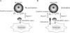
There is a report that membrane-associated IL-2 is expressed in certain population of T cells, but its functional meaning was not determined yet (53). A cancer vaccine anchoring recombinant IL-2 via diphtheria toxin T domain induced tumor specific CTL activity (54). Through this approach they could observe a reduced toxicity of IL-2. As a different approach to express membrane-bound form of IL-2 (mbIL-2), glycosylphosphatidylinositol (GPI)-linked form of IL-2 was expressed as a chimeric form with DAF or CD59, GPI-linked membrane proteins (55,56). The growth of the B16 tumor cells expressing GPI-linked IL-2 was inhibited in vivo, but the growth inhibition was not observed with the tumor cells transduced with secretory form of IL-2. The GPI-linked protein also has other advantage that the protein is incorporated into cell membrane by mixing tumor cells with the prepared GPI-linked proteins without involving gene transfer and extended in vitro tumor cell culture. One of disadvantage of these approaches is that production of IL-2 in vivo is no longer happen, so that the benefit of higher immunogenicity of live tumor call vaccine should be sacrificed. In our laboratory, we prepared tumor vaccines expressing mbIL-2 in other way. IL-2 gene was fused with those of type I and II transmembrane proteins (CD4 or TNFα, respectively) considering orientation of IL-2 on cell surface (57,58). In this approach we postulated that, if tumor cells express IL-2 as a costimulatory molecule on tumor cells surface, CTL may get two signals to fulfill activation; signal 1 from TAA peptide/MHC class I complex, and signal 2 from mbIL-2. This approach intended selective CTL activation, which is specific for TAAs (Fig. 1). The cells will be stimulated through IL-2 costimulatory signal only when they are in cell-to-cell physical contacts through T cell receptors (TCR) and TAA/MHC class I molecules. The expression level of IL-2 as a chimeric protein was higher with TNFα than with CD4. In MethA fibrosarcoma model, the tumor cell clone expressing mbIL-2 was superior to the tumor cells expressing secretory form of IL-2 in supporting the growth of IL-2 dependent CTLL-2 cells. Moreover, the mbIL-2 expressing MethA clones (L-d positive) were better stimulatory for the spleen cells from the 2C TCR transgenic mice, which TCR is responsive to L-d allogeneic MHC class I molecule, indicating that the mbIL-2 on tumor cells provide signal 2 as costimulatory molecule and the L-d molecules provide signal 1. The tumor clones expressing mbIL-2 lost tumorigenicity. and the mice once rejected the tumor clones showed resistance to re-challenge with wild type B16 tumor cells (57). This stimulation of antitumor immunity was better in tumor clones expressing mbIL-2 than the tumor clone expressing secretory form of IL-2. In a mixed cell culture of spleen cells with inactivated tumor clones showed that the mbIL-2 clone was effective in amplification of CD8+ T cells. Furthermore, the mbIL-2 clone produced in B16 melanoma model efficiently inhibited metastasis to lung by involving CD8+ T cell activation (58). Another trial to accelerate CTL recruitment into tumor growing site, SDF-1 was transduced into B16 melanoma cells with mbIL-2 (59). The additional expression of SDF-1 could enhance the antitumor immunity of mbIL-2 expressing tumor cells.
TUMOR THERAPY USING MEMBRANE-BOUND FORM OF IL-4
IL-4 is a Th2 cytokine, and mice lacking either IL-4 or IL-4 receptors fail to develop Th2 cells. IL-4 producing tumor cell clones generated by transfection with mouse IL-4 gene caused tumor rejection when co-inoculated into nude mice with a variety of non-IL-4 producing tumor cell lines. Rejection was apparently mediated by macrophages and eosinophils which could not have been entirely predicted from the previously known functions of IL-4. More significantly, IL-4 producing tumor cells grew in mice treated with anti-IL-4 antibody, but regressed when the antibody therapy was stopped, and local IL-4 production can cause regression of established tumors (60). IL-4 is a multifunctional cytokine produced by Th2 cells and also involved in the generation of CTL. Of several cytokines whose presence affects tumor growth, IL-4 appears to be very potent. Tumor cells secreting a substantial amount of IL-4 are unable to form tumors and induce systemic immunity. The IL-4 activated tumor inhibition is a consequence of macrophage and eosinophil infiltration. Host T cells are also involved in the induction and effector phase of IL-4-activated tumor rejection with the antigen non-specific cells predominating.
Initial studies have shown that expression of IL-4 in different tumor types induced an eosinophil-mediated tumor rejection. Further studies demonstrated that IL-4 has pleiotropic effects on immune cells of multiple lineages and can paradoxically play a role as an inducer of type I T cell immunity. Nave CD8+ T cells are activated in vitro in the presence of IL-4 but not IL-2 or IL-12 and developed into functional long-term memory cells (61). Moreover, IL-4 from CD8+ T cells are required for the generation of tumor-associated CTL (62) and CTL- mediated tumor immunity is impaired in IL-4 deficient mice, suggesting tumor immunity requires IL-4 in the priming phase for the generation of effector cells (63).
Prud'homme and colleagues investigated antitumor immune responses against human carcinoembryonic antigen (CEA) with plasmids encoding secretory form of IL-4, IL-12, or IFN-γ (64). Intramuscular injection of the IL-12 or IFN-γ-encoding plasmid with that of encoding CEA elicited effective antitumor immune responses to syngeneic CEA-positive P815 tumor cells (CEA/P815). In contrast, coinjection of the CEA vector with an IL-4 vector increased IgG1 production, suppressed the Th1 and CTL activity (high Th2 and low CTL activity) and antitumor response. In the following study, they intended to enhance adjuvant effects of cytokines and restrict the localization of the cytokine to the site of injection by expressing membrane-bound form of IL-4 (mbIL-4) or membrane-bound form of IL-12 (mbIL-12) (65). Surprisingly, the mbIL-4 plasmid was more effective in antitumor immune responses than that of mbIL-12 or secretory form of IL-12. These results propose that the function of IL-4 in Th1 immune response should be investigated further. We also prepared a tumor cell vaccine expressing mbIL-4 in a chimeric form with transmembrane region of TNFα (66). The mbIL-4 tumor clones supported the growth of D10 Th2 cells and CT.4S, an IL-4 dependent T cell line, in mixed cell cultures. Moreover, the mbIL-4 clone stimulated spleen cells from 2C TCR transgenic mice. The tumor clone expressing mbIL-4 failed to form tumor, and the mice once rejected the live mbIL-4 clone acquired systemic antitumor immunity. Though IL-4 is expressed secretory form only naturally, the observed significant antitumor effect of the mbIL-4 expressing tumor cell vaccine opens the gate of application for other secretory cytokines. On the other hand, in virology area, it was reported that incorporation of mbCytokines (IL-2, GM-CSF, and IL-4) into influenza viral vaccines boosted immunogenicity (67), implying that the mbCytokine is applicable for therapeutic development against viral infection, at least to enveloped virus.
TUMOR THERAPY USING MEMBRANE-BOUND FORM OF IL-12
IL-12 is a cytokine identified as a NK cell stimulatory factor and a CTL maturation factor (68,69), and normally released by professional antigen-presenting cells. IL-12 also induces IFN-γ production (70,71), and plays its role as an efficient molecule for the initiation of a Th1 response (72). There's a report that IL-12 is also expressed as membrane-bound form as well as secretory form, but the report has not been confirmed conclusively by others yet (73). IL-12 is considered as a potent antitumor and antimetastatic cytokine (74). The administration of recombinant human IL-12 is associated with severe toxicity, and hampers its development as an anticancer drug (75-77). Side effects of systemically administered IL-12 are fever, fatigue, nausea, headache, and sometimes leads to death. To avoid the toxicity of IL-12, local delivery methods were adopted using plasmid or viral vectors (41,78-80). The circulating cytokine was suspected to have some adverse physiological effects, such as allergy, immunosuppression, systemic mastocytosis, severe hepatic and renal dysfunction and cardiotoxicity (65). Tumor cell vaccines prepared by IL-12 gene transfer into autologous cells also injected intradermal or peritumoral sites, and observed reduction of tumor size in limited number of patients (78,81). To confine the effective range of IL-12 and for convenience of practical preparation of tumor cell vaccine expressing cytokine, Prud'homme and colleagues tested effectiveness of the DNA vaccines secreting IL-12 or expressing mbIL-12 in chimeric form with B7.1 (65). The DNA vaccine expressing mbIL-12 also enhanced the antitumor response, but to a slightly lesser extent than secretory IL-12 plasmids. The mbIL-12 effect was not much distinct to that of secretory form of IL-12 in the model system. Tumor cells engineered to express GPI-anchored form of IL-12 also induced antitumor immunity (82,83). Moreover, the tumor cell vaccine expressing both GPI-linked IL-2 and GPI-linked IL-12 showed synergistic antitumor effect in subcutaneous and intravenous tumor models (55). In our laboratory, we prepared a tumor vaccine expressing membrane-bound form of IL- 12p35 (mbIL-12p35) in a chimeric form with transmembrane domain of TNFα. Tumor cells expressing mbIL-12p35 induced potent antitumor effects in MethA mouse fibrosarcoma model. Tumorigenicity of mbIL-12p35 clone was reduced and the mice once rejected the mbIL-12p35 clone acquired systemic antitumor immunity to the wild type tumor cells (unpublished data). Analysis of effector cells involved in the antitumor effect of mbIL-4 and mbIL-12 expressing tumor cell vaccines as representative Th2 and Th1 cytokines would provide information explaining the effective CTL induction by mbIL-4 expressing tumor cell vaccine.
Tumor Therapy Using Membrane-Bound Form Of Flt3-ligand, FRACTALKINE, AND IFN-γ
Fms-like tyrosine kinase 3 ligand (Flt3-L) is a cytokine with potent costimulatory activity and has a synergistic effect with other murine cytokines, especially a growth-stimulatory effect on DCs (84) and NK cells (85). Flt3-L is expressed both membrane-bound form and secretory form by alternative splicing (10), but functional differentiation of the two types of Flt3-L has not been studied yet. The Flt3-L plays an important role in the differentiation and maturation of early murine and human haematopoietic precursor and stem cells. The tumor vaccine expressing membrane-bound form of Flt3-L (mbFlt3-L) was investigated in murine breast tumor model to investigate whether the Flt3-L expression induces DC activation as mbGM-CSF does. This study was intended to take advantage of the DC growth-stimulatory effect of Flt3-L (86). In tumor cell vaccine trials with both membrane-bound form and secretory form, the protective effect was comparable each other (86-88). Furthermore, the effector cells involved in antitumor effect was discernable, that is, both the DC and NK cells were affected.
Fractalkine (CX3CL1) is a CX3C chemokine with a CXXXC motif at its N-terminus. Fractalkine (FK) is expressed on DCs, mast cells, and the IL-1β or TNFα-activated endothelial cells, either as a 95 kDa membrane-bound form or as a secreted chemokine upon cleavage by ADAM 10 and ADAM17 proteases (89). The membrane-bound form of FK (mbFK) functions as an effective adhesion molecule by interacting with CX3C chemokine receptor 1 (CX3CR1). Experimentally expressed mbFK on tumor cell surface could activate NK cells (90). After its release from cell surface, FK also functions as a chemoatttractant for monocytes, NK cells and T lymphocytes. The B16-F0 melanoma cells also express endogenous FK and knock down of FK gene by RNA interference reduced tumor growth by inhibiting tumor angiogenesis in mice (91). On the other hand, to take advantage of chemoattractive and adhesion functions of the FK, Cao and colleagues prepared tumor cells expressing secretory FK and mbFK by gene transfer into 3LL lung carcinoma cells (92). The 3LL-FK tumor cells induced protective immunity and CTL activity. They also observed infiltration of CD4+ T cells, CD8 + T cells, and DCs. Adhesion of DCs by 3LL-FK upregulated IL-12 production by activated DCs, and production of IL-2 and IFN-γ in 3LL-FK tumor tissues was increased. In mouse hapatocellular carcinoma model, the tumor cells expressing native form of FK induced similar antitumor activity (93). To dissociate the function of secretory form from the membrane-bound form, Schmid-Antomarch and colleagues compared tumorigenicity of three different transfectants of C26 colon carcinoma cells; natural form, secretory form, and membrane-bound form (94). They found that both secretory and membrane-bound form have similar antitumor effect in a subcutaneous model of tumor implantation, but only secretroy form was effective in liver and metastatic lung model of the colon cancer. The mbFK even enhanced tumor growth in the model system. The induction of angiogenesis and chemoattraction and adhesion functions of FK should be reconciled for successful tumor therapy.
IFN-γ is a potent immunoregulatory molecule secreted by NK and T cells. The IFN-γ is detected on the cell surface of activated T cells expressing IFN-γ, so that it may function as a Th1 marker (15). It abrogated tumor growth by inhibiting the proliferation of tumor cells and by activating host immune cells including T cells, B cells, macrophages, and NK cells. Eisenbach and colleagues expressed IFN-γ as a membrane-bound form (mbIFN-γ) in the low immunogenic and highly metastatic clone of 3LL Lewis lung carcinoma and found comparable effects to the mbGM-CSF tumor vaccine (34). The responsible effector cells of the antitumor immunity by mbIFN-γ producing tumor cell vaccine were not analyzed yet.
CONCLUSION
The functional differentiation of the secretory and the membrane-bound forms of cytokine has not been studied thoroughly yet, but tumor cell vaccines expressing mbCytokines showed at least comparable levels of immune stimulatory effects to secretory cytokine expressing vaccines. As a mechanism of induction of antitumor immunity, indirect priming of T cells via professional antigen presenting cells seems to be potently functional as seen with tumor vaccines expressing mbGM-CSF and mbFK. The toxicity and side effects of systemically injected recombinant cytokines and secretory cytokines from cytokine gene transduced tumor cell vaccines could be avoided by minimizing incorporation of cytokines into circulation, as seen in mbTNFα. In addition, direct priming of T cells by mbCytokine expressing tumor cell vaccine itself has been intended to selectively activate TAA-specific CTL effector cells as in the cases of mbIL-2 or mbIL-4 expressing tumor cell vaccines. Expression of chimeric cytokine with certain transmembrane protein or GPI-linkage signal sequences, and chemical cross-linking of recombinant cytokines on cell surface could be choices of the tumor cell vaccine preparation. Understanding of required costimulatory molecules for sufficient CTL activation are indispensible for successful tumor therapy using CTL-targeted tumor vaccines, because different origin of tumor cells express different repertoire of costimulatory molecules. The effector cells involved in the antitumor immunity induction by various mbCytokines are also waiting for further analysis.
 XML Download