Journal List > Immune Netw > v.9(4) > 1033800

Lee and Kim: Further Characterization of Activin A-induced IgA Response in Murine B Lymphocytes

Abstract

We have recently shown that activin A, a member of TGF-β superfamily, stimulates mouse B cells to express IgA isotype but other isotypes. In the present study, we further characterized effects of activin A on B cell growth and IgA expression. We found that activin A did not have effect on LPS-stimulated cell viability. In parallel, CFSE staining analysis revealed that activin A did not alter cell division. An increase of IgA secretion by activin A was completely abrogated by anti-activin A Ab but not by anti-TGFβ1 Ab. In the same conditions, no other isotypes are significantly affected by each antibody treatment. Finally, activin A, as similar to TGF-β1, increased IgA secretion by mesenteric lymph node cells. These results suggest that activin A can specifically stimulate IgA response, independent of TGF-β in the gut.

INTRODUCTION

IgA is the major Ig isotype in mucosal secretions and constitutes the great majority of the Ig synthesized in mucosal tissues (1,2). IgA class-switch recombination (CSR) mainly takes place in Peyer's patches (PP) and then, IgA+ B cells migrate through the lymph and blood circulation and eventually home to lamina propria of the intestine (3). TGF-β1 is known to induce IgA and IgG2b CSR in murine B cells (4-9). In addition, TGF-β1 inhibits B cell proliferation and cell cycle progression in the mid to late G1 phase of the cell cycle (10-12).
Activin A, a member of the TGF-β superfamily, is a local regulator of cell growth and differentiation (13). Activin A is known to be produced by Th2 but not by Th1 cells upon activation (14). We have recently shown that activin A stimulates IgA expression in mouse B cells (15). Thus, activin A increased the expression of Ig germ-line α transcripts, an indicative of IgA CSR, via Smad3 and Smad4 leading to increased IgA secretion.
Since both activin A and TGF-β1 use the same proteins (Smad3 and/or Smad4) in signal transduction and enhances IgA response, it was necessary to determine whether the activity of activin A is related to that of TGF-β1. We found that activin A little affected B cell proliferation, which was contrasted to anti-proliferative activity of TGF-β1. In addition, we found that activin A can independently modulate B cells to express IgA and that this may occur in mesenteric lymph nodes of the gut.

MATERIALS AND METHODS

Animals

BALB/c mice were purchased from Orient. Co. Ltd. (Gyeonggi-do, Korea) and maintained in an animal environmental control chamber (Myung Jin Inst. Co., Seoul, Korea). Animals were fed Purina Laboratory Rodent Chow 5001 ad libitum. Eight to twelve weeks age of mice were used in this study. Animal care was in accordance of with the institutional guidelines set forth by Kangwon National University.

B cell preparations and cell culture

The murine B lymphoma cell line, CH12F3-2A was provided by Dr. T. Honjo (Osaka University, Japan) (16). These cells were cultured at 37℃ in a humidified atmosphere containing 5% CO2 in RPMI 1,640 medium (Sigma) supplemented with 10% FBS, 50µM 2-ME, 5 mM HEPES, penicillin (100 U/ml)/streptomycin (100µg/ml). Mouse spleen B cell suspensions were prepared as described before (4) and whole mesenteric lymph node cells were prepared as teasing the tissues with forceps.

Isotype-specific ELISA

ELISAs were performed as described previously (17). The reaction products were measured at 405 nm with an ELISA reader (Molecular Devices VERSAMAX reader, Molecular Devices, Sunnyvale, CA, USA).

CFSE staining and analysis

Isolated mouse spleen B cells were labeled with a CFSE kit (Invitrogen Life Technologies, Carlsbad, CA, USA), according to the manufacture's instruction, and added with LPS, activin A (10 ng/ml), and TGF-β1 (0.2 ng/ml). Dilution of CFSE was measured by counting 30,000 viable cells with a FACSCalibur (BD Biosciences).

RESULTS AND DISCUSSION

Activin A do not alter the cell proliferation and viability

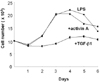
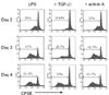
We have recently shown that activin A selectively stimulates IgA expression (15). However, the relationship between actions of activin A and TGF-β1 in the context of IgA CSR is unclear at the moment. In this regard, we have demonstrated in the earlier study that anti-proliferative activity of TGF-β facilitates IgA CSR leading to an increase of IgA production (18). Therein, we first examined the effect of activin A on the growth of murine spleen B cells. LPS is generally adopted for the non-specific stimulation of mouse B cells. As shown in Fig. 1, LPS increased cell viability up to 4 day of culture. Effect of Activin A on the cell viability was marginal while TGF-β1 substantially inhibited it. Subsequently, we tested the effect of activin A on B cell proliferation. In parallel to cell viability, Activin A had no influence on cell proliferation though anti-proliferative activity of TGF-β1 was evident (Fig. 2). These results suggest that activin A increases IgA expression through the mechanism different from that of TGF-β1 and that activin A has such activity without inhibiting cell proliferation.

Activin A specifically enhances IgA expression independent of the activity of TGF-β1

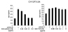
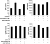
Then, how does activin A stimulate B cells to express IgA isotype expression? To assess its underlying mechanism, we examined the possible involvement of TGF-β in the activin A-induced IgA secretion. In the culture of CH12F3-2A B cell lymphoma, the activin A-induced IgA production was abrogated by anti-activin A Ab in a dose dependent manner (Fig. 3). In contrast, anti-TGFβ1 Ab failed to eliminate the IgA response enhanced by activin A. Moreover, in normal spleen B cell cultures, a similar pattern of IgA secretion was observed when anti-activin A Ab and anti-TGFβ1 Ab were treated as shown in Fig. 4. On the other hand, anti-activin A Ab or anti-TGFβ1 Ab little affected secretion patterns of IgM, IgG2b, and IgG3. These results indicate that activin A selectively induces IgA expression and that this activity of activin A is independent of that of TGF-β1. Furthermore, these results exclude the possibility that activin A has such activity through induction of TGF-β1 production.

Effect of activin A on IgA secretion by mesenteric lymph node B cells

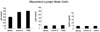
Peyer's patch (PP) is the most important gut-associated lymphoid tissue, where most of IgA B cells are committed (19). Therein, it is difficult to isolate surface IgA negative B cells from PP. On the other hand, mesenteric lymph node (MLN) B cells are also known to be the site for IgA B cell commitment (20,21). Here, we were interested if activin A affects IgA production by MLN B cells. As shown in Fig. 5, activin A, as similar to TGF-β1, increased IgA secretion by MLN cells but neither IgG1 nor IgG2b.
In summary, we demonstrate in this study that activin A, as similar to TGF-β1, selectively increases IgA expression. Nevertheless, Activin A had no influence on cell proliferation, which is in contrast to the anti-proliferative property of TGF-β1. We have recently shown that activin A, like TGF-β1 adopts Smad3 and Smad4 as the main signaling mediators (15). Therefore, it still remains to determine the mechanism by which activin A causes IgA isotype switching, distinguishing from that of TGF-β1. Finally we observed that activin A can stimulate mesenteric lymph node B cells to produce IgA. These results raise the possibility that activin A may have important effects on the expression of IgA in the gut.

Figures and Tables

Figure 1
Effect of activin A and TGF-β1 on the growth kinetics of splenic B cells. Normal spleen B cells (1×10⁶) stimulated with LPS were cultured in the presence of TGF-β1 (0.2 ng/ml) and activin A (10 ng/ml). Numbers of viable cells were enumerated by trypan blue exclusion. Data are means±SEM (bars) of triplicate culture wells.
in-9-133-g001
Figure 2
Effect of activin A on B cell proliferation. CFSE-labeled splenic B cells (1×10⁶) were cultured as in Fig. 1. After 48 h, 72 h, and 96 h incubation, B cell proliferation was assessed by analyzing the dilution of CFSE in the same number of viable cells.
in-9-133-g002
Figure 3
Effect of anti-activin A Ab on IgA secretion by B lymphoma cells. B lymphoma cells were stimulated with LPS (12.5µg/ml) and activin A (10 ng/ml). Titrated anti-activin A Ab and anti-TGF-β Ab were pre-incubated for 1 h at room temperature before addition to culture. Supernatants were harvested after 3 days of culture. IgA secretion was determined by ELISA. Data are means of triplicate cultures±SEM.
in-9-133-g003
Figure 4
Effect of anti-activin A Ab on Ig secretion by normal spleen B cells. Mouse spleen B cells were stimulated with LPS (12.5 g/ml) and activin A (10 ng/ml). As in Fig. 3, anti-activin A Ab (0.5µg/ml) and anti-TGF-β Ab (5µg/ml) were added to cultures. Supernatants were harvested after 7 days of culture. Secretion of Ig isotypes was determined by ELISA. Data are means of triplicate cultures±SEM.
in-9-133-g004
Figure 5
Activin A increases IgA secretion by mesenteric lymph node cells. Mesenteric lymph node (1×10⁶) cells were treated 10 ng/ml of activin A, and 0.2 ng/ml of TGF-β1. Supernatants were harvested after 7 days of culture. IgA, IgG1, and IgG2b secretion was determined by ELISA. Data are means of triplicate cultures±SEM.
in-9-133-g005

ACKNOWLEDGEMENTS

This work was supported by the Regional Core Research Program funded by the Korea Ministry of Education, Science and Technology (Medical & Bio-material Research Center). It was carried out in the facilities of the Vascular System Research Center and Institute of Bioscience & Biotechnology at Kangwon National University.

Notes

The authors have no financial conflict of interest.

References

1. Mestecky J. The common mucosal immune system and current strategies for induction of immune responses in external secretions. J Clin Immunol. 1987. 7:265–276.
crossref
2. van der Heijden PJ, Stok W, Bianchi AT. Contribution of immunoglobulin-secreting cells in the murine small intestine to the total 'background' immunoglobulin production. Immunology. 1987. 62:551–555.
3. Fagarasan S, Honjo T. Intestinal IgA synthesis: regulation of front-line body defences. Nat Rev Immunol. 2003. 3:63–72.
crossref
4. Kim PH, Kagnoff MF. Transforming growth factor-beta 1 is a costimulator for IgA production. J Immunol. 1990. 144:3411–3416.
5. Sonoda E, Matsumoto R, Hitoshi Y, Ishii T, Sugimoto M, Araki S, Tominaga A, Yamaguchi N, Takatsu K. Transforming growth factor beta induces IgA production and acts additively with interleukin 5 for IgA production. J Exp Med. 1989. 170:1415–1420.
crossref
6. Coffman RL, Lebman DA, Shrader B. Transforming growth factor beta specifically enhances IgA production by lipopolysaccharide-stimulated murine B lymphocytes. J Exp Med. 1989. 170:1039–1044.
crossref
7. van Vlasselaer P, Punnonen J, de Vries JE. Transforming growth factor-beta directs IgA switching in human B cells. J Immunol. 1992. 148:2062–2067.
8. Spieker-Polet H, Yam PC, Arbieva Z, Zhai SK, Knight KL. In vitro induction of the expression of multiple IgA isotype genes in rabbit B cells by TGF-beta and IL-2. J Immunol. 1999. 162:5380–5388.
9. McIntyre TM, Klinman DR, Rothman P, Lugo M, Dasch JR, Mond JJ, Snapper CM. Transforming growth factor beta 1 selectivity stimulates immunoglobulin G2b secretion by lipopolysaccharide-activated murine B cells. J Exp Med. 1993. 177:1031–1037.
crossref
10. Kehrl JH, Taylor A, Kim SJ, Fauci AS. Transforming growth factor-beta is a potent negative regulator of human lymphocytes. Ann N Y Acad Sci. 1991. 628:345–353.
crossref
11. Smeland EB, Blomhoff HK, Holte H, Ruud E, Beiske K, Funderud S, Godal T, Ohlsson R. Transforming growth factor type beta (TGF beta) inhibits G1 to S transition, but not activation of human B lymphocytes. Exp Cell Res. 1987. 171:213–222.
crossref
12. Cross D, Cambier JC. Transforming growth factor beta 1 has differential effects on B cell proliferation and activation antigen expression. J Immunol. 1990. 144:432–439.
13. Luisi S, Florio P, Reis FM, Petraglia F. Expression and secretion of activin A: possible physiological and clinical implications. Eur J Endocrinol. 2001. 145:225–236.
crossref
14. Ogawa K, Funaba M, Chen Y, Tsujimoto M. Activin A functions as a Th2 cytokine in the promotion of the alternative activation of macrophages. J Immunol. 2006. 177:6787–6794.
crossref
15. Lee HJ, Seo GY, Kim HA, Kim PH. Activin A stimulates IgA expression in mouse B cells. Biochem Biophys Res Commun. 2008. 366:574–578.
crossref
16. Nakamura M, Kondo S, Sugai M, Nazarea M, Imamura S, Honjo T. High frequency class switching of an IgM+ B lymphoma clone CH12F3 to IgA+ cells. Int Immunol. 1996. 8:193–201.
crossref
17. Park SR, Lee JH, Kim PH. Smad3 and Smad4 mediate transforming growth factor-beta1-induced IgA expression in murine B lymphocytes. Eur J Immunol. 2001. 31:1706–1715.
crossref
18. Kim PH, Kagnoff MF. Transforming growth factor beta 1 increases IgA isotype switching at the clonal level. J Immunol. 1990. 145:3773–3778.
19. Craig SW, Cebra JJ. Peyer's patches: an enriched source of precursors for IgA-producing immunocytes in the rabbit. J Exp Med. 1971. 134:188–200.
crossref
20. Santiago AF, Fernandes RM, Santos BP, Assis FA, Oliveira RP, Carvalho CR, Faria AM. Role of mesenteric lymph nodes and aging in secretory IgA production in mice. Cellular immunology. 2008. 253:5–10.
crossref
21. Kawanishi H, Saltzman L, Strober W. Mechanisms regulating IgA class-specific immunoglobulin production in murine gut-associated lymphoid tissues. II. Terminal differentiation of postswitch sIgA-bearing Peyer's patch B cells. J Exp Med. 1983. 158:649–669.
crossref
TOOLS
Similar articles