Journal List > Immune Netw > v.17(3) > 1033547

Lee, Kang, and Choi: TLR/MyD88-mediated Innate Immunity in Intestinal Graft-versus-Host Disease

Abstract

Graft-versus-host disease (GHVD) is a severe complication after allogeneic hematopoietic stem cell transplantation. The degree of inflammation in the gastrointestinal tract, a major GVHD target organ, correlates with the disease severity. Intestinal inflammation is initiated by epithelial damage caused by pre-conditioning irradiation. In combination with damages caused by donor-derived T cells, such damage disrupts the epithelial barrier and exposes innate immune cells to pathogenic and commensal intestinal bacteria, which release ligands for Toll-like receptors (TLRs). Dysbiosis of intestinal microbiota and signaling through the TLR/myeloid differentiation primary response gene 88 (MyD88) pathways contribute to the development of intestinal GVHD. Understanding the changes in the microbial flora and the roles of TLR signaling in intestinal GVHD will facilitate the development of preventative and therapeutic strategies.

Abbreviations

GVHD

graft-versus host disease

allo

allogeneic

HSCT

hematopoietic stem cell transplantation

GI

gastrointestinal

MDSCs

myeloidderived suppressor cells

BM

bone marrow

INTRODUCTION

Allogeneic (allo) hematopoietic stem cell transplantation (HSCT) is an effective treatment for hematological disorders, including lymphoma and leukemia (1234). Graft-versus-leukemia (GVL) effects, which are derived from the activation of donor T cells that recognize the allo-antigens expressed by the recipient's tumor cells, contribute to the eradication of malignant host cells (5). However, donor T cells are also reactive to allo-antigens expressed by the recipient's tissues and parenchymal cells in the gastrointestinal (GI) tract, liver, lung, and skin, and induce graft-versus-host disease (GVHD), a life-threatening complication of allo-HSCT (67). The suppression of severe GVHD is important for the success of allo-HSCT.
GI tract damage is a critical event in the pathogenesis of GVHD (89). The integrity of the GI tract and innate immunity to the intestinal microbiome both contributes to the maintenance of intestinal homeostasis; disruption of intestinal homeostasis during allo-HSCT provokes intestinal GVHD, which leads to exacerbation of the disease and systemic GVHD (9). Signaling through Toll-like receptors (TLRs) and myeloid differentiation factor 88 (MyD88), a signaling adaptor downstream of TLRs, is pivotal in innate immunity that controls response to microbial stimulation; evidence supporting the significances of their signaling in GVHD is accumulating (1011). In this article, we will review recent research into the role of TLR/MyD88-mediated innate immunity in acute intestinal GVHD.

ACUTE AND CHRONIC GVHD

GVHD is broadly classified into acute and chronic GVHD, depending on the timing of disease incidence after allo-HSCT. Chronic GVHD was classically defined as a late complication of allo-BMT that occurs in 100 days post-transplantation. Chronic GVHD is similar to autoimmune and other immunological diseases, such as scleroderma (1213), systemic lupus-like diseases (14), primary biliary cirrhosis (15), and immune cytopenia (16); it is characterized by tissue inflammation and fibrosis, and is mediated by cellular and CD4 T helper cell type 2-dependent humoral immunity (1718). In 2014, revised chronic GVHD criteria were proposed, which facilitate distinction of chronic and acute GVHD, that include diagnostics in the skin (e.g., poikiloderma and sclerotic features including lichen planus-like features), mouth (e.g., lichen planus-like changes), lung (e.g., bronchiolitis obliterans), and GI tract (e.g., esophageal web, strictures or stenosis in the upper to middle third of the esophagus) (19).
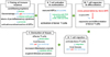
Development of acute GVHD is observed within 100 days post-HSCT, with symptoms indicating damage to the skin (e.g., maculopapular rash on the palms, soles and ears, and diffuse erythematous rash over the entire body), liver (e.g., hyperbilirubinemia, jaundice, and elevated transaminases), GI tract (e.g., nausea, vomiting, abdominal cramps, anorexia, bleeding, and diarrhea), and, occasionally, lungs, eyes and oral mucosa (20). Although donor T cell-mediated adaptive immunity is an essential component of the development of acute GVHD, innate immunity also plays significant roles (62122). Chemoirradiation conditioning of recipients prior to HSCT provokes apoptosis of epithelial cells and tissue inflammation in several organs, including the intestines. The release of inflammatory cytokines activates antigen-presenting cells (APCs), which promote the activation and effector differentiation of allo-reactive donor T cells. Activated of T cells mediate cytotoxicity against allo-antigen-bearing recipient cells in damaged tissues, which increase inflammation in the target organs (Fig. 1). In particular, intestinal inflammation initiated by epithelial cell damage disrupts the epithelial barrier, which exposes innate immune cells to intestinal microbial stimuli. This innate cell stimulation by microbial antigens enhances the recruitment of activated T cells to the intestines, where they kill GI epithelial cells and cause cryptic cell degeneration, resulting in heightened intestinal inflammation and nutrient malabsorption. The degree of intestinal inflammation is associated with the severity of acute GVHD. Acute intestinal GVHD occurs in more than 50% of allo-HSCT patients (23).

GUT MICROBIOME AND INNATE IMMUNITY IN ACUTE INTESTINAL GVHD

The gut microbiome consists of diverse sets of bacteria, fungi, archaea, and viruses (24). Under physiological conditions, 1014 bacteria from 200 to 1500 species are approximated to exist in the colon (2526). Alterations to or loss of intestinal microbiome diversity is related to the aggravation of acute GVHD (2728). In a murine acute GVHD model, distinct microbes in the ileum were highly decreased (e.g., Clostridiales and phylum Firmicutes) or increased (e.g., Lactobacillus johnsonii) compared to bone marrow transplanted control mice without GVHD counterparts. L. johnsonii participated in the amelioration of acute GVHD by suppressing Enterococcus spp. (27). Inhibition of the production of the antimicrobial peptide α-defensin by Paneth cells reduced the physiological diversity of the microflora and permitted expansion of Escherichia coli in GVHD mice (28). Antibiotic treatment to reduce gram-negative bacteria in the GI tract ameliorated acute GVHD severity (29). Shifts in the gut microbiota towards enterobacteria, enterococci, and Bacteroides/Prevotella spp. are associated with increased inflammatory responses in intestinal GVHD (11). Thus, the intestinal microbiota could potentially be manipulated to improve allo-HSCT outcomes.
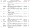
Innate pattern recognition receptors (PRRs), such as TLRs and nucleotide oligomerization domain (NOD)-like receptors (NLRs), recognize intestinal bacterial pathogens and/or pathogenic molecules. Ligand binding by the TLRs and NLRs expressed on host and/or donor-derived APCs substantially amplifies the release of inflammatory mediators (30). The transfer of HoxB8 neutrophils that lack expression of TLR 2, 3 4, 7, and 9 reduced GVHD severity compared with the transfer of WT HoxB8 neutrophils, indicating that TLR signals promote GVHD development (31). Conditioning-induced GI damage allows the translocation of outer membrane-derived endotoxins from gram-negative bacteria (e.g., lipopolysaccharide (LPS)) into systemic circulation (113233). The binding of LPS to TLR4 accelerated lethal intestinal GVHD by stimulating the production of inflammatory cytokines (e.g., TNFα, IL-1, IL-6, IL-10, IL-12, and TGFβ) from gut-associated lymphoid tissues (GALTs) and macrophages, and IFN-γ from activated donor T cells (934). The endogenous TLR4 agonist heparan sulfate activated dendritic cells (DCs) and aggravated acute GVHD (35). Unexpectedly, however, Tlr4–/– mic developed fulminant GVHD, and allogeneic hosts with a TLR4 mutation (C3H/HeJ mice) had increased intestinal damage compared to wild type counterparts (3637). TLR4 signaling mediated protective effects during GVHD, characterized by reduced intestinal cell apoptosis compared to that in hosts that did not undergo TLR4 signaling (36). In addition, TLR4 ligands were not necessary for the maturation of host APCs for GVHD induction (37). Collectively, these finding suggest that TLR4 signaling is involved in both positive and negative regulation of GVHD. Tlr9–/– mice developed less severe acute GVHD post-HSCT than controls (1138). Consistent with these findings, treatment of wild type mice with a synthetic TLR9 agonist (CpG oligonucleotides) markedly accelerated GVHD severity (39), and treatment with the TLR9-inhibitory oligonucleotide (iODN) 2088 reduced apoptosis of colonic cells in intestinal GVHD (1139). Thus, TLR9 signaling is associated with the induction of intestinal GVHD.
Application of the TLR7/8 agonist R-848 (resiquimod) promoted substantial innate immune activation and T cell migration into target organs (40). Another TLR7/8 agonist, 3M-011, caused differential effects on GVHD depending on the timing of the treatment. Administration of 3M-011 after allogenic transplant increased GVHD mortality, but pre-treatment with 3M-011 reduced the damage to target organs by inducing IDO expression in the colon (394142). Alterations to TLR2 expression on recipient lymphoid and myeloid cells from splenocytes had little effect on acute GVHD (43) (Table I). Thus, each of the TLRs is involved in acute GVHD to a different extent (43444546). Reports on the functional associations of TLRs and their adaptor molecules with GVHD are summarized in Table I and Table II.

MYD88-DEPENDENT EXPANSION OF MYELOID-DERIVED SUPPRESSOR CELLS (MDSCS) IN ACUTE INTESTINAL GVHD

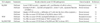
MyD88 is an adaptor molecule that activates inflammatory responses downstream of TLR ligand ligation (Table II) (474849). All TLRs, except TLR3, transduce signals through MyD88 (50). In MyD88-deficient recipient mice, the infiltration of donor T cells into the intestines and the apoptosis of colon cells were reduced, resulting in improved survival and clinical scoring for acute intestinal GVHD (11). However, MyD88-deficiency in donor bone marrow (BM) cells aggravated GVHD, resulting in increased intestinal pathology (51). The exacerbation of intestinal GVHD in recipients of MyD88-deficient BM cells was associated with insufficient expansion of MDSCs from the transplanted MyD88-deficient stem cells. These findings indicate that MyD88 signaling in donor cells promotes MDSC expansion and immune suppression in acute GVHD. The transfer of WT MDSCs into recipients of MyD88-deficient BM cells ameliorated intestinal GVHD, which supports a role for MyD88 in driving MDSC expansion in GVHD. Thus, MyD88 signaling has opposite impacts on intestinal GVHD, depending on whether MyD88 is expressed by host or donor cells.
MDSCs consist of two main subtypes: granulocytic/polymorphonuclear MDSCs and monocytic MDSCs. The phenotypes CD11b+LyG6+Ly6Clow and CD11b+LyG6lowLy6Chigh are used to identify the respective populations in mice. MDSCs expand robustly in various pathological conditions, such as cancers (52), autoimmune diseases (53), inflammation (54), infectious diseases (55565758), and GVHD (49515960). Most MDSC biology has been studied in tumor microenvironments, and preclinical and clinical tumor therapies have been tested for their ability to block MDSC expansion and function. Inhibitors of vascular endothelial growth factor (VEGF; bevacizumab) (61), signal transducer and activator of transcription 3 (STAT3; sunitinib) (62), arginase (NOHA) (52), inducible nitric oxide synthase (iNOS; nitroaspirin) (63), and cyclooxygenase-2 (COX2; celecoxib) (64), as well as agents that induce MDSC apoptosis and necrosis (gemcitabine and IL4Rα aptamer), have been shown to decrease MDSC expansion and tumor growth (6566). The expansion and functional enhancement of MDSCs are required for the control of acute intestinal GVHD. Arginase-1, iNOS, reactive oxygen species (ROS), and nitric oxide (NO) are mediators of the suppressive functions of MDSCs (52). Inflammatory mediators such as COX-2, G-CSF, GM-CSF, IFN-γ, IL-6, IL-10, VEFG, and prostaglandin E2 induce the differentiation and expansion of MDSCs, and inhibit the differentiation of mature myeloid cells in pathogenic environments (6768). These mediators could be targeted to enhance the suppressive functions of MDSCs to ameliorate GVHD. The selective modulation or exploitation of MyD88-mediated signaling to induce MDSC expansion and functional enhancement could be a strategy to suppress acute intestinal GVHD.

CONCLUSION

The dysregulation of microbial homeostasis and TLR signaling-mediated inflammatory responses are involved in the pathogenesis of intestinal GVHD. Understanding the effects and cellular/molecular mechanisms of TLR/MyD88 signaling on innate immune regulation of gut bacteria and MDSCs would aid the development of specific immune modulators to treat intestinal GVHD.

Figures and Tables

Figure 1

Schematic diagram of the development of acute GVHD. Acute GVHD can be classified into five distinct phases. Conditioning regimens (radiation or chemotherapy) induce tissue damage (I), and increase production of inflammatory cytokines, which cause the activation and maturation of APCs (II), leading to allo-reactive donor T cell priming and expansion (III). Activated donor T cells migrate to damaged host tissues (IV), where they amplify inflammatory responses and worsen GVHD (V). DC, dendritic cell; MΦ, macrophage.

in-17-144-g001
Table I

Studies of GVHD associated with innate immune responses through TLRs

in-17-144-i001
TLRs Treatments Results related to acute GVHD pathogenesis Donor/recipient References
TLR1 SNP genotyping SNPs in the TLR1 showed significant association with acute GVHD (e. g., SNP id: rs483307) Donor (human) 44
SNP genotyping No effect on the incidence of acute GVHD by polymorphisms of the TLR1 Donor (human) 45
TLR2 Deficient No effect on apoptosis/proliferation/neutrophilic granulocytes/survival in intestinal GVHD, donor T cells ↓ Recipient (mouse) 11
SNP genotyping Four SNPs in the TLR2 showed association with acute GVHD (e.g., SNP id: rs6535927) Donor (human) 44
SNP genotyping No effect on the incidence of acute GVHD by polymorphisms of the TLR2 Donor (human) 45
Deficient No effect on acute GVHD by upregulation of TLR2 expression in G-CSF-mobilized donor grafts Donor (mouse) 43
Deficient GVHD severity ↓, translocating bacteria ↓ (in TLR2/3/4/7/9– in HoxB8 neutrophils) Recipient (mouse) 31
TLR3 Deficient GVHD severity ↓, translocating bacteria ↓ (in TLR2/3/4/7/9– in HoxB8 neutrophils) Recipient (mouse) 31
SNP genotyping No effect on the incidence of acute GVHD by polymorphisms of the TLR3 Donor (human) 45
TLR4 Deficient No effects on apoptosis/proliferation/neutrophilic granulocytes/survival in intestinal GVHD, donor T cells ↓ Recipient (mouse) 11
Agonist GVHD severity ↑, alloreactive donor T cell proliferation ↑ Recipient (mouse) 35
Deficient Protection against intestinal cell apoptosis during acute GVHD by induction of tissue protective factors Recipient (mouse) 36
Mutation No difference in GVHD in HLA-matched HCT with mutation in donor Both (human) 46
Deficient No effect on GVHD severity Donor (mouse) 37
TLR5 SNP genotyping SNP in the TLR5 showed no sufficient evidence for the TLR5 importance in GVHD Donor (human) 44
TLR6 SNP genotyping SNP in the TLR6 showed association with acute GVHD (e.g., SNP id: rs6531656) Both (human) 44
TLR7 Deficient GVHD severity ↓, translocating bacteria ↓ (in TLR2/3/4/7/9– in HoxB8 neutrophils) Recipient (mouse) 31
Agonist Localized GVHD ↑ , infiltration of donor T cells ↑ Recipient (mouse) 39
TLR8 SNP genotyping No effect on the incidence of acute GVHD by polymorphisms of the TLR8 Donor (human) 45
TLR9 Deficient GVHD severity ↓, translocating bacteria ↓ (in TLR2/3/4/7/9– in HoxB8 neutrophils) Recipient (mouse) 31
Deficient Intestinal GVHD severity ↓ (dependent on MyD88 signaling), survival rates ↑ in-17-144-i001 11
Agonist GVHD severity ↑ Recipient (mouse) 11
Deficient GVHD severity ↓, apoptotic cells , proliferation of cells in colon ↑ Recipient (mouse) 44
SNP genotyping Associated with the risk of acute GVHD by TLR9 SNPs in the donors of allogeneic HSCT Donor (human) 45
TLR10 SNP genotyping SNP in the TLR10 showed significant association with acute GVHD (e.g., SNP id: rs337629) Both (human) 44

TLR, toll-like receptor; GVHD, graft-versus host disease; SNP, small nucleotide polymorphism; HSCT, hematopoietic stem cell transplantation; HoxB8, Homeobox B8.

Table II

Studies of GVHD associated with innate immune responses through TLR adaptor molecules

in-17-144-i002
TLR adaptors Treatments Results related to acute GVHD pathogenesis Donor/recipient References
MyD88 Deficient Acute GVHD severity ↓, apoptotic cell ↓, proliferation of cells in colon ↓ Recipient (mouse) 11
Deficient Intestinal GVHD ↑ , myeloid cell apoptosis ↑, donor T cells , expansion/function of MDSCs ↓ Donor (mouse) 49
Deficient Hepatic GVHD severity ↓, infiltration of T cells into the liver of the recipients ↓ Donor (mouse) 51
Deficient No effect on acute GVHD (lack of MyD88 in donor APC) Donor (mouse) 37
TRIF Deficient No effect on acute GVHD, neutrophil infiltration in to colon ↑ Recipient (mouse) 11
Deficient No effect on acute GVHD (lack of TRIF in donor APC) Donor (mouse) 37
MyD88/TRIF Deficient No effect on acute GVHD (lack of MyD88 and TRIF in donor APC) Donor (mouse) 11

GVHD, graft-versus host disease; APC, antigen-presenting cell; MyD88, myeloid differentiation primary response 88; TRIF, TIR-domain-containing adaptor-inducing Interferon-β.

Abbreviations

GVHD

graft-versus host disease

allo

allogeneic

HSCT

hematopoietic stem cell transplantation

GI

gastrointestinal

MDSCs

myeloidderived suppressor cells

BM

bone marrow

ACKNOWLEDGEMENTS

This work was supported by the Education and Research Encouragement Fund of Seoul National University Hospital (2017).

Notes

CONFLICTS OF INTEREST There is no conflict of interest.

References

1. Mohty M, de LH, Ladaique P, Faucher C, Vey N, Coso D, Stoppa AM, Gastaut JA, Blaise D. The role of reduced intensity conditioning allogeneic stem cell transplantation in patients with acute myeloid leukemia: a donor vs no donor comparison. Leukemia. 2005; 19:916–920.
crossref
2. Poon LM, Bassett R Jr, Rondon G, Hamdi A, Qazilbash M, Hosing C, Jones RB, Shpall EJ, Popat UR, Nieto Y, Worth LL, Cooper L, De LM, Champlin RE, Kebriaei P. Outcomes of second allogeneic hematopoietic stem cell transplantation for patients with acute lymphoblastic leukemia. Bone Marrow Transplant. 2013; 48:666–670.
crossref
3. Demirer T, Barkholt L, Blaise D, Pedrazzoli P, Aglietta M, Carella AM, Bay JO, Arpaci F, Rosti G, Gurman G, Niederwieser D, Bregni M. Transplantation of allogeneic hematopoietic stem cells: an emerging treatment modality for solid tumors. Nat Clin Pract Oncol. 2008; 5:256–267.
crossref
4. Dvorak CC, Cowan MJ. Hematopoietic stem cell transplantation for primary immunodeficiency disease. Bone Marrow Transplant. 2008; 41:119–126.
crossref
5. Warren EH, Fujii N, Akatsuka Y, Chaney CN, Mito JK, Loeb KR, Gooley TA, Brown ML, Koo KK, Rosinski KV, Ogawa S, Matsubara A, Appelbaum FR, Riddell SR. Therapy of relapsed leukemia after allogeneic hematopoietic cell transplantation with T cells specific for minor histocompatibility antigens. Blood. 2010; 115:3869–3878.
crossref
6. Blazar BR, Murphy WJ, Abedi M. Advances in graft-versus-host disease biology and therapy. Nat Rev Immunol. 2012; 12:443–458.
crossref
7. Shlomchik WD. Graft-versus-host disease. Nat Rev Immunol. 2007; 7:340–352.
crossref
8. Washington K, Jagasia M. Pathology of graft-versus-host disease in the gastrointestinal tract. Hum Pathol. 2009; 40:909–917.
crossref
9. Hill GR, Ferrara JL. The primacy of the gastrointestinal tract as a target organ of acute graft-versus-host disease: rationale for the use of cytokine shields in allogeneic bone marrow transplantation. Blood. 2000; 95:2754–2759.
crossref
10. Fukata M, Vamadevan AS, Abreu MT. Toll-like receptors (TLRs) and Nod-like receptors (NLRs) in inflammatory disorders. Semin Immunol. 2009; 21:242–253.
crossref
11. Heimesaat MM, Nogai A, Bereswill S, Plickert R, Fischer A, Loddenkemper C, Steinhoff U, Tchaptchet S, Thiel E, Freudenberg MA, Gobel UB, Uharek L. MyD88/TLR9 mediated immunopathology and gut microbiota dynamics in a novel murine model of intestinal graft-versus-host disease. Gut. 2010; 59:1079–1087.
crossref
12. Marcellus DC, Altomonte VL, Farmer ER, Horn TD, Freemer CS, Grant J, Vogelsang GB. Etretinate therapy for refractory sclerodermatous chronic graft-versus-host disease. Blood. 1999; 93:66–70.
crossref
13. Pai CC, Chen M, Mirsoian A, Grossenbacher SK, Tellez J, Ames E, Sun K, Jagdeo J, Blazar BR, Murphy WJ, Abedi M. Treatment of chronic graft-versus-host disease with bortezomib. Blood. 2014; 124:1677–1688.
crossref
14. Rozendaal L, Pals ST, Gleichmann E, Melief CJ. Persistence of allospecific helper T cells is required for maintaining autoantibody formation in lupus-like graft-versus-host disease. Clin Exp Immunol. 1990; 82:527–532.
crossref
15. Wakae T, Takatsuka H, Seto Y, Iwata N, Mori A, Okada M, Fujimori Y, Okamoto T, Kakishita E, Hara H. Similarity between hepatic graft-versus-host disease and primary biliary cirrhosis. Hematology. 2002; 7:305–310.
crossref
16. Shono Y, Shiratori S, Kosugi-Kanaya M, Ueha S, Sugita J, Shigematsu A, Kondo T, Hashimoto D, Fujimoto K, Endo T, Nishio M, Hashino S, Matsuno Y, Matsushima K, Tanaka J, Imamura M, Teshima T. Bone marrow graft-versus-host disease: evaluation of its clinical impact on disrupted hematopoiesis after allogeneic hematopoietic stem cell transplantation. Biol Blood Marrow Transplant. 2014; 20:495–500.
crossref
17. Silva R, Morgado JM, Freitas A, Couceiro A, Orfao A, Regateiro F, Paiva A. Influence of pro- and anti-inflammatory cytokines in Th1 polarization after allogeneic stimulation. Int J Biomed Sci. 2005; 1:46–52.
18. Biedermann BC, Sahner S, Gregor M, Tsakiris DA, Jeanneret C, Pober JS, Gratwohl A. Endothelial injury mediated by cytotoxic T lymphocytes and loss of microvessels in chronic graft versus host disease. Lancet. 2002; 359:2078–2083.
crossref
19. Jagasia MH, Greinix HT, Arora M, Williams KM, Wolff D, Cowen EW, Palmer J, Weisdorf D, Treister NS, Cheng GS, Kerr H, Stratton P, Duarte RF, McDonald GB, Inamoto Y, Vigorito A, Arai S, Datiles MB, Jacobsohn D, Heller T, Kitko CL, Mitchell SA, Martin PJ, Shulman H, Wu RS, Cutler CS, Vogelsang GB, Lee SJ, Pavletic SZ, Flowers ME. National institutes of health consensus development project on criteria for clinical trials in chronic graft-versus-host disease: I. The 2014 diagnosis and staging working group report. Biol Blood Marrow Transplant. 2015; 21:389–401.
crossref
20. Ferrara JL, Deeg HJ. Graft-versus-host disease. N Engl J Med. 1991; 324:667–674.
crossref
21. Reddy P. Pathophysiology of acute graft-versus-host disease. Hematol Oncol. 2003; 21:149–161.
crossref
22. Ju JM, Lee H, Oh K, Lee DS, Choi EY. Kinetics of IFN-gamma and IL-17 production by CD4 and CD8 T cells during acute graft-versus-host disease. Immune Netw. 2014; 14:89–99.
crossref
23. Ross WA, Ghosh S, Dekovich AA, Liu S, Ayers GD, Cleary KR, Lee JH, Couriel D. Endoscopic biopsy diagnosis of acute gastrointestinal graft-versus-host disease: rectosigmoid biopsies are more sensitive than upper gastrointestinal biopsies. Am J Gastroenterol. 2008; 103:982–989.
crossref
24. Mai V, Draganov PV. Recent advances and remaining gaps in our knowledge of associations between gut microbiota and human health. World J Gastroenterol. 2009; 15:81–85.
crossref
25. Sender R, Fuchs S, Milo R. Revised estimates for the number of human and bacteria cells in the body. PLoS Biol. 2016; 14:e1002533.
crossref
26. Savage DC. Microbial ecology of the gastrointestinal tract. Annu Rev Microbiol. 1977; 31:107–133.
crossref
27. Jenq RR, Ubeda C, Taur Y, Menezes CC, Khanin R, Dudakov JA, Liu C, West ML, Singer NV, Equinda MJ, Gobourne A, Lipuma L, Young LF, Smith OM, Ghosh A, Hanash AM, Goldberg JD, Aoyama K, Blazar BR, Pamer EG, van den Brink MR. Regulation of intestinal inflammation by microbiota following allogeneic bone marrow transplantation. J Exp Med. 2012; 209:903–911.
crossref
28. Eriguchi Y, Takashima S, Oka H, Shimoji S, Nakamura K, Uryu H, Shimoda S, Iwasaki H, Shimono N, Ayabe T, Akashi K, Teshima T. Graft-versus-host disease disrupts intestinal microbial ecology by inhibiting Paneth cell production of alpha-defensins. Blood. 2012; 120:223–231.
crossref
29. Beelen DW, Elmaagacli A, Muller KD, Hirche H, Schaefer UW. Influence of intestinal bacterial decontamination using metronidazole and ciprofloxacin or ciprofloxacin alone on the development of acute graft-versus-host disease after marrow transplantation in patients with hematologic malignancies: final results and long-term follow-up of an open-label prospective randomized trial. Blood. 1999; 93:3267–3275.
crossref
30. Murphy S, Nguyen VH. Role of gut microbiota in graft-versus-host disease. Leuk Lymphoma. 2011; 52:1844–1856.
crossref
31. Schwab L, Goroncy L, Palaniyandi S, Gautam S, Triantafyllopoulou A, Mocsai A, Reichardt W, Karlsson FJ, Radhakrishnan SV, Hanke K, Schmitt-Graeff A, Freudenberg M, von Loewenich FD, Wolf P, Leonhardt F, Baxan N, Pfeifer D, Schmah O, Schonle A, Martin SF, Mertelsmann R, Duyster J, Finke J, Prinz M, Henneke P, Hacker H, Hildebrandt GC, Hacker G, Zeiser R. Neutrophil granulocytes recruited upon translocation of intestinal bacteria enhance graft-versus-host disease via tissue damage. Nat Med. 2014; 20:648–654.
crossref
32. Hill GR, Crawford JM, Cooke KR, Brinson YS, Pan L, Ferrara JL. Total body irradiation and acute graft-versus-host disease: the role of gastrointestinal damage and inflammatory cytokines. Blood. 1997; 90:3204–3213.
crossref
33. Cooke KR, Gerbitz A, Crawford JM, Teshima T, Hill GR, Tesolin A, Rossignol DP, Ferrara JL. LPS antagonism reduces graft-versus-host disease and preserves graft-versus-leukemia activity after experimental bone marrow transplantation. J Clin Invest. 2001; 107:1581–1589.
crossref
34. Holler E. Cytokines, viruses, and graft-versus-host disease. Curr Opin Hematol. 2002; 9:479–484.
crossref
35. Brennan TV, Lin L, Huang X, Cardona DM, Li Z, Dredge K, Chao NJ, Yang Y. Heparan sulfate, an endogenous TLR4 agonist, promotes acute GVHD after allogeneic stem cell transplantation. Blood. 2012; 120:2899–2908.
crossref
36. Imado T, Iwasaki T, Kitano S, Satake A, Kuroiwa T, Tsunemi S, Sano H. The protective role of host Toll-like receptor-4 in acute graft-versus-host disease. Transplantation. 2010; 90:1063–1070.
crossref
37. Li H, Matte-Martone C, Tan HS, Venkatesan S, McNiff J, Demetris AJ, Jain D, Lakkis F, Rothstein D, Shlomchik WD. Graft-versus-host disease is independent of innate signaling pathways triggered by pathogens in host hematopoietic cells. J Immunol. 2011; 186:230–241.
crossref
38. Calcaterra C, Sfondrini L, Rossini A, Sommariva M, Rumio C, Menard S, Balsari A. Critical role of TLR9 in acute graft-versus-host disease. J Immunol. 2008; 181:6132–6139.
crossref
39. Taylor PA, Ehrhardt MJ, Lees CJ, Panoskaltsis-Mortari A, Krieg AM, Sharpe AH, Murphy WJ, Serody JS, Hemmi H, Akira S, Levy RB, Blazar BR. TLR agonists regulate alloresponses and uncover a critical role for donor APCs in allogeneic bone marrow rejection. Blood. 2008; 112:3508–3516.
crossref
40. Spranger S, Javorovic M, Burdek M, Wilde S, Mosetter B, Tippmer S, Bigalke I, Geiger C, Schendel DJ, Frankenberger B. Generation of Th1-polarizing dendritic cells using the TLR7/8 agonist CL075. J Immunol. 2010; 185:738–747.
crossref
41. Hemmi H, Kaisho T, Takeuchi O, Sato S, Sanjo H, Hoshino K, Horiuchi T, Tomizawa H, Takeda K, Akira S. Small anti-viral compounds activate immune cells via the TLR7 MyD88-dependent signaling pathway. Nat Immunol. 2002; 3:196–200.
crossref
42. Jasperson LK, Bucher C, Panoskaltsis-Mortari A, Mellor AL, Munn DH, Blazar BR. Inducing the tryptophan catabolic pathway, indoleamine 2,3-dioxygenase (IDO), for suppression of graft-versus-host disease (GVHD) lethality. Blood. 2009; 114:5062–5070.
crossref
43. Lee WS, Kim JY, Won HJ, Lee SM, Suh YS, Joo YD, Lee JY, Jang WH, Kang SW, Kang MS, Park SG, Choi IW, Choi I, Seo SK. Effect of upregulated TLR2 expression from G-CSF-mobilized donor grafts on acute graft-versus-host disease. Int Immunopharmacol. 2015; 29:488–493.
crossref
44. Sivula J, Cordova ZM, Tuimala J, Jaatinen T, Partanen J, Volin L, Turpeinen H. Toll-like receptor gene polymorphisms confer susceptibility to graft-versus-host disease in allogenic hematopoietic stem cell transplantation. Scand J Immunol. 2012; 76:336–341.
crossref
45. Xiao HW, Luo Y, Lai XY, Shi JM, Tan YM, He JS, Xie WZ, Zheng WY, Ye XJ, Yu XH, Cai Z, Lin MF, Huang H. Donor TLR9 gene tagSNPs influence susceptibility to aGVHD and CMV reactivation in the allo-HSCT setting without polymorphisms in the TLR4 and NOD2 genes. Bone Marrow Transplant. 2014; 49:241–247.
crossref
46. Lorenz E, Schwartz DA, Martin PJ, Gooley T, Lin MT, Chien JW, Hansen JA, Clark JG. Association of TLR4 mutations and the risk for acute GVHD after HLA-matched-sibling hematopoietic stem cell transplantation. Biol Blood Marrow Transplant. 2001; 7:384–387.
crossref
47. Akira S, Takeda K. Toll-like receptor signalling. Nat Rev Immunol. 2004; 4:499–511.
crossref
48. Takeda K, Kaisho T, Akira S. Toll-like receptors. Annu Rev Immunol. 2003; 21:335–376.
crossref
49. Lim JY, Lee YK, Lee SE, Ju JM, Park G, Choi EY, Min CK. Attenuation of hepatic graft-versus-host disease in allogeneic recipients of MyD88-deficient donor bone marrow. Immune Netw. 2015; 15:125–134.
crossref
50. Yamamoto M, Sato S, Hemmi H, Hoshino K, Kaisho T, Sanjo H, Takeuchi O, Sugiyama M, Okabe M, Takeda K, Akira S. Role of adaptor TRIF in the MyD88-independent toll-like receptor signaling pathway. Science. 2003; 301:640–643.
crossref
51. Lim JY, Lee YK, Lee SE, Ju JM, Eom KS, Kim YJ, Chung NG, Jeong DC, Park G, Choi EY, Min CK. MyD88 in donor bone marrow cells is critical for protection from acute intestinal graft-vs.-host disease. Mucosal Immunol. 2016; 9:730–743.
crossref
52. Gabrilovich DI, Ostrand-Rosenberg S, Bronte V. Coordinated regulation of myeloid cells by tumours. Nat Rev Immunol. 2012; 12:253–268.
crossref
53. Crook KR, Liu P. Role of myeloid-derived suppressor cells in autoimmune disease. World J Immunol. 2014; 4:26–33.
crossref
54. Smith AR, Reynolds JM. Editorial: the contribution of myeloid-derived suppression to inflammatory disease. J Leukoc Biol. 2014; 96:361–364.
crossref
55. Chandra D, Jahangir A, Quispe-Tintaya W, Einstein MH, Gravekamp C. Myeloid-derived suppressor cells have a central role in attenuated Listeria monocytogenes-based immunotherapy against metastatic breast cancer in young and old mice. Br J Cancer. 2013; 108:2281–2290.
crossref
56. Schmid M, Zimara N, Wege AK, Ritter U. Myeloid-derived suppressor cell functionality and interaction with Leishmania major parasites differ in C57BL/6 and BALB/c mice. Eur J Immunol. 2014; 44:3295–3306.
crossref
57. Terrazas LI, Walsh KL, Piskorska D, McGuire E, Harn DA Jr. The schistosome oligosaccharide lacto-N-neotetraose expands Gr1(+) cells that secrete anti-inflammatory cytokines and inhibit proliferation of naive CD4(+) cells: a potential mechanism for immune polarization in helminth infections. J Immunol. 2001; 167:5294–5303.
crossref
58. Gabrilovich DI, Nagaraj S. Myeloid-derived suppressor cells as regulators of the immune system. Nat Rev Immunol. 2009; 9:162–174.
crossref
59. Koehn BH, Apostolova P, Haverkamp JM, Miller JS, McCullar V, Tolar J, Munn DH, Murphy WJ, Brickey WJ, Serody JS, Gabrilovich DI, Bronte V, Murray PJ, Ting JP, Zeiser R, Blazar BR. GVHD-associated, inflammasome-mediated loss of function in adoptively transferred myeloid-derived suppressor cells. Blood. 2015; 126:1621–1628.
crossref
60. Messmann JJ, Reisser T, Leithauser F, Lutz MB, Debatin KM, Strauss G. In vitro-generated MDSCs prevent murine GVHD by inducing type 2 T cells without disabling antitumor cytotoxicity. Blood. 2015; 126:1138–1148.
crossref
61. Kusmartsev S, Eruslanov E, Kubler H, Tseng T, Sakai Y, Su Z, Kaliberov S, Heiser A, Rosser C, Dahm P, Siemann D, Vieweg J. Oxidative stress regulates expression of VEGFR1 in myeloid cells: link to tumor-induced immune suppression in renal cell carcinoma. J Immunol. 2008; 181:346–353.
crossref
62. Ko JS, Zea AH, Rini BI, Ireland JL, Elson P, Cohen P, Golshayan A, Rayman PA, Wood L, Garcia J, Dreicer R, Bukowski R, Finke JH. Sunitinib mediates reversal of myeloid-derived suppressor cell accumulation in renal cell carcinoma patients. Clin Cancer Res. 2009; 15:2148–2157.
crossref
63. De SC, Serafini P, Marigo I, Dolcetti L, Bolla M, Del SP, Melani C, Guiducci C, Colombo MP, Iezzi M, Musiani P, Zanovello P, Bronte V. Nitroaspirin corrects immune dysfunction in tumor-bearing hosts and promotes tumor eradication by cancer vaccination. Proc Natl Acad Sci U S A. 2005; 102:4185–4190.
crossref
64. Veltman JD, Lambers ME, van NM, Hendriks RW, Hoogsteden HC, Aerts JG, Hegmans JP. COX-2 inhibition improves immunotherapy and is associated with decreased numbers of myeloid-derived suppressor cells in mesothelioma. Celecoxib influences MDSC function. BMC Cancer. 2010; 10:464.
crossref
65. Wu H, Tao N, Liu X, Li X, Tang J, Ma C, Xu X, Shao H, Hou B, Wang H, Qin Z. Polysaccharide from Lentinus edodes inhibits the immunosuppressive function of myeloid-derived suppressor cells. PLoS One. 2012; 7:e51751.
crossref
66. Roth F, De La Fuente AC, Vella JL, Zoso A, Inverardi L, Serafini P. Aptamer-mediated blockade of IL4Ralpha triggers apoptosis of MDSCs and limits tumor progression. Cancer Res. 2012; 72:1373–1383.
crossref
67. Sawanobori Y, Ueha S, Kurachi M, Shimaoka T, Talmadge JE, Abe J, Shono Y, Kitabatake M, Kakimi K, Mukaida N, Matsushima K. Chemokine-mediated rapid turnover of myeloid-derived suppressor cells in tumor-bearing mice. Blood. 2008; 111:5457–5466.
crossref
68. Sinha P, Clements VK, Fulton AM, Ostrand-Rosenberg S. Prostaglandin E2 promotes tumor progression by inducing myeloid-derived suppressor cells. Cancer Res. 2007; 67:4507–4513.
crossref
TOOLS
Similar articles