Journal List > Immune Netw > v.15(2) > 1033489

Kim, Kim, Pyo, Lee, Hong, Kim, and Han: Adoptive Cell Therapy of Melanoma with Cytokine-induced Killer Cells

Abstract

Melanoma is the most aggressive skin cancer and its incidence is gradually increasing worldwide. Patients with metastatic melanoma have a very poor prognosis (estimated 5-year survival rate of <16%). In the last few years, several drugs have been approved for malignant melanoma, such as tyrosine kinase inhibitors and immune checkpoint blockades. Although new therapeutic agents have improved progression-free and overall survival, their use is limited by drug resistance and drug-related toxicity. At the same time, adoptive cell therapy of metastatic melanoma with tumor-infiltrating lymphocytes has shown promising results in preclinical and clinical studies. In this review, we summarize the currently available drugs for treatment of malignant melanoma. In addition, we suggest cytokine-induced killer (CIK) cells as another candidate approach for adoptive cell therapy of melanoma. Our preclinical study and several previous studies have shown that CIK cells have potent anti-tumor activity against melanomas in vitro and in an in vivo human tumor xenograft model without any toxicity.

INTRODUCTION

Skin cancer, the most common malignancy worldwide, is classified into two main categories, non-melanoma skin cancer (NMSC) and melanoma (1). Basal cell cancer (80%) and squamous cell cancer (16%) are the major subtypes of NMSC (1). Although NMSC is the most common skin cancer, it is rarely lethal (2). Melanoma, which originates from melanocytes, accounts for <4% of all skin cancer cases but is the major cause of death from skin cancer (3). Melanocytes are melanin-producing cells found mainly in the skin and eyes. In the skin, melanocytes reside in the bottom layer of the epidermis. Exposure to ultraviolet (UV) radiation increases melanin production by melanocytes. The biological function of melanogenesis is to protect the tissues under the skin from DNA-damaging UV exposure (3). Although the causes of melanoma are unknown, DNA damage because of UV exposure and genetic predisposition may play a role (4,5,6). It is estimated that approximately 76,100 patients in USA will be diagnosed with melanoma in 2014 and 9,710 will die of malignant melanoma (7). While the incidence rates of most cancer types are declining, the incidence of melanoma is gradually increasing (7). Although localized melanoma may be curable by surgical resection, patients with distant metastases have an extremely poor prognosis with an estimated 5-year survival rate of <16% (7). In this review, we summarize the currently available drugs and therapies of malignant melanoma and suggest the possible use of adoptive cell therapy (ACT) with cytokine-induced killer (CIK) cells as an additional therapeutic approach for malignant melanoma.

CURRENT TREATMENT OF MALIGNANT MELANOMA

Before 2011, there were only two agents approved by the US Food and Drug Administration (FDA) for the treatment of metastatic melanoma: the chemotherapeutic agent dacarbazine (an alkylating compound) and interleukin-2 (IL-2) (8). However, both drugs show a low response rate (5%~20%) and have considerable adverse effects (9). Over the last decade, a better understanding of genetic and molecular pathogenesis of melanoma, in particular mutations that increase the risk of this cancer, has led to new treatment approaches. Hyper-activation of mitogen-activated protein kinase (MAPK) signaling (namely, the RAS-RAF-MEK-ERK pathway), is the main characteristic of malignant melanoma detected in up to 75% of human melanomas. The most common mutations in human melanomas are found in neuroblastoma RAS viral oncogene homolog (NRAS, 15~25%) and v-raf murine sarcoma viral oncogene homolog B (BRAF, 50~70%) (10,11). FDA has approved several selective inhibitors of BRAF (vemurafenib and dabrafenib) and MEK (trametinib). Vemurafenib and dabrafenib target V600E-mutated BRAF, which is the most common BRAF mutation (approximately 90%) (9). All chemical inhibitors improve progression-free survival and overall survival of metastatic melanoma patients with the BRAF V600E or V600K mutation (12,13). However, targeted therapy of malignant melanoma is often limited by the emergence of acquired drug resistance 6~7 months after administration (14). Drug resistance occurs via reactivation of the MAPK pathway or activation of an alternative signaling pathway such as PI3K-AKT-mTOR (13,15). As a result, most patients relapse with lethal drug resistance.
In addition, FDA also have approved monoclonal antibody (mAb) therapeutics, such as ipilimumab targeting cytotoxic T lymphocyte-associated antigen 4 (CTLA-4) receptor and pembrolizumab targeting programmed death 1 (PD-1) receptor. The purpose of immune checkpoint blockade with anti-CTLA4 and anti-PD-1 mAb is to restore the immune response. CTLA-4 and CD28 share B7 ligands, for example B7-1 (CD80) and B7-2 (CD86). CD28 provides the second signal required for T-cell activation after the first signal of antigen recognition through T cell receptor (TCR), whereas CTLA-4 inhibits T cell activation (16,17). Although the mechanisms of CTLA-4 action are still not fully understood, CTLA-4 may inhibit T-cell activation by competing with B7 ligand binding to CD28 or by interfering with CD28 signaling. Thus, CTLA-4 blockade with mAb can restore antitumor T cell immunity (18). A recent study shows that ipilimumab can bind CTLA-4 expressed on melanoma cells, which induce antibody-dependent cell-mediated cytotoxicity by natural killer (NK) cells and γδT lymphocytes (19). Another immune checkpoint molecule is PD-1, a T-cell co-inhibitory molecule. PD-1 is expressed on activated T cells and negatively regulates T-cell response (16,20,21,22). Most melanoma and solid tumor cells express PD-1 ligand (PD-L1) on their surfaces (23). PD-1 blockade can enhance the antitumor activity of T cells by inhibiting the PD-1-PD-L1 interaction (24). Pembrolizumab (anti-PD-1 mAb) was approved by the FDA on September 4, 2014. Although immune checkpoint blockades have more durable responses than MAPK inhibitors, grade 3 or 4 immune-related adverse events were observed in ~15~25% of patients; these events included dermatitis, colitis, hypophysitis, hepatitis, pneumonitis, and neuromuscular toxicity (25,26,27,28). In addition, clinical responses to immune checkpoint blockades are generally slow (18).

ADOPTIVE CELL THERAPY OF MELANOMA WITH TUMOR-INFILTRATING LYMPHOCYTES

Adoptive cell therapy (ACT) is a form of cancer immunotherapy for malignant melanoma, which entails the transfer of immune cells such as tumor-infiltrating lymphocytes (TILs), genetically modified T cells, or NK cells. After host lymphodepletion, adoptive transfer of TILs increases the objective response rate by >51% and durable response more than conventional chemotherapy and targeted therapy do (29). Lymphodepletion prior to ACT has various advantages, including the elimination of Treg or myeloid-derived suppressor cells and increase of the cytokine levels in the serum (30,31,32). So far, TIL therapy has been the most effective ACT treatment for patients with metastatic melanoma. However, it is difficult to obtain tumor tissue to isolate TILs, and TILs from many patients fail to expand sufficiently in culture. Therefore, TIL therapy has been applied to only a limited number of patients (33). To increase the clinical application of TILs, genetically modified T cells are being developed. T lymphocytes expressing αβTCRs recognize tumor-associated antigens on the surface of MHC molecules on tumor cells. Some tumor-associated antigens have been identified, including melanoma antigen recognized by T cells 1 (MART-1) and cancer testis antigen (NY-ESO-1) (34,35). T cells expressing antigen-specific TCR can be made by genetic modification by using viral or non-viral transduction of peripheral blood lymphocytes (PBLs). Although genetic modification might increase the clinical application of T cell therapy, objective responses (19~30% of patients) were still lower than with autologous TILs (36,37). In addition, MART-1 and gp100-specific TCRs cause on-target toxicity in approximately half of patients, which leads to destruction of normal melanocytes in the skin (rash), eye (uveitis), and/or ear (hearing loss) (37). This suggests that safer and more effective antigens are needed. In addition, the mechanisms of action restricted to MHC I are a limitation for antigen-specific T cell therapy for MHC I-negative tumor cells.

ADOPTIVE CELL THERAPY OF MELANOMA WITH NATURAL KILLER CELLS

NK cells are a type of cytotoxic lymphocytes that function in innate immunity. They can lyse tumor cells without prior immunization or MHC-restriction (38). NK cell-mediated tumor cell killing depends on the balance between inhibitory and activating receptors and their signaling. The inhibitory receptors, which bind to MHC-I molecules, are present on most normal cells and prevent their killing by NK cells. Tumor cells often decrease the expression of MHC molecules, which allows them to avoid T cell-mediated killing (39,40) but makes such tumor cells potential targets of NK cells. However, tumor cells expressing MHC-I molecules suppress autologous NK cell-mediated killing (41). For this reason, haploidentical allogeneic NK cells have been successfully used for cancer immunotherapy without causing graft-versus-host disease (42). Although allogeneic NK cells eventually lead to immune-mediated rejection by host immunity, NK cell-based therapy has shown promising results in hematological cancer patients (42). However, in a clinical trial with melanoma patients, although a high level of circulating autologous NK cells persisted for several months, they failed to mediate tumor regression (43).

ADOPTIVE CELL THERAPY OF MELANOMA WITH CYTOKINE-INDUCED KILLER CELLS

Cytokine-induced killer cells are ex vivo-activated lymphocytes generated by culturing peripheral blood mononuclear cells (PBMCs) with the timed addition of IFN-γ, anti-CD3 antibodies, and IL-2 for >2 weeks (44). Anti-CD3 antibodies and IL-2 are essential for proliferation and activation of CIK cells (45). Heterogeneous CIK cells consist of two major populations, CD3+CD56- and CD3+CD56+ cells, and a relatively minor fraction of CD3-CD56+ cells. The CD3+CD56+ cells are the most potent cytotoxic cells. Interestingly, during ex vivo expansion, they originate from CD3+CD8+CD56- cells but not from CD3+CD56+ cells (46). Cytotoxicity of CIK cells is mainly mediated by perforin (47) and depends on the activating receptors such as NKG2D (48), NKp30, and DNAM-1 (49). CIK cells have the following advantages for cancer immunotherapy in comparison with other ACT approaches: 1) they can be easily generated and produced in large quantities by expansion from PBMCs ex vivo (50); 2) they exhibit non-MHC-restricted cytotoxic activity (49); and 3) they are effective in eliminating multidrug-resistant tumor cell lines (51,52,53,54). In addition, the antitumor activity of CIK cells can be increased by combination therapy without an increase in adverse effects and by co-culturing them with tumor lysate-pulsed dendritic cells (55,56).
In the last decade, many preclinical studies have demonstrated the antitumor activity of CIK cells against various tumor cells such as hepatoma (57), leukemia (58), as well as lung (59), ovarian (60), renal (61)and gastric (62) cancers. However, only a few studies have been reported on melanoma. Gammaitoni and colleagues reported that patient-derived CIK cells are able to kill not only autologous metastatic melanoma cells in vitro and in an in vivo xenograft mouse model, but also putative melanoma cancer stem cells (63). Such cells, which have stemness characteristics, are well known to cause relapse and drug resistance (64). This implies that CIK-cell therapy may be effective for treatment of patients with metastatic melanoma and also for prevention of relapse and metastasis.
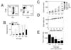
Here, we provide additional evidence that CIK-cell therapy can effectively eliminate melanoma in an in vivo xenograft mouse model. CIK cells were generated from PBMCs of healthy volunteers. PBMCs were isolated by Ficoll-Hypaque density centrifugation, washed three times with PBS and cultured in the presence of immobilized anti-CD3 antibody (5 µg/ml) and recombinant human IL-2 (700 U/ml) for 5 days. The cell suspension was further incubated in complete medium containing IL-2 only (170 U/ml) for 9 days. IL-2 and medium were replenished every 2 or 3 days (60). Cell density was maintained at ~1×10⁶ cells/ml. On day 14, the viability of expanded cell populations was 85~90%. Cell phenotypes were examined by flow cytometry. CIK cell populations contained 93% CD3+, 5% CD3-CD56+, 47% CD3+CD56+, 11% CD4+, and 74% CD8+ cells (Fig. 1A), which was typical for a heterogeneous CIK cell population.
To determine the anti-tumor activity of CIK cells, two cell lines were used. LOX-IMVI cell lines are human malignant melanoma and have been used widely utilized for drug screening and molecular target identification (65,66,67). K-562 cell lines are human leukemic cells and used as reference target cells. Both cell lines were obtained from American Type Culture Collection (Manassas, VA, USA) and were grown in the RPMI-1640 medium supplemented with 10% fetal bovine serum, 100 U/ml penicillin and 100 mg/ml streptomycin [20]. A 4-h 51Cr release assay revealed that CIK cells destroyed 11%, 16%, and 38% of LOX-IMVI cells at the effector:target ratios of 10:1, 30:1 and 100:1, respectively (Fig. 1B). Strong CIK cell cytotoxicity was also observed against K562 cells.
The nude mouse xenograft assay was used to examine the in vivo antitumor activity of CIK cells. LOX-IMVI cells (1.5×10⁶) in 300 µl of PBS were injected subcutaneously on day 0, followed by intravenous once-a-week injection of CIK cells (see below for doses) and adriamycin (used as a reference drug; 2 mg/kg). On day 15, mice were sacrificed and the tumor mass and body weight (to determine toxicity) were measured. In control mice, LOX-IMVI cells grew to a tumor volume of 254±43 mm3 (n=7) (Fig. 1C). A strong anti-tumor effect of CIK cells was observed (Fig. 1E). CIK cells injected at doses of 1×10⁶, 3×10⁶, and 10×106 cells per mouse inhibited tumor growth by 34%, 57%, and 76%, respectively. Adriamycin also strongly inhibited the growth of LOX-IMVI cells (Fig. 1C). In control mice, the weight of LOX-IMVI cells reached 1,676±530 mg at 15 days after implantation. CIK cells injected at doses of 3×10⁶ and 10×106 cells per mouse reduced tumor weight by 52% and 76%, respectively. Adriamycin also strongly inhibited tumor growth. CIK cells did not affect the body weight gain of nude mice, indicating that CIK cells are not toxic (Fig. 1D). Thus, our preclinical efficacy data show that CIK cells can destroy LOX-IMVI cells in vitro and in vivo, and suggest that CIK cells might be a good candidate immunotherapy of melanoma.

CONCLUSION

Patients with metastatic melanoma have a high risk of metastases leading to death. So far, conventional chemo- and targeted therapies have been the first-line treatments, but they are often limited by drug resistance. Although combination therapy delays drug resistance, an increase in side effects needs to be carefully managed or treatments need to be discontinued. Over the last few years, CIK cell therapy has been evaluated in many clinical studies in patients with hepatocellular carcinoma, renal cell carcinoma, non-small cell lung cancer, and gastric cancer (68). CIK cell therapy improved the prognosis of these cancer patients (increased overall survival and progression-free survival) without significant adverse events. In most cancer patients, combination of CIK cell therapy with conventional treatment options shows better clinical outcomes than standard therapy alone. Unfortunately, there are no reports of clinical studies of the use of CIK cells for melanoma treatment. Preclinical data obtained by us and others, which demonstrate that CIK cells have sufficient antitumor activity against melanoma in vitro and in in vivo animal models, warrant clinical studies on adoptive cell therapy of melanoma with CIK cells.

Figures and Tables

Figure 1

Efficacy of cytokine-induced killer (CIK) cells for melanoma treatment in a mouse model. Generation of CIK cells from human peripheral blood mononuclear cells (PBMCs) was induced by culturing them in the presence of IL-2 and anti-CD3 antibody for 14 days. The resulting CIK cells were stained with human monoclonal antibodies against CD3, CD56, CD4, and CD8 (A). in vitro cytotoxicity of CIK cells was examined by a 51Cr-release assay that used LOX-IMVI and K562 as target cells (B). Effector (E) and target (T) cells (1×10⁴ cells/100µl/well) were mixed at different E:T ratios (1:1 to 100:1). The percentage of cytotoxicity was calculated as following: cytotoxicity=[(sample-spontaneous)/(maximum-spontaneous)] ×100. Spontaneous release was measured upon target cell incubation in medium alone, whereas maximal release was measured after treatment with 2% Nonidet P-40. Nude mice (n=7) were implanted subcutaneously with 1.5×10⁶ LOX-IMVI cells. CIK cells (1 to 10×106 cells/mouse) were injected intravenously once a week. Adriamycin (ADR) was injected intravenously at 2 mg/kg. Tumor size was estimated as length (mm)×width (mm)×height (mm)/2 (C). Body weight was measured to estimate toxicity (D). Mice were sacrificed on day 15 and tumors were weighed (E). Statistical significance was determined by Student's t-test versus the PBS-treated control group (*p<0.01). All experimental procedures were approved by the Animal Experimentation Ethics Committee and by the Institutional Ethics Committee of Chungbuk National University. Informed consents have been obtained from volunteers.

in-15-58-g001

ACKNOWLEDGEMENTS

This work was supported by the research grant of the Chungbuk National University in 2013.

Notes

CONFLICTS OF INTEREST The authors have no financial conflict of interest.

References

1. Gordon R. Skin cancer: an overview of epidemiology and risk factors. Semin Oncol Nurs. 2013; 29:160–169.
2. Lomas A, Leonardi-Bee J, Bath-Hextall F. A systematic review of worldwide incidence of nonmelanoma skin cancer. Br J Dermatol. 2012; 166:1069–1080.
crossref
3. Gray-Schopfer V, Wellbrock C, Marais R. Melanoma biology and new targeted therapy. Nature. 2007; 445:851–857.
crossref
4. Marks R. Epidemiology of melanoma. Clin Exp Dermatol. 2000; 25:459–463.
crossref
5. Whiteman DC, Whiteman CA, Green AC. Childhood sun exposure as a risk factor for melanoma: a systematic review of epidemiologic studies. Cancer Causes Control. 2001; 12:69–82.
6. Pollock PM, Harper UL, Hansen KS, Yudt LM, Stark M, Robbins CM, Moses TY, Hostetter G, Wagner U, Kakareka J, Salem G, Pohida T, Heenan P, Duray P, Kallioniemi O, Hayward NK, Trent JM, Meltzer PS. High frequency of BRAF mutations in nevi. Nat Genet. 2003; 33:19–20.
crossref
7. Siegel R, Ma J, Zou Z, Jemal A. Cancer statistics. CA Cancer J Clin. 2014; 64:9–29.
8. Ko JM, Fisher DE. A new era: melanoma genetics and therapeutics. J Pathol. 2011; 223:241–250.
crossref
9. Tsao H, Goel V, Wu H, Yang G, Haluska FG. Genetic interaction between NRAS and BRAF mutations and PTEN/MMAC1 inactivation in melanoma. J Invest Dermatol. 2004; 122:337–341.
crossref
10. Davies H, Bignell GR, Cox C, Stephens P, Edkins S, Clegg S, Teague J, Woffendin H, Garnett MJ, Bottomley W, Davis N, Dicks E, Ewing R, Floyd Y, Gray K, Hall S, Hawes R, Hughes J, Kosmidou V, Menzies A, Mould C, Parker A, Stevens C, Watt S, Hooper S, Wilson R, Jayatilake H, Gusterson BA, Cooper C, Shipley J, Hargrave D, Pritchard-Jones K, Maitland N, Chenevix-Trench G, Riggins GJ, Bigner DD, Palmieri G, Cossu A, Flanagan A, Nicholson A, Ho JW, Leung SY, Yuen ST, Weber BL, Seigler HF, Darrow TL, Paterson H, Marais R, Marshall CJ, Wooster R, Stratton R, Futreal PA. Mutations of the BRAF gene in human cancer. Nature. 2002; 417:949–954.
crossref
11. Curtin JA, Fridlyand J, Kageshita T, Patel HN, Busam KJ, Kutzner H, Cho KH, Aiba S, Brocker B, LeBoit PE, Pinkel D, Bastian BC. Distinct sets of genetic alterations in melanoma. N Engl J Med. 2005; 353:2135–2147.
crossref
12. Chapman PB, Hauschild A, Robert C, Haanen JB, Ascierto P, Larkin J, Dummer R, Garbe C, Testori A, Maio M, Hogg D, Lorigan P, Lebbe C, Jouary T, Schadendorf D, Ribas A, O'Day SJ, Sosman JA, Kirkwood JM, Eggermont AM, Dreno B, Nolop K, Li J, Nelson B, Hou J, Lee RJ, Flaherty KT, McArthur GA. BRIM-3 Study Group. Improved survival with vemurafenib in melanoma with BRAF V600E mutation. N Engl J Med. 2011; 364:2507–2516.
crossref
13. Straussman RT, Morikawa T, Shee K, Barzily-Rokni M, Qian ZR, Du J, Davis A, Mongare MM, Gould J, Frederick DT, Cooper ZA, Chapman PB, Solit DB, Ribas A, Lo RS, Flaherty KT, Ogino S, Wargo JA, Golub TR. Tumour micro-environment elicits innate resistance to RAF inhibitors through HGF secretion. Nature. 2012; 487:500–504.
crossref
14. Sosman JA, Kim KB, Schuchter L, Gonzalez R, Pavlick AC, Weber JS, McArthur GA, Hutson TE, Moschos SJ, Flaherty KT, Hersey P, Kefford R, Lawrence D, Puzanov I, Lewis KD, Amaravadi RK, Chmielowski B, Lawrence HJ, Shyr Y, Ye F, Li J, Nolop KB, Lee RJ, Joe AK, Ribas A. Survival in BRAF V600-mutant advanced melanoma treated with vemurafenib. N Engl J Med. 2012; 366:707–714.
crossref
15. Sullivan RJ, Flaherty KT. Resistance to BRAFtargeted therapy in melanoma. Eur J Cancer. 2013; 49:1297–1304.
crossref
16. Parry RV, Chemnitz JM, Frauwirth KA, Lanfranco AR, Braunstein I, Kobayashi SV, Linsley PS, Thompson CB, Riley JL. CTLA-4 and PD-1 receptors inhibit T-cell activation by distinct mechanisms. Mol Cell Biol. 2005; 25:9543–9553.
crossref
17. Schneider H, Downey J, Smith A, Zinselmeyer BH, Rush C, Brewer JM, Wei B, Hogg N, Garside P, Rudd CE. Reversal of the TCR stop signal by CTLA-4. Science. 2006; 313:1972–1975.
crossref
18. Pardoll DM. The blockade of immune checkpoints in cancer immunotherapy. Nat Rev Cancer. 2012; 12:252–264.
crossref
19. Laurent S, Queirolo P, Boero S, Salvi S, Piccioli P, Boccardo S, Minghelli S, Morabito A, Fontana V, Pietra G, Carrega P, Ferrari N, Tosetti F, Chang LJ, Mingari MC, Ferlazzo G, Poggi A, Pistillo MP. The engagement of CTLA-4 on primary melanoma cell lines induces antibody-dependent cellular cytotoxicity and TNF-alpha production. J Transl Med. 2013; 11:108.
20. Ishida Y, Agata Y, Shibahara K, Honjo T. Induced expression of PD-1, a novel member of the immunoglobulin gene superfamily, upon programmed cell death. EMBO J. 1992; 11:3887–3895.
crossref
21. Freeman GJ, Long AJ, Iwai Y, Bourque K, Chernova T, Nishimura H, Fitz LJ, Malenkovich N, Okazaki T, Byrne MC, Horton HF, Fouser L, Carter L, Ling V, Bowman MR, Carreno BM, Collins M, Wood CR, Honjo T. Engagement of the PD-1 immunoinhibitory receptor by a novel B7 family member leads to negative regulation of lymphocyte activation. J Exp Med. 2000; 192:1027–1034.
crossref
22. Keir ME, Liang SC, Guleria I, Latchman YE, Qipo A, Albacker LA, Koulmanda M, Freeman GJ, Sayegh MH, Sharpe AH. Tissue expression of PD-L1 mediates peripheral T cell tolerance. J Exp Med. 2006; 203:883–895.
crossref
23. Chapon M, Randriamampita C, Maubec E, Badoual C, Fouquet S, Wang SF, Marinho E, Farhi D, Garcette M, Jacobelli S, Rouquette A, Carlotti A, Girod A, Prevost-Blondel A, Trautmann A, Avril MF, Bercovici N. Progressive upregulation of PD-1 in primary and metastatic melanomas associated with blunted TCR signaling in infiltrating T lymphocytes. J Invest Dermatol. 2011; 131:1300–1307.
crossref
24. Hamid O, Robert C, Daud A, Hodi FS, Hwu WJ, Kefford R, Wolchok JD, Hersey P, Joseph RW, Weber JS, Dronca R, Gangadhar TC, Patnaik A, Zarour H, Joshua AM, Gergich K, Elassaiss-Schaap J, Algazi A, Mateus C, Boasberg P, Tumeh PC, Chmielowski B, Ebbinghaus SW, Li XN, Kang SP, Ribas A. Safety and tumor responses with lambrolizumab (anti-PD-1) in melanoma. N Engl J Med. 2013; 369:134–144.
crossref
25. Kirkwood JM, Lorigan P, Hersey P, Hauschild A, Robert C, McDermott D, Marshall MA, Gomez-Navarro J, Liang JQ, Bulanhagui CA. Phase II trial of tremelimumab (CP-675,206) in patients with advanced refractory or relapsed melanoma. Clin Cancer Res. 2010; 16:1042–1048.
crossref
26. Topalian SL, Hodi FS, Brahmer JR, Gettinger SN, Smith DC, McDermott DF, Powderly JD, Carvajal RD, Sosman JA, Atkins MB, Leming PD, Spigel DR, Antonia SJ, Horn L, Drake CG, Pardoll DM, Chen L, Sharfman WH, Anders RA, Taube JM, McMiller TL, Xu H, Korman AJ, Jure-Kunkel M, Agrawal S, McDonald D, Kollia GD, Gupta A, Wigginton JM, Sznol M. Safety, activity, and immune correlates of anti-PD-1 antibody in cancer. N Engl J Med. 2012; 366:2443–2454.
crossref
27. Kwon ED, Drake CG, Scher HI, Fizazi K, Bossi A, van den Eertwegh AJ, Krainer M, Houede N, Santos R, Mahammedi H, Ng S, Maio M, Franke FA, Sundar S, Agarwal N, Bergman AM, Ciuleanu TE, Korbenfeld E, Sengelov L, Hansen S, Logothetis C, Beer TM, McHenry MB, Gagnier P, Liu D, Gerritsen WR. CA184-043 Investigators. Ipilimumab versus placebo after radiotherapy in patients with metastatic castration-resistant prostate cancer that had progressed after docetaxel chemotherapy (CA184-043): a multicentre, randomised, double-blind, phase 3 trial. Lancet Oncol. 2014; 15:700–712.
crossref
28. Teply BA, Lipson EJ. Identification and management of toxicities from immune checkpoint-blocking drugs. Oncology (Williston Park). 2014; Suppl 3. 30–38.
29. Rosenberg SA. Cell transfer immunotherapy for metastatic solid cancer--what clinicians need to know. Nat Rev Clin Oncol. 2011; 8:577–585.
crossref
30. Dudley ME, Yang JC, Sherry R, Hughes MS, Royal R, Kammula U, Robbins PF, Huang J, Citrin DE, Leitman SF, Wunderlich J, Restifo NP, Thomasian A, Downey SG, Smith FO, Klapper J, Morton K, Laurencot C, White DE, Rosenberg SA. Adoptive cell therapy for patients with metastatic melanoma: evaluation of intensive myeloablative chemoradiation preparative regimens. J Clin Oncol. 2008; 26:5233–5239.
crossref
31. Heylmann D, Bauer M, Becker H, van GS, Bacher N, Steinbrink K, Kaina B. Human CD4+CD25+ regulatory T cells are sensitive to low dose cyclophosphamide: implications for the immune response. PLoS One. 2013; 8:e83384.
crossref
32. Suzuki E, Kapoor V, Jassar AS, Kaiser LR, Albelda SM. Gemcitabine selectively eliminates splenic Gr-1+/CD11b+ myeloid suppressor cells in tumor-bearing animals and enhances antitumor immune activity. Clin Cancer Res. 2005; 11:6713–6721.
crossref
33. Phan GQ, Rosenberg SA. Adoptive cell transfer for patients with metastatic melanoma: the potential and promise of cancer immunotherapy. Cancer Control. 2013; 20:289–297.
crossref
34. Cormier JN, Abati A, Fetsch P, Hijazi YM, Rosenberg SA, Marincola FM, Topalian SL. Comparative analysis of the in vivo expression of tyrosinase, MART-1/Melan-A, and gp100 in metastatic melanoma lesions: implications for immunotherapy. J Immunother. 1998; 21:27–31.
crossref
35. van der Bruggen P, Traversari C, Chomez P, Lurquin C, De PE, Van den Eynde BJ, Knuth A, Boon T. A gene encoding an antigen recognized by cytolytic T lymphocytes on a human melanoma. J Immunol. 2007; 178:2617–2621.
crossref
36. Morgan RA, Dudley ME, Wunderlich JR, Hughes MS, Yang JC, Sherry RM, Royal RE, Topalian SL, Kammula US, Restifo NP, Zheng Z, Nahvi A, de Vries CR, Rogers-Freezer LJ, Mavroukakis SA, Rosenberg SA. Cancer regression in patients after transfer of genetically engineered lymphocytes. Science. 2006; 314:126–129.
crossref
37. Johnson LA, Morgan RA, Dudley ME, Cassard L, Yang JC, Hughes MS, Kammula US, Royal RE, Sherry RM, Wunderlich JR, Lee CC, Restifo NP, Schwarz SL, Cogdill AP, Bishop RJ, Kim H, Brewer CC, Rudy SF, VanWaes C, Davis JL, Mathur A, Ripley RT, Nathan DA, Laurencot CM, Rosenberg SA. Gene therapy with human and mouse T-cell receptors mediates cancer regression and targets normal tissues expressing cognate antigen. Blood. 2009; 114:535–546.
crossref
38. Miller JS. The biology of natural killer cells in cancer, infection, and pregnancy. Exp Hematol. 2001; 29:1157–1168.
crossref
39. Seliger B. Different regulation of MHC class I antigen processing components in human tumors. J Immunotoxicol. 2008; 5:361–367.
crossref
40. Seliger B, Cabrera T, Garrido F, Ferrone S. HLA class I antigen abnormalities and immune escape by malignant cells. Semin Cancer Biol. 2002; 12:3–13.
crossref
41. Geller MA, Miller JS. Use of allogeneic NK cells for cancer immunotherapy. Immunotherapy. 2011; 3:1445–1459.
crossref
42. Miller JS, Soignier Y, Panoskaltsis-Mortari A, McNearney SA, Yun GH, Fautsch SK, McKenna D, Le C, Defor TE, Burns LJ, Orchard PJ, Blazar BR, Wagner JE, Slungaard A, Weisdorf DJ, Okazaki IJ, McGlave PB. Successful adoptive transfer and in vivo expansion of human haploidentical NK cells in patients with cancer. Blood. 2005; 105:3051–3057.
crossref
43. Parkhurst MR, Riley JP, Dudley ME, Rosenberg SA. Adoptive transfer of autologous natural killer cells leads to high levels of circulating natural killer cells but does not mediate tumor regression. Clin Cancer Res. 2011; 17:6287–6297.
crossref
44. Schmidt-Wolf GD, Negrin RS, Schmidt-Wolf IG. Activated T cells and cytokine-induced CD3+CD56+ killer cells. Ann Hematol. 1997; 74:51–56.
45. Schmidt-Wolf IG, Negrin RS, Kiem HP, Blume KG, Weissman IL. Use of a SCID mouse/human lymphoma model to evaluate cytokine-induced killer cells with potent antitumor cell activity. J Exp Med. 1991; 174:139–149.
crossref
46. Franceschetti M, Pievani A, Borleri G, Vago L, Fleischhauer K, Golay J, Introna M. Cytokine-induced killer cells are terminally differentiated activated CD8 cytotoxic T-EMRA lymphocytes. Exp Hematol. 2009; 37:616–628.
47. Verneris MR, Baker J, Edinger M, Negrin RS. Studies of ex vivo activated and expanded CD8+ NK-T cells in humans and mice. J Clin Immunol. 2002; 22:131–136.
48. Verneris MR, Karami M, Baker J, Jayaswal A, Negrin RS. Role of NKG2D signaling in the cytotoxicity of activated and expanded CD8+ T cells. Blood. 2004; 103:3065–3072.
crossref
49. Pievani A, Borleri G, Pende D, Moretta L, Rambaldi A, Golay J, Introna M. Dual-functional capability of CD3+CD56+ CIK cells, a T-cell subset that acquires NK function and retains TCR-mediated specific cytotoxicity. Blood. 2011; 118:3301–3310.
crossref
50. Alvarnas JC, Linn YC, Hope EG, Negrin RS. Expansion of cytotoxic CD3+ CD56+ cells from peripheral blood progenitor cells of patients undergoing autologous hematopoietic cell transplantation. Biol Blood Marrow Transplant. 2001; 7:216–222.
crossref
51. Schmidt-Wolf IG, Lefterova P, Johnston V, Scheffold C, Csipai M, Mehta BA, Tsuruo T, Huhn D, Negrin RS. Sensitivity of multidrug-resistant tumor cell lines to immunologic effector cells. Cell Immunol. 1996; 169:85–90.
crossref
52. Zhang YS, Yuan FJ, Jia GF, Zhang JF, Hu LY, Huang L, Wang J, Dai ZQ. CIK cells from patients with HCC possess strong cytotoxicity to multidrug-resistant cell line Bel-7402/R. World J Gastroenterol. 2005; 11:3339–3345.
crossref
53. Liu P, Chen L, Huang X. The antitumor effects of CIK cells combined with docetaxel against drug-resistant lung adenocarcinoma cell line SPC-A1/DTX in vitro and in vivo. Cancer Biother Radiopharm. 2009; 24:91–98.
crossref
54. Zhao Q, Zhang H, Li Y, Liu J, Hu X, Fan L. Anti-tumor effects of CIK combined with oxaliplatin in human oxaliplatin-resistant gastric cancer cells in vivo and in vitro. J Exp Clin Cancer Res. 2010; 29:118.
crossref
55. Zhu HH, Xu KL, Pan XY, Liu JQ, Chen FX, Huang YH. [Specific anti-leukemic cell effect mediated by dendritic cells pulsed with chronic myelogenous leukemia lysate antigen in vitro]. Zhongguo Shi Yan Xue Ye Xue Za Zhi. 2003; 11:278–281.
56. Zhang S, Jiang SJ, Zhang CQ, Wang HM, Bai CX. Antitumour activities of cytokine-induced killer cells and dendritic cells in vitro andin vivo. Chin Med J (Engl). 2005; 118:1308–1312.
57. Wang FS, Liu MX, Zhang B, Shi M, Lei ZY, Sun WB, Du QY, Chen JM. Antitumor activities of human autologous cytokine-induced killer (CIK) cells against hepatocellular carcinoma cells in vitro and in vivo. World J Gastroenterol. 2002; 8:464–468.
crossref
58. Kornacker M, Moldenhauer G, Herbst M, Weilguni E, Tita-Nwa F, Harter C, Hensel M, Ho AD. Cytokine-induced killer cells against autologous CLL: direct cytotoxic effects and induction of immune accessory molecules by interferon-gamma. Int J Cancer. 2006; 119:1377–1382.
crossref
59. Kim HM, Lim J, Park SK, Kang JS, Lee K, Lee CW, Lee KH, Yun MJ, Yang KH, Han G, Kwon SW, Kim Y, Han SB. Antitumor activity of cytokine-induced killer cells against human lung cancer. Int Immunopharmacol. 2007; 7:1802–1807.
crossref
60. Kim HM, Kang JS, Lim J, Park SK, Lee K, Yoon YD, Lee CW, Lee KH, Han G, Yang KH, Kim YJ, Kim Y, Han SB. Inhibition of human ovarian tumor growth by cytokine-induced killer cells. Arch Pharm Res. 2007; 30:1464–1470.
crossref
61. Kim JS, Chung IS, Lim SH, Park Y, Park MJ, Kim JY, Kim YG, Hong JT, Kim Y, Han SB. Preclinical and clinical studies on cytokine-induced killer cells for the treatment of renal cell carcinoma. Arch Pharm Res. 2014; 37:559–566.
crossref
62. Sun S, Li XM, Li XD, Yang WS. Studies on inducing apoptosis effects and mechanism of CIK cells for MGC-803 gastric cancer cell lines. Cancer Biother Radiopharm. 2005; 20:173–180.
crossref
63. Gammaitoni L, Giraudo L, Leuci V, Todorovic M, Mesiano G, Picciotto F, Pisacane A, Zaccagna A, Volpe MG, Gallo S, Caravelli D, Giacone E, Venesio T, Balsamo A, Pignochino Y, Grignani G, Carnevale-Schianca F, Aglietta M, Sangiolo D. Effective activity of cytokine-induced killer cells against autologous metastatic melanoma including cells with stemness features. Clin Cancer Res. 2013; 19:4347–4358.
crossref
64. Dean M, Fojo T, Bates S. Tumour stem cells and drug resistance. Nat Rev Cancer. 2005; 5:275–284.
crossref
65. Tailor NK, Lee HB, Sharma M. Effective melanoma inhibition by synthetic pentacyclic triterpenoid 2-(3-phenylprop-2-en-1-ylidene)-22beta-hydroxy-3-oxoolean12-en-28-oic acid: an in vitro and in vivo study. J Environ Pathol Toxicol Oncol. 2013; 32:59–72.
crossref
66. Schott S, Niessner H, Sinnberg T, Venturelli S, Berger A, Ikenberg K, Villanueva J, Meier F, Garbe C, Busch C. Cytotoxicity of new duplex drugs linking 3'-C-ethynylcytidine and 5-fluor-2'-deoxyuridine against human melanoma cells. Int J Cancer. 2012; 131:2165–2174.
crossref
67. Choi WK, El-Gamal MI, Choi HS, Baek D, Oh CH. New diarylureas and diarylamides containing 1,3,4-triarylpyrazole scaffold: Synthesis, antiproliferative evaluation against melanoma cell lines, ERK kinase inhibition, and molecular docking studies. Eur J Med Chem. 2011; 46:5754–5762.
crossref
68. Jakel CE, Schmidt-Wolf IG. An update on new adoptive immunotherapy strategies for solid tumors with cytokine-induced killer cells. Expert Opin Biol Ther. 2014; 14:905–916.
crossref
TOOLS
Similar articles