

 PDF
PDF ePub
ePub Citation
 Print
Print


INTRODUCTION
The mucosal immune system consists of immune inductive sites including Peyer's patches (PP), mesenteric lymph nodes, and isolated lymphoid follicles, and immune effector sites including the lamina propria (LP) (1). Although a single layer of epithelial cells in the gastrointestinal tract tightly blocks the influx of luminal contents, luminal antigens can be introduced into mucosal immune inductive sites through specialized epithelial microfold (M) cells, which occupy approximately 10% of the epithelium overlying lymphoid follicles of PP (2). M cells are characterized by low expression of alkaline phosphatase, short and irregular microvilli, a thin glycocalyx layer, and internal pockets containing antigen-presenting cells (3). In M cells, apical proteins can play a role not only as mediators of the transport of luminal antigens, but also as indicators to distinguish these cells from other enterocytes (4). For example, glycoprotein 2 (GP2), which is specifically expressed on M cells, can selectively transport FimH+ bacteria into M cells (5). Complement 5a receptor is also closely associated with the infiltration of Yersinia enterocolitica into M cells (6). In addition, antibody specific to this receptor can be utilized not only to distinguish M cells in PP, but also to achieve antigen targeting to M cells (5). In fact, many recent studies have concentrated on understanding the mechanism of transcytosis of luminal antigens and pathogens into M cells. Importantly, M cells have been suggested as a potential target for constructing a specific microenvironment in PP that can be mediated by luminal molecules, although it was poorly defined.
Numerous microorganisms including bacteria, virus, and fungi colonize the single epithelial cell layer of the gut, and their direct interaction with epithelial cells is closely associated with regulation of mucosal homeostasis (7). For instance, commensal microbes induce the production of thymic stromal lymphopoietin by epithelial cells. This cytokine modulates dendritic cells (DCs), which are required for priming and Th2 polarization of naïve T cells (8). Epithelial cell-derived TGF-β and retinoic acid produced by colonization of commensal microbes can promote the conditioning of tolerogenic CD103+ DCs for mucosal homeostasis (9). However, the molecules and regulating mechanisms involved in the interaction between gut microbes and epithelial cells are still largely unknown. The nature of the molecules and intercellular signaling mechanisms induced by the interaction between gut microbes and epithelial cells is one of the most important topics of research in mucosal immunity. Recent findings have highlighted that microbe-derived molecules, such as short chain fatty acids, flagellin, and ATP, may function as immune mediators between gut microbiota and the mucosal immune system (10). Although ATP is best known as an energy source for living cells, its role as an immune mediator via ATP-gated ionotropic P2Xs receptors (P2X1-7) in the plasma membrane of eukaryotic cells was recently identified (11). For example, luminal ATP can promote Th17 cell development in colonic LP through colonic CD70highCD11c+ LP DCs expressing ATP-gated P2X and P2Y receptors (12). One of these receptors, pore-forming receptor P2X7 (P2X7R), can elicit inflammatory responses, such as the induction of nitric oxide, IL-1β, IL-2, IL-6, and TNF-α (13). It has been reported that P2X7R activation in intestinal epithelial cells of villi is closely associated with regulation of inflammatory bowel disease (14). However, its role in the follicle-associated epithelium (FAE) of ileum PP has yet to be explored.
In this study, we concentrated our efforts on the unraveling the role of luminal ATP in M cells and investigated the expression of P2X7R in M cells. We report for the first time that P2X7R is highly localized in the apical area of M cells. In addition, stimulation of P2X7R with ATP or LL-37 evoked the production of proinflammatory cytokine IL-6 in M-like cells. Therefore, it is conceivable that stimulation of M cells by luminal ATP via P2X7R may contribute to constructing a proinflammatory microenvironment in PP. In addition, we suggest a novel role of P2X7R as an M cell-targeting receptor, discovered by using a recombinant EGFP protein fused with LL-37, which is a ligand of P2X7R. We conclude that P2X7R in M cells not only plays a role as an immune modulator upon stimulation by luminal ATP, but also can be utilized as an antigen-targeting receptor in mucosal vaccine delivery.
MATERIALS AND METHODS
Experimental materials
All chemicals were purchased from Sigma Chemical Co. (St. Louis, MO, USA) unless otherwise specified. Transwell® polycarbonate filter inserts (12 wells, pore diameter of 3 µm) were from Corning Costar (New York, NY, USA), and rhodamine-conjugated UEA-1 and Alexa Flour 350-conjugated WGA were from Vector Laboratories (Burlingame, CA, USA) and Invitrogen (Carlsbad, CA, USA), respectively. Synthetic LL-37 (LLGDFERKSKEKIGKEFKRIVQRIKDFLRNLVPRTES) were purchased from Peptron (Daejon, Korea). Syngeneic BALB/c mice were purchased from Charles River Technology through Orient Bio (Sungnam, Korea).
In vitro M-like cell culture system
A human colon carcinoma cell line, Caco-2, and Burkitt's lymphoma cell line, Raji, were purchased from American Type Culture Collection (Manassas, VA, USA). The Caco-2 cells were cultured in Dulbecco's modified Eagle's minimal essential medium (25 mM glucose) with 10% (v/v) FBS (Hyclone, Logan, UT, USA), and the Raji cells were cultured in RPMI-1640 medium supplemented with 10% (v/v) FBS. The in vitro M-like cell co-culture model was assembled as described by Kerneis et al. and modified by Gullberg et al. (15).
Production of the ligand-fused recombinant antigens
To produce LL-37-conjugated EGFP, complementary oligomers of the LL-37 sequence were annealed in a buffer (10 mM Tris HCl (pH7.5), 100 mM NaCl, and 1 mM EDTA) by heating in boiling water for 2 minutes followed by slow cooling. Sequences for the complementary oligomers of LL-37 were 5'-GAT CCG ACC CGC TGC TGG GTG ATT TCT TCC GGA AAT CTA AAG AGA AGA TTG GCA AAG AGT TTA AAA GAA TTG TCC AGA GAA TCA AGG ATT TTT TGC GGA ATC TTG TAC CCA GGA CAG AGT CCT AGG-3' and 5'-AAT TCT AGG ACT CTG TCC TGG GTA CAA GAT TCC GCA AAA AAT CCT TGA TTC TCT GGA CAA TTC TTT TAA ACT CTT TGC CAA TCT TCT CTT TAG ATT TCC GGA AGA AAT CAC CCA GCA GCG GGT CG-3'. The EGFP gene was amplified by PCR from pEGFP-1 (Clontech, Mountain View, CA, USA) using primers (F:5'-AGC TGA GCT CAT GGT GAG CAA GGG CGA GGA G-3', R:5'-GGA TCC CTT GTA CAG CTC GTC GAT, where the underlined letters represent SacI or BamHI sites). Amplified gene products were cloned into a pCold expression vector containing a double-strand LL-37 oligomer. Recombinant proteins were expressed using a BL21 (DE3) expression host and purified as described previously (6,15).
Immunofluorescent staining
To characterize the expression of P2X7R in M cells and FAE, frozen section (10 µM in thickness) or whole-mount sample of PPs were stained with rhodamine-conjugated UEA-1, Alexa Fluor 350-conjugated WGA, NKM 16-2-4 (MBLI, Woburn, MA, USA), anti-P2X7R (Abcam, Cambridge, UK) followed by FITC-conjugated anti-rabbit IgG (Abcam) and then monitored by confocal laser scanning microscopy (CLSM) (LSM 510 META; Carl Zeiss, Thornwood, NY, USA).
To analyze the in vivo antigen targeting in whole mount sample, BALB/c mice were sacrificed 30 min after oral administration with EGFP-LL37 and PPs were excised from the small intestine. The PPs were washed with ice-cold PBS and whole mounted using 4% paraformaldehyde. After blocking with 1% BSA in 0.1% glycine, specimens were stained with NKM 16-2-4 followed by Alex Fluor 700-conjugated secondary antibody, and Alex Fluor 350-conjugated WGA and then analyzed by CLSM.
ELISA assay
To measure the level of IL-6, ELISA was performed. Briefly, MaxiSorp 96-well plates (Nunc, Roskilde, Denmark) were coated with antigen or anti-IL-6 antibody (BD Biosciences, Franklin Lakes, NJ, USA). After adding each sample or recombinant standard protein, they were detected with biotin-conjugated anti-IL-6 antibody followed by HRP-conjugated streptavidin (BD Biosciences). Finally, substrate solution was added, and color development was measured using an ELISA reader (Packard Instrument, Meriden, CT, USA). Antibody titers were expressed as the reciprocal log2 titer of the highest sample dilution that gave an OD430 of 0.08, the value of the PBS blank.
RESULTS
PP M cells express ATP-gated P2X7R
Luminal ATP can serve as a mucosal regulator to maintain homeostasis through induction of Th17 cell differentiation and inhibition of luminal bacteria (10). However, the role of luminal ATP in M cells is not clearly understood. Accordingly, we monitored the expression of the P2X7 receptor in sectioned slices containing FAE of mouse PP (Fig. 1). As shown in Fig. 1, ATP-gated P2X7R was detected in FAE of PP. In addition, the expression pattern of this receptor was similar to the binding pattern of UEA-1 to FAE. UEA-1 can bind to both goblet cells and M cells. To further identify the expression pattern of P2X7R, whole mount PP were stained with wheat germ agglutinin (WGA), or the M cell-specific antibody NKM 16-2-4 or anti-P2X7R, followed by secondary antibody (Fig. 2). As shown in the figure, P2X7R appeared to be densely expressed in FAE upon staining with NKM 16-2-4, but not with WGA which does not stain M cells. Interestingly, this result was obtained without permeabilization, suggesting that P2X7R is expressed in the apical area of M cells. We thus conclude that ATP-gated P2X7R is specifically expressed on M cells.
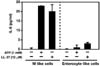
Luminal ATP can induce expression of a proinflammatory cytokine in M cells
To identify the function of P2X7R in M cells, we utilized M-like cells differentiated from Caco-2-derived enterocyte-like cells by co-culturing them with Raji cells. When M-like cells were stimulated with ATP, the proinflammatory cytokine IL-6 was highly secreted into the basolateral chamber from M-like cells (Fig. 3). In addition, treatment with LL-37, which is a P2X7R agonist, can also evoke the production of IL-6 in M-like cells. However, enhancement of IL-6 production by stimulation with ATP or LL-37 was not detected in control enterocyte-like cells that were not co-cultured with Raji cells. Therefore, we assume that IL-6 production upon treatment with ATP or LL-37 is closely related with P2X7R specifically expressed on M cells. Collectively, we conclude that luminal ATP can modulate the tolerogenic PP microenvironment toward a stimulatory microenvironment through IL-6 production by M cells.
ATP-gated P2X7R can play a role as an M cell-targeting receptor
In previous studies, it has been suggested that receptors expressed in the apical area of M cells could be utilized as M cell-targeting receptors for mucosal vaccine antigens (16). Consequently, we speculated that P2X7R might also serve as an M cell-targeting receptor for delivery of mucosal vaccine antigens. In order to test this assumption, we prepared a recombinant EGFP protein fused with LL-37 and tested for M cell-targeting of the protein after oral administration to mice (Fig. 4). As shown in Fig. 4, it was found that orally-introduced EGFP-LL-37 was localized to P2X7R-expressing M cells in PP. Consequently, we suggest that P2X7R on M cells could serve as an M cell-targeting receptor for mucosal vaccines.
DISCUSSION
In the present study, we have identified for the first time that P2X7R is expressed at the apical area of M cells, and that ATP stimulation promotes the production of the proinflammatory cytokine IL-6 via P2X7R signaling. These results suggest that P2X7R is a potential immune modulator that can induce an antigen-specific immune response in PP, which are mucosal immune inductive sites. In addition, M cell-localization of EGFP-LL-37 after oral administration suggests that P2X7R can be utilized as an antigen-targeting receptor for oral mucosal vaccines to enhance antigen-specific immune response induction.
Purinergic receptors are divided into P2X and P2Y receptors. P2X receptors consist of seven P2X subtypes and are ionotropic ligand-gated non-selective cation channel receptors, while P2Y receptors are G protein-coupled receptors (13). P2X7R is distinguished from other P2X subclass members through having a long intracellular C-terminus compared with others, and is closely associated with a proinflammatory response cascade (17). Although its expression was first reported in macrophages and monocytes, it has been identified in several cell types. In order to activate P2X7R, at least a 1 mM concentration of ATP is required, while in contrast, low levels of ATP can activate the other P2 receptors (13). Continuous stimulation of P2X7R by ATP can create an irreversible pore in the plasma membrane. This pore permits the free entrance of ions, hydrophilic solutes of up to 900 Da, and bacterial products (18). Consequently, we predicted the expression of P2X7R in M cells, because these characteristics are similar to the non-specific transcytotic activity of M cells that are continuously exposed to luminal ATP. Therefore, our findings may suggest a possible mechanism for the entrance of luminal contents into M cells.
Although cathelicidin LL-37 is a known antimicrobial peptide, it can also play a role as an immune modulator via its interaction with P2X7R (19). For instance, LPS-primed monocytes stimulated with LL-37 highly express IL-1β via P2X7R signaling. Production of IL-1β via ATP-P2X7R signaling in intestinal epithelial cells is promoted by the addition of LL-37 (20). In this study, we found that ATP or LL-37 can promote the expression of IL-6 by M-like cells. We feel this is a potentially important finding because IL-6 can act as an immune response initiator in a tolerogenic microenvironment through enhancing Th2 and Th17 cell polarization. The effective activation of immune responses is a major concern in mucosal vaccine development because the tolerogenic environment in the oral mucosal compartment must be overcome in order to induce antigen-specific immune responses (21). M cell-targeting of antigen is also an ideal strategy to deliver vaccine material into the mucosal inductive area (5). Given that most vaccines must be introduced to recipients in combination with adjuvants, we believe that identification of an interaction between EGFP-LL-37 and P2X7R on M cells may indicate a new target receptor for antigen delivery in the development of mucosal vaccine adjuvants. Collectively, we conclude that P2X7R is expressed at the apical area of M cells, and that its activation by ATP or LL-37 can modulate the tolerogenic microenvironment into a stimulatory microenvironment through production of IL-6. In addition, we suggest that this receptor can be exploited for the development of oral vaccines to induce antigen-specific immune responses.


 XML Download