

 PDF
PDF ePub
ePub Citation
 Print
Print


INTRODUCTION
Minor histocompatibility antigens are MHC-bound peptides. Differences in minor H antigens due to polymorphism at the loci between donors and recipients contribute to generation of T-cell responses after allogeneic transplantation (1). Thus, controlling T cells reactive to allogeneic minor H antigens is necessary for the success of allogeneic transplantation. The biology of T cells specific for minor H antigens, such as thymic selection and differentiation and response development, has not been fully investigated, although this information would be the basis for the development of strategies to control allo-responses following transplantation. Existence of a TCR transgenic mouse whose T cells have specificity to a minor H antigen would be helpful in understanding the response of T cells reactive to minor H antigens and the development of such strategies.
H60 is a dominant minor H antigen that induces strong CD8 T-cell activation in C57BL/6 (B6) anti-BALB.B settings (23). Previous studies showed that H60-specific CD8 T-cell response recruits diverse TCRs to the response and that the diversity of recruited TCRs is influenced by CD4 helper T cells (45). This wide breadth of the T-cell repertoire of H60-reactive CD8 T cells is ascribed to the presence of naïve precursors with high frequency in the periphery (6), which is ascribed to the lack of negative selection against the H60-reactive CD8 T cells in the thymus of B6 mice due to H60 not being expressed in B6 mice (78).
Our previous characterization of several different CTL clones specific for H60 showed differences in the CTL activity of each clone, including proliferation capacity and avidity to H60 peptide/H-2Kb complex (9). Among the CTL clones tested, clone #5 was found to have moderate CTL activity, proliferation capacity, and avidity to H60 peptide/H-2Kb complex. Here, we report generation of a TCR transgenic mouse line expressing TCRs specific for H60 using recombined genomic sequences originated from CTL clone #15. Using this TCR transgenic mouse line, we show that thymocytes expressing TCRs specific for H60 undergo different selection and fates according to antigenic environments within the thymus, and that a single amino-acid change in the cognate H60 peptide influences the fate of TCR-expressing thymocytes.
Go to : 
MATERIALS AND METHODS
Mice
B6, C57BL/6-Tg (TcraTcrb) 1100Mjb/J (OT-I), and C57BL/6-Tg (CAG-OVA) 916Jen/J (OVATg) mice were obtained from the Jackson Laboratory (Bar Harbor, ME, USA). J15 TCR transgenic mice (J15), H60 transgenic mice (H60Tg) (10), J15×H60Tg F1 mice (J15-/Tg H60-/Tg), H60H transgenic mice (H60HTg) (11), and J15×H60HTg F1 mice (J15-/Tg H60H-/Tg) were housed under specific pathogen-free conditions at the Biomedical Center for Animal Resource Development of Seoul National University College of Medicine in Korea. All experiments were approved by the Seoul National University Institutional Animal Care and Use Committee (IACUC) and performed in accordance with guidelines.
Generation of a TCR transgenic mouse line
To generate a transgenic mouse line that expresses TCRs recognizing H60 peptide (LTFNYRNL) in association with H-2Kb, genomic DNA fragments, including rearranged TCRVα-Jα and TCRVβ-Dβ-Jβ sequences, were obtained from CTL clone #15, which has TCR complexes composed of Vα13D-1 and Jα34-02 for the α chain, and Vβ 13-1-02, Dβ2-01, and Jβ2-7-01 for the β chain (9). Cassette vectors pTα and pTβ, each containing V-region (Vα and Vβ, respectively) promoter and the complete constant-region (Cα and Cβ, respectively) gene sequences (12), were subcloned with the recombined genomic sequences of Vα13D-1/Jα34-02 for the α chain using XmaI and NotI restriction sites to generate a #15-pTα DNA plasmid and of Vβ13-1-02/Dβ2-01/Jβ2-7-01 for the β chain using XhoI and SacII restriction sites to generate the #15-pTβ plasmid. DNA fragments devoid of prokaryotic regions from the subcloned #15-pTα and #15-pTβ DNA plasmids were then co-microinjected into fertilized B6 eggs.
Cytotoxic assays with various peptides
Splenocytes isolated from B6 mice were labeled with 2.5 µM CFSE (Molecular Probes, Eugene, OR, USA). A total of 5×10⁶ splenocytes labeled with CFSE were incubated with altered peptide ligands at different concentrations for 1 h at 37℃. After washing twice to remove free peptides, 1×10⁵ H60-specific CTLs (1:1) were added and the mixtures (1:1) of target cells and CTLs incubated for 4 h. Flow cytometry was performed to analyze CTL cytotoxicity and detect the CFSE-labeled target cells.
Cell staining and flow cytometry
Thymocytes, splenocytes, and peripheral blood lymphocytes from mice were incubated at 4℃ for 30 min in FACS buffer (1X PBS with 0.1% bovine calf serum and 0.05% sodium azide) containing phycoerythrin-cyanine 5-conjugated anti-CD4 (GK1.5, eBioscience, San Diego, CA, USA), allophycocyanin-Cy7-conjugated anti-CD8 (53-6.7, eBioscience), PE-conjugated H60-tetramer, fluorescein isothiocyanate (FITC)-conjugated anti-TCR Vβ8.3 (1B3.3, BD Pharmingen, San Diego, CA, USA), or anti-TCR Vβ 5.2 (MR9-4, BD Pharmingen). Tetramers of biotinylated H60/H2-Kb complexes were prepared by mixing four parts of peptide/MHC class I complexes with one part of streptavidin-PE (Molecular Probes). The stained cells were acquired on a FACS LSRII (BD Bioscience, San Jose, CA, USA), and data were analyzed with FlowJo software (TreeStar, Ashland, OR, USA).
Statistical analysis
All data are presented as mean±standard deviation (SD). Statistical analyses were prepared using GraphPad Prism (GraphPad Software Inc., La Jolla, CA, USA). The unpaired Student's t-test was used for comparisons between groups, with a p<0.05 considered significant.
Go to :
RESULTS
Generation of J15 TCR transgenic mice
In order to develop a transgenic mouse line where T cells express TCRs recognizing H60 peptide (LTFNYRNL) in association with H-2Kb, genomic DNA fragments encompassing rearranged TCRVα-Jα and TCRVβ-Dβ-Jβ sequences from an established H60-specific CTL clone were obtained for co-microinjection into fertilized B6 eggs. The clone was characterized to express TCR complexes composed of Vα13D-1 and Jα34-02 for the α chain, and Vβ 13-1-02, Dβ2-01 and Jβ2-7-01 for the β chain (Fig. 1A) (9). Each of the rearranged α and β chain DNA segments was subcloned into cassette vectors pTα and pTβ, which have V-region (Vα and Vβ, respectively) promoter and the complete constant-region (Cα and Cβ, respectively) gene sequences (12) for the microinjection (Fig. 1A). Transgene-positive founders were screened by performing PCR with genomic-tail DNA (data not shown) and flow cytometric analysis of PBLs after staining with anti-CD8 mAb and anti-Vβ8.3 mAb, since Vβ13-1-02 corresponds to Vβ8.3 in the international immunogenetics information system (Fig. 1B). Expression of the transgenic TCR specific for H60 was further confirmed by staining the PBLs with H60-tetramers. Among three different transgene-positive founder lines, we chose a founder line for which TCR levels on peripheral-blood CD8 T cells were comparable to those of wild type (WT) B6 T cells and named it J15 (13).
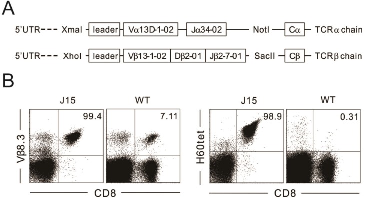 | Figure 1Generation and evaluation of J15 TCR transgenic mice. (A) Diagram depicting cassette vectors pTα and pTβ. Each vector has V-region (Vα and Vβ, respectively) promoter and the complete constant-region (Cα and Cβ, respectively) gene sequences and were subcloned with Vα13D-1 and Jα34-02 for the α chain (using XmaI and NotI), as well as Vβ13-1-02, Dβ2-01, and Jβ2-7-01 for the β chain (using XhoI and SacII). (B) J15 TCR transgenic mice were screened by staining the peripheral blood lymphocytes with anti-V β8.3 mAb, anti-CD8 mAb, and H60-tetramer, followed by flow-cytometric analysis. Vβ8.3 by CD8 and H60-tetramer by CD8 profiles of peripheral-blood lymphocytes from J15 and WT mice. Numbers in plots indicate the frequency of Vβ8.3 expression or H60-tetramer binding of CD8 T cells in the peripheral blood from J15 and WT mice.
|
Positive selection of J15 TCR transgenic T cells in the B6 thymus
We then investigated thymic profiles of J15 to confirm the positive selection of thymocytes expressing J15 TCR on the B6 background, where H60 is not expressed (7), and determined the stage of transgenic TCR α- and β-chain expression. Thymocytes obtained from 8-week-old J15 mice were stained with anti-CD4 and anti-CD8 mAbs for flow-cytometric analysis. Age-matched OT-I TCR Tg and WT B6 thymocytes were analyzed in parallel. Cellularity of J15 thymus was 31.3×10⁶ cells, on average, which was comparable to that of OT-I thymus (25.3×10⁶), but was a little lower than that observed for WT thymus (88.1×10⁶) (Fig. 2A). Proportions of CD4+CD8+ double positive (DP) cells (38%, on average) and CD4+ single positive (SP) cells (6%) were significantly reduced, however, those of CD8+ SP cells were significantly enhanced (25%) within the J15 thymus relative to those in normal B6 thymocytes (85%, 9%, and 2%, on average, respectively). This profile indicated the presence of positive selection and skewed differentiation of thymocytes expressing the J15 TCRs into CD8+ SP cells on the B6 antigenic background. The differentiation into CD8+ SP cells was enhanced for J15 thymocytes relative to that observed in OT-I thymocytes.
 | Figure 2Positive selection of J15 TCR transgenic T cells specific for H60. (A) CD4 by CD8 profiles (left) of thymocytes and numbers (right) of each thymic population from J15, OT-I, and WT mice on the B6 background. Numbers at the top of plots indicate total number of thymocytes. (B) Surface expression of Vβ8.3 and Vβ5.2, as well as (C) the binding of the H60- and OVA-tetramer of DN, DP, and CD8 SP thymocytes from J15, OT-I, and WT mice. (D) CD4 by CD8 profiles (left) of splenocytes and numbers (right) of CD4 and CD8 T cells in the spleen from J15, OT-I, and WT mice. (E) Surface expression of Vβ8.3 and Vβ5.2, as well as the binding of the H60- and OVA-tetramer of CD8 T cells in the spleen from J15, OT-I, and WT mice. Numbers in plots indicate the percentage of cells in each quadrant. ***p<0.001; **p<0.01; *p<0.05. Data are representative of three independent experiments and depict the mean±SD.
|
We then compared the expression patterns of transgenic TCR β chain between J15 and OT-I thymocytes. Staining with anti-Vβ8.3 mAb demonstrated that transgenic β chain was expressed at the CD4-CD8- double negative (DN) stage of J15 thymocytes, as was the transgenic Vβ 5.2+ β chain expression of OT-I thymocytes (Fig. 2B). H60- and OVA-tetramers were binding specifically to DP and CD8 SP stages of J15 and OT-I thymocytes, respectively (Fig. 2C). Therefore, although we were not able to identify the expression stage of transgenic TCRα of J15 TCR complexes for the lack of antibody to detect Vα 13D-1, we were able to determine that expression of the α and β chains of J15 TCR was completed before the DP stage based upon the tetramer-staining data. By comparing the flow-cytometric profiles of J15 and OT-1 thymocytes, we found that differentiation into CD8+ SP cells was more efficient in the J15 thymus than in OT-1 thymus.
In accordance with thymic characteristics, splenic profiles demonstrated that proportions (34%, on average) and numbers (31×106) of CD8+ T cells were higher in the spleen of J15 mice than those of WT (14% and 12×106) and even OT-I (23% and 19×106) mice (Fig. 2D). More than 98% of CD8 T cells from J15 splenocytes were bound to H60-tetramers specifically (Fig. 2E). Thus, T cells in the J15 transgenic mice were positively selected and selectively differentiated into CD8+ T cells with specificity for H60/H-2Kb on a B6 background.
Negative selection of J15 TCR transgenic T cells in the thymus expressing cognate H60
We then assessed whether thymocytes expressing the transgenic J15 TCR would be deleted in the thymus where cognate antigen H60 is expressed. To this end, we crossed J15 mice with H60 transgenic mice (H60Tg), wherein H60 is expressed ubiquitously under control of the actin promoter (10), and analyzed thymocytes of the F1 progenies expressing both of the J15 TCR and H60 transgenes (J15-/Tg H60-/Tg; J15-H60 Tg). When the thymic profiles of the double-transgenic J15-H60 Tg mice were compared with those of single-transgenic J15 mice, the proportions of CD8+ SP cells (3%, on average), as well as of CD4+CD8+ DP cells (8%), were obviously reduced in the J15-H60 Tg thymus (Fig. 3A), indicating the presence of negative selection of thymocytes expressing J15 within the thymus where cognate antigen H60 is expressed. This was supported by lower cellularity (5.10×10⁶, on average) of the J15-H60 Tg thymus, >7-fold lower, than that of J15 single-transgenic thymus (37.7×10⁶). When the thymic profiles of J15-H60 Tg mice were compared with those of OT-I-OVA Tg mice, F1 progenies (OT-I-/Tg OVA-/Tg) from crossing between OT-I-transgenic and mOVA-transgenic mice ubiquitously expressing membrane-bound forms of chicken ovalbumin under control of the actin promoter (14), loss of DP cells was more obvious within the J15-H60 Tg thymus than the OT-I-OVA Tg thymus. This implied that deletion of J15 thymocytes within the H60 Tg thymic environment might have occurred earlier during thymic development relative to that of OT-I thymocytes within the OVA Tg thymic environment.
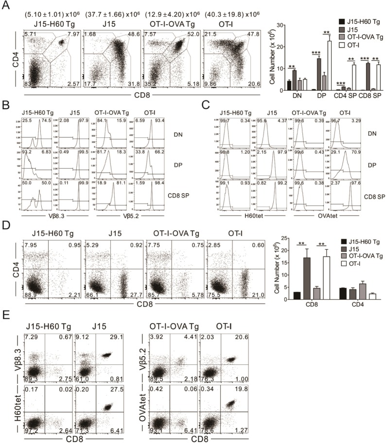 | Figure 3Negative selection of J15 TCR transgenic T cells specific for H60. (A) CD4 by CD8 profiles (left) of thymocytes and numbers (right) of each thymic population from J15-H60 Tg, J15, OT-I-OVA Tg, and OT-I mice on the B6 background. Numbers at the top of the plots indicate total number of thymocytes. (B) Surface expression of Vβ8.3 and Vβ5.2, as well as (C) the binding of the H60- and OVA-tetramer of DN, DP, and CD8 SP thymocytes from J15-H60 Tg, J15, OT-I-OVA Tg, and OT-I mice. (D) CD4 by CD8 profiles (left) of splenocytes and numbers (right) of CD4 and CD8 T cells in the spleen from J15-H60 Tg, J15, OT-I-OVA Tg, and OT-I mice. (E) Surface expression of Vβ8.3 and Vβ5.2, as well as the binding of the H60- and OVA-tetramer of CD8 T cells in the spleen from J15-H60 Tg, J15, OT-I-OVA Tg, and OT-I mice. Numbers in plots indicate the percentage of cells in each quadrant. ***p<0.001; **p<0.01. Data are representative of three independent experiments and depict the mean±SD.
|
Staining the J15-H60 Tg thymocytes with anti-Vβ8.3 mAb and H60-tetramer demonstrated the scarcity of DP cells positive for Vβ8.3-staining, indicating that the deletion of thymocytes expressing transgenic J15 TCR occurred at the DP stage. Although some of the survivors at the DP and CD8 SP stages were positive for Vβ8.3, their binding of H60-tetramers was rare, implying that the Vβ8.3-positive survivors from the deletion were expressing endogenous (non-transgenic) Vα (Fig. 3B and C). A similar phenomenon was observed with OT-I-OVA-Tg thymocytes. In the OT-I-OVA-Tg thymus, Vβ5.2-positive cells were decreased in DP cells compared to those of OT-I. OVA-tetramer-binding cells were rare in CD8+ SP cells, even though most of the CD8+ SP cells were expressing Vβ5.2. This suggested that the Vβ5.2-positive escapees from negative selection were also expressing endogenous Vα chains. Thus, the negative selection of thymocytes expressing transgenic OT-I TCR was also considered to occur at the DP stage.
Examination of splenic profiles confirmed significant contractions in proportions and numbers of mature CD8+ T cells in the J15-H60 Tg and OT-I-OVA-Tg mice, in comparison with corresponding single-transgenic mice, J15, and OT-I Tg mice, respectively (Fig. 3D). In accordance, H60- and OVA-tetramer-binding cells were not detected in the mature CD8+ T cells of the corresponding double-transgenic mice (Fig. 3E).
Altogether, these data demonstrate negative selection of thymocytes expressing J15 TCR within the thymus where cognate H60 is expressed, confirming again the specificity of J15 transgenic TCR for H60.
A single amino-acid change in the H60 epitope rescues J15 TCR transgenic T cells from negative selection
Peptides present in the thymus influence the fate of developing thymocytes (1516). Peptides with intermediate-tohigh affinities to a TCR participate in negative selection of thymocytes expressing the TCR, while those with low affinity select the thymocytes positively. Generally, MHC I molecules present peptides that are 8~10 amino acids long (17). Residues at P2, P3, P5, and P8 in octameric peptides presented by H2-Kb interact with MHC I molecules, while those at P1, P4, P6, and P7 potentially interact with TCR (1819). We tested the affinities of several different single amino-acid variants of the H60 epitope by performing cytotoxicity assays, specifically, changing the amino acid asparagine (N) residue at P4 of the H60 epitope to different amino acids. In cytotoxicity assays, cognate peptide H60N showed the highest cytotoxicity at the 10-5 M peptide concentration (Fig. 4A), indicating that peptide concentration of 10-5 M was the optimal concentration for elucidating maximum cytotoxicity from H60-specific CTL cells established after mixed leukocyte reaction of the splenocytes from H60-immunized B6 mice with irradiated H60-expressing splenocytes originated from H60-congenic mice (9). Among the different peptides tested, alteration of N at P4 to histidine (H) [H60H (LTFHYRNL)] elucidated the next highest cytotoxicity activity from the H60-specific CTL cells, suggesting that the H60H variant might be the optimal candidate to manipulate the fate of thymocytes expressing J15 TCR.
 | Figure 4Lack of negative selection of thymocytes expressing J15 TCR in the presence of H60H. (A) Cytotoxicity assays (left) tested affinities of several different single-amino-acid variants of the H60 epitope by changing the amino acid asparagine (N) residue at P4 of the H60 epitope to different amino acids. The amino acid sequences of the normal H60 and H60H epitope (right), LTFNYRNL and LTFHYRNL, respectively. (B) CD4 by CD8 profiles (left) of thymocytes and numbers (right) of each thymic population from J15-H60H Tg, J15, and H60HTg mice on the B6 background. Numbers at the top of plots indicate the total number of thymocytes. (C) H60-tetramer binding of DN, DP, and CD8 SP thymocytes from J15-H60H Tg, J15, and H60HTg mice. (D) CD4 by CD8 profiles (left) of splenocytes and numbers (right) of CD4 and CD8 T cells in the spleen from J15-H60H Tg, J15, and H60HTg mice. (E) Surface expression of Vβ8.3 and the binding of the H60-tetramer of CD8 T cells in the spleen from J15-H60H Tg, J15, and H60HTg mice. Numbers in plots indicate the percentage of cells in each quadrant. **p< 0.01; *p<0.05; ns, non-significant. Data are representative of two independent experiments and depict the mean±SD.
|
We then investigated whether presence of the H60H epitope within the thymus could influence the fate of thymocytes expressing J15 TCR by using transgenic mice expressing H60 with the H mutation at the P4 residue of the epitope region under control of the actin promoter (11). Thymic profiles of F1 progenies (J15-/Tg H60H-/Tg; J15-H60H Tg) generated by crossing J15 with H60HTg mice were analyzed and compared with those of J15 or H60HTg littermates by flow cytometry. Cellularity (27.7×10⁶, on average) of the J15-H60H Tg thymus was slightly reduced, however, was not significantly lower compared to that (36.6×10⁶) of J15 thymus (Fig. 4B). Moreover, proportions and numbers of DP (58%, on average, and 13.9×10⁶) and CD8+ SP (24% and 8.3×10⁶) cells in the J15-H60H Tg thymus were also comparable to those in the J15 thymus control (DP: 43% and 13.7×10⁶; CD8 SP: 31% and 11.1× 10⁶), indicating that the thymocytes expressing J15 TCR were positively selected within the thymus expressing the H60H epitope (Fig. 4B). Most of DP and CD8+ SP cells in the J15-H60H Tg thymus were able to bind to the H60-tetramer, confirming the positive selection of thymocytes expressing the J15 TCR (Fig. 4C). Such thymic events were further solidified by the presence of mature CD8+ T cells positive for Vβ 8.3 and H60-tetramers in the spleens of J15-H60H Tg mice (Fig. 4D and E).
Altogether, the results obtained from analyses on the T cells of J15-H60H Tg mice demonstrated that H60H was the peptide that induced positive selection of thymocytes expressing J15 TCRs. Thus, the avidity of H60H was enough to induce some levels of cytotoxic activity by H60-specific CTLs, however, was not high enough to delete the thymocytes expressing the J15 TCRs.
Go to :
DISCUSSION
In this study, we demonstrated the generation of a transgenic mouse line wherein T cells express TCRs specific for H60, a dominant minor histocompatibility antigen. J15 TCR transgenic T cells express transgenic TCR β chains containing variable segments composed of Vβ8.3 (Vβ 13-1-02 according to the nomenclature defined in the international immunogenetics information system). Although expression of the transgenic TCR α chain could not be confirmed directly due to the lack of a suitable antibody to stain the transgenic Vα13D-1, staining with H60-tetamer confirmed expression of transgenic TCR by recognizing the H60 peptide/H-2Kb complex. Furthermore, negative selection of thymocytes expressing the J15 TCR in the thymus where H60 is ubiquitously expressed verified the specificity of the J15 TCRs.
Negative selection eliminates T cells with high avidity to antigens expressed in the thymus or the periphery (20) and occurs in the thymus where cognate antigen is presented by thymic epithelial cells and dendritic cells (212223). Antigen-driven negative selection of thymocytes in a TCR transgenic mouse is influenced by the expression by cortical epithelial cells of the selecting antigen (24252627) and also by the timing of TCR α-chain expression and supposedly by the timing of expression of a rearranged TCRαβ complex (2728). We determined that J15 TCR β chain is expressed from the DN stage. Based upon the deletion of DP cells in the J15-H60 Tg thymus, we presume that the expression of transgenic α and β chains and formation of intact J15 TCR would be completed before the DP stage.
We showed that the H variant of H60 (H60H) was not able to induce negative selection of thymocytes expressing J15 TCRs. This result indicates that although cytotoxicity of H60-specific CTLs reached 50% of their original activity following incubation with the H60H peptide, this range of avidity is not responsible for the deletion of the J15 TCRs, suggesting that the negative selection threshold for J15 TCRs requires higher avidity than that observed here. This failure also suggests that several aspects of CD8 T-cell functionalities in response to peptide stimulation, such as proliferation and cytokine production, as well as cytotoxic activity, may all cooperate in determining the fate of thymocytes expressing specific TCRs. Examining the various peptides tested in this study for their influences on the functionalities of H60-specific CD8 CTLs and the fates of T cells expressing J15 TCR provides more detailed information regarding the peptides selecting thymocytes expressing the J15 TCR and for controlling the J15 TCR-positive T cells during allo-responses.
In conclusion, we report here the generation of TCR transgenic mice (J15) whose T cells express TCRs specific for H60 in complex with H-2Kb and exhibit different selection of J15-expressing thymocytes within different antigenic environments. These results and the systems established in this study will be useful for dissecting T-cell biology against minor histocompatibility antigens.
Go to :
 XML Download