

 PDF
PDF ePub
ePub Citation
 Print
Print


INTRODUCTION
Ag challenge can activate B cells to proliferate and differentiate into plasma cells. Plasma cells are a unique population of specialized cells that can produce Abs through T-dependent (TD) and T-independent (TI) pathways (1). Abs elicit a variety of effects, such as complement activation and opsonization. Therefore, plasma cells act as key players during the humoral immune response. However, the development program of plasma cell populations is not clearly understood.
The early growth response (Egr) protein family comprises Cys2-His2-type zinc finger transcription factors, which share 90% homology in their DNA binding region (2). Egr-1, which is also known as NGFI-A, Zif268, and Krox24, is expressed ubiquitously by many cell types and binds to a consensus DNA motif (GCGGTGGGCG) in many genes, including c-Myc, cyclins D2 and G2, and p19 (345). Other Egr-1 target genes identified in B cells include genes encoding TNF, IL-2, CD44, and ICAM-1, which are important for growth and functions of B cells (6).
Many studies have shown that Egr-1 participates in B cell maturation as a positive regulator. Egr-1 is crucial for B lymphopoiesis, especially in the development of pre-B cells and marginal zone B cells (78). Induction of Egr-1 expression following engagement with the BCR is associated with the growth and proliferation of mature B cells, suggesting that Egr-1 plays a role in their activation (29). However, it has also been observed that a deficiency in Egr-1 failed to inhibit BCR crosslinking-induced proliferation of mature B cells, due to the compensatory functions of Egr-2 and -3 (8). Egr-1 can exert anti- and pro-apoptotic roles, depending on the cell type. Its anti-apoptotic role is important for the survival of immature B cells during BCR-induced growth inhibition (1011), while its pro-apoptotic role contributes to the elimination of bortezomib-resistant multiple myeloma cells (12). Although transgenic mice expressing a dominant negative form of Egr-1 develop lower numbers of plasma cells than normal mice (8), it remains unclear whether this effect stems from the role of Egr-1 as none of the other Egr family members were expressed in these transgenic mice.
We investigated the functions of Egr-1 in B cells, and what cellular events in B cells are affected when Egr-1 is deficient. We used mice in which Egr1 was knocked out (Egr1-/-), and found that Egr-1 is involved in the differentiation of naive B cells into plasma cells. However, Egr-1 did not appear to be involved in the proliferation and apoptosis of B cells. Our results highlight the non-redundant role of Egr-1, which acts as a positive regulator of humoral immunity.
MATERIALS AND METHODS
Mice and immunization
Egr1+/- mice on a C57BL/6 background were purchased from The Jackson Laboratory (Bar Harbor, ME, USA) and maintained in a pathogen-free barrier facility at Hanyang University (Seoul, South Korea). Egr1+/- mice were incross to generate Egr1-/- mice. When Egr1-/- mice and their wild-type (WT) littermates were 6~12-weeks old, they were injected i.p. with a mixture of 4-hydroxy-3-nitrophenylacetyl-keyhole limpet hemocyanin (NP-KLH; 100 µg/mouse; Biosearch Technologies) and alum (Thermo Scientific). Our study was approved by the Institutional Animal Care and Use Committee (HY-IACUC-13-053).
RT-PCR assays
Erythrocyte-depleted spleen cell fractions from WT and Egr1-/- mice were stimulated with 20 ng/ml PMA and 1 µM ionomycin (Sigma-Aldrich) for either 1 or 3 h. Total RNA was isolated using Trizol reagent (Life Technologies). Semi-quantitative and quantitative RT-PCR assays were conducted as described previously (13). The mRNA expression levels of each gene were normalized to that for the gene encoding β2-microglobulin (B2m). Differences between samples were normalized using the cycle threshold method (ΔCt). We used specific oligonucleotide primers targeting Egr1 (5'-AAC CGG CCC AGC AAG ACA CC-3' and 5'-TGG CAA ACT TCC TCC CAC AAA T-3'), Egr2 (5'-CTT CAG CCG AAG TGA CCA CC-3' and 5'-GCT CTT CCG TTC CTT CTG CC-3'), Egr3 (5'-CAA CGA CAT GGG CTC CAT TC-3' and 5'-GGG CAG GCT GCC GAA TCC CG-3'), and B2m (5'-CAG TGT GAG CCA GGA TAT AG-3' and 5'-TGA CCG GCT TGT ATG CTA TC-3').
Abs and FACS
Splenic single-cell suspensions from WT and Egr1-/- mice were stained and analyzed using a FACSCanto II (BD Biosciences) as described previously (14). The following mAbs and reagents were purchased from BD Biosciences or eBioscience: anti-GL7-FITC, anti-CD4-PE, anti-B220-PerCP, anti-CD11C-allophycocyanin, anti-CD138-allophycocyanin, 7-AAD, and Annexin V-FITC.
B cell cultures
Naive GL7-CD138-CD11c-B220+ B cells from the spleens of WT and Egr1-/- mice were sorted using a FACSAria III (BD Biosciences) and stained with 3 µM CFSE (Molecular Probes). Cells at 1×10⁶/ml were cultured in the presence of 20 µg/ml LPS (Sigma-Aldrich) and 10 ng/ml IL-4 (Peprotech), or 5 µg/ml goat anti-mouse IgM (Jackson Immune Research) and 10 ng/ml IL-4. After culturing for 4 days, cells were subjected to flow cytometry.
ELISA
Sera were collected from pre- and post-immunized mice and assayed by ELISA to determine levels of NP-specific Abs. In brief, sera were diluted 1:100,000 in PBS (WelGene) and dispensed into 96-well immunosorbent plates (Nunc) pre-coated with 10 µg/ml NP8 conjugated to BSA (Biosearch Technologies). A serum sample containing the highest titer of anti-NP8 IgG was serially diluted and used as a standard. Plates were incubated with an anti-mouse IgG conjugated to biotin (Sigma-Aldrich), followed by streptavidin conjugated to HRP (BD Biosciences).
RESULTS AND DISCUSSION
Egr-1 expression was selectively abolished in Egr1-/- B cells
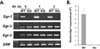
We examined whether depletion of Egr1 induces compensatory expression of other Egr genes. Egr1 mRNA were detected in unstimulated WT B cells, and were more abundant 1 h later in response to stimulation with PMA and ionomycin. We failed to detect Egr1 mRNA in Egr1-/- B cells, regardless of whether cells were stimulated (Fig. 1). We also found that Egr2 and Egr3 mRNA were expressed at similar levels in WT and Egr1-/- B cells. Our findings show that deficient Egr1 expression did not induce compensatory expression of Egr2 and Egr3. Thus, our Egr1-/- knockout model appears to be appropriate for investigating the role(s) of Egr-1.
Normal development of naive and germinal center B cells in Egr1-/- mice
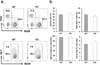
Given that a deficiency in Egr-1 affects normal development of B-lineage cells (78), we assessed whether Egr1-/- mice have an abnormal composition of spleen cells. We observed similar proportions of B220+ and CD4+ cells among WT and Egr1-/- mice (Fig. 2). The proportions of B220+GL7- naive B cells and B220+GL7+ germinal center B cells within the whole B cell population were similar between WT and Egr1-/- mice. Therefore, Egr-1 activity does not appear to be necessary for the development of naive B cells, CD4+ T cells, or germinal center B cells.
Differentiation of Egr1-/- B cells to plasma cells is defective, while proliferation and apoptosis of Egr1-/- B cells is unaffected
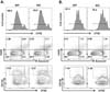
We sought to determine whether a deficiency in Egr-1 affects the proliferation and apoptosis of Egr1-/- B cells, and whether naive Egr1-/- B cells can effectively differentiate into plasma cells. Naive B cells sorted from WT and Egr1-/- mice were labeled with CFSE and cultured with LPS plus IL-4 or anti-IgM Ab plus IL-4, and then subjected to flow cytometry. The vast majority of cells proliferated, as determined by the decrease in CFSE intensity (CFSEdim cells), when treated with both types of stimulants (Fig. 3). The proportions of CFSEdim cells were not significantly different between WT and Egr1-/- mice, regardless of the stimulants used (88.7±1.2 for LPS/IL-4-treated WT versus 89.5±2.1 for LPS/IL-4-treated KO, p=0.67; 88.2±1.4 for anti-IgM/IL-4-treated WT versus 82.9±4.5 for anti-IgM/IL-4-treated KO, p=0.46). In addition, the proportions of early apoptotic (Annexin V+7-AAD-) and late apoptotic (Annexin V+7-AAD+) cells were equivalent between WT and Egr1-/- mice. However, the proportion of CD138+CFSEdim cells among Egr1-/- cells was approximately 50% of that seen for WT cells. These results demonstrate that a deficiency in Egr-1 impaired the differentiation of B cells into plasma cells, while proliferation and apoptosis of these cells were unaffected. Thus, Egr-1 appears to play an important role in plasma cell differentiation, and its depletion cannot be adequately compensated for by other Egr family members. Results from previous studies have shown that Egr-1 activity is associated with the proliferation of mature B lymphoma cell lines (29). Therefore, it is likely that, rather than not participating in proliferation, the functions of Egr-1 are redundant and compensated for by other Egr family members, as suggested previously (8).
Egr1-/- mice produce less Abs than their WT littermates
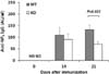
To validate the in vitro effects of Egr-1 deficiency on plasma cell development, we immunized Egr1-/- mice and their WT littermates with NP-KLH and alum. We then determined serum titers of Abs with high affinity for NP at 14 and 21 days post-immunization (dpi). The anti-NP8 IgG was detected at both dpi; its titer was significantly lower in Egr1-/- mice at 21, but not 14, dpi than that in WT mice (Fig. 4). Our findings reveal that Egr-1 is involved in the production of Abs, possibly by promoting the development of plasma cells through a TD pathway.
Results from several studies have indicated that Egr-1 is possibly associated with plasma cell development. When chronic lymphocytic leukemia B cells were stimulated with phorbol esters, Egr-1 was upregulated, giving rise to cells with a plasmacytic phenotype (15). Egr-1 could bind to the GC-box element of human PRDM1, which encodes Blimp-1, a master regulator of plasma cell differentiation (16). In the light of this result, we speculate that the effect of Egr-1 in the differentiation of B cells into plasma cells may be mediated by the induction of Blimp-1 expression. Transgenic mice lacking all Egr family members contain lower numbers of Ab-secreting cells than their WT counterparts (8). However, these studies have provided relatively little information regarding the role of Egr-1 during plasma cell differentiation.
In summary, we found that the proliferation and death of B cells was unaffected by the absence of Egr-1, indicating that Egr-1 is not required for these processes. We also showed that Egr1-/- B cells could not efficiently differentiate into plasma cells in vitro and in vivo, which was a strong indicator of the crucial and non-redundant role of Egr-1 in the commitment of B cells to a plasma cell fate.
 XML Download