

 PDF
PDF ePub
ePub Citation
 Print
Print


INTRODUCTION
Glucocorticoids have been effectively used for several decades as potent immunosuppressive agents in the treatment of inflammatory and autoimmune diseases. The immunosuppressive activities of glucocorticoids are primarily attributed to their influence on T cells and monocytes/macrophages (1,2,3). Glucocorticoids inhibit the secretion of cytokines from T cells and their proliferation induced by various stimuli (4,5,6,7). Glucocorticoids also inhibit several immunologically relevant activities of monocytes and macrophages. They block the production of cytokines, the expression of surface receptors for complement and immunoglobulins, phagocytosis and pinocytosis, and bactericidal and fungicidal activities of monocyte/macrophages (8,9,10,11).
Dexamethasone (Dex) is a synthetic glucocorticoid exerting 25 times more potent immunosuppressive activity than cortisol. Dex is particularly interesting because it promotes tolerance in vivo by enriching tolerogenic macrophages, while inducing apoptosis of effector T cells (12,13,14). Dex was also shown to severely impair the differentiation, maturation, and function of dendritic cells (DCs) and macrophages (15,16,17). The effects of Dex on DCs and macrophages, however, were investigated in cells cultured in vitro in the presence of Dex for two to several days.
In the present study, we examined the direct effects of Dex on the MHC-restricted presentation of exogenous antigens. Macrophages were generated from mouse bone marrow cells and allowed to phagocytose microencapsulated ovalbumin (OVA) in the presence of Dex for 2 h. The efficacy of OVA peptide presentation was evaluated using OVA-specific CD8 and CD4 T cells. Our results show that Dex inhibits the intracellular processing events of phagocytosed antigens in macrophages. We also discovered that immature macrophages are much more sensitive to the Dex-induced inhibition of MHC-restricted antigen processing than mature macrophages.
MATERIALS AND METHODS
Cell lines and reagents
The T-cell hybridoma cell lines B3Z86/90.14 (B3Z) and DOBW were kindly provided by Dr. Nilabh Shastri (University of California, Berkeley, CA, USA) and Dr. Clifford V. Harding (Case Western Reserve University, Cleveland, OH, USA), respectively (18,19). Recombinant human M-CSF was purchased from PeproTech (Rocky Hill, NJ, USA). Dexamethasone was purchased from Sigma-Aldrich (St. Louis, MO, USA).
Generation of macrophages from bone marrow cells
Macrophages were generated from mouse bone marrow using recombinant human macrophage colony stimulating factor (rhM-CSF). Briefly, bone marrow cells obtained from femurs of C57BL/6 or Balb/c mice were cultured in a 6-well plate (5×10⁶/well) in culture media supplemented with 20 U/ml rhM-CSF. At days 3 and 4 after the initiation of the culture, non-adherent cells were discarded by gentle shaking and replacement of the culture medium with fresh medium containing rhM-CSF. Immature macrophages were harvested on day 6 using cell stripper solution. Lipopolysaccharide (100 ng/ml) was added to immature macrophage cultures for maturation. Cells were cultured for 2 additional days and then harvested using cell stripper solution.
Preparation of OVA-nanospheres
Nanospheres containing OVA were prepared using a homogenization/solvent evaporation method with 400µl of OVA-containing water (50 mg/ml OVA) and 2 ml of ethyl acetate containing poly(lactic-co-glycolic acid) (100 mg/ml, Sigma-Aldrich) as described previously (Lee et al., 2010). Fluorescein isothiocyanate (FITC)-containing PLGA-nanospheres were prepared by adding FITC to the ethyl acetate phase together with PLGA. The OVA content was determined using a micro-bicinchoninic acid assay kit (Pierce, Rockford, IL, USA) after lysis of the nanospheres with a lysis buffer containing 0.1% SDS and 0.1 N NaOH.
MHC class I-restricted presentation assay
Class I MHC-complexed OVA peptide quantities on macrophages were assessed using B3Z cells (20). Briefly, macrophages (1×10⁵/well) generated from bone marrow cells of C57BL/6 mice (H-2b) were incubated with the indicated amounts of Dex for 2 h, and then OVA-nanospheres were added (50µg as OVA). After 2 h incubation at 37℃, the plate was washed twice with pre-warmed PBS (300µl/well) and then fixed with ice-cold 1.0% paraformaldehyde (100µl/well) for 5 min at room temperature, followed by washing of the plate three times with PBS (300µl/well). Class I MHC-complexed OVA peptide quantities were assessed by IL-2 secretion assays after culturing the paraformaldehyde-fixed macrophages with CD8.OVA cells (2×10⁴/well) for 18 h as described previously (20).
MHC class II-restricted presentation assay
Class II MHC-complexed OVA peptide quantities on macrophages were assessed using DOBW cells (20). Briefly, macrophages (1×10⁵/well) generated from bone marrow cells of BALB/C mice (H-2d) were incubated with the indicated amounts of Dex for 2 h, and then OVA-nanospheres were added (50µg as OVA). After 2 h incubation at 37℃, unphagocytosed nanospheres were removed by suction and then fixed with ice-cold 1.0% paraformaldehyde (100µl/well) for 5 min at room temperature. Class II MHC-complexed OVA peptide quantities were assessed by IL-2 secretion assays after culturing the paraformaldehyde-fixed macrophages with DOBW cells (2×10⁴/well) for 18 h as described previously (20).
Phagocytosis activity
Twenty minutes after nanospheres containing both OVA and FITC (1 mg/well as OVA) were added to macrophages, unphagocytosed nanospheres were removed by washing with pre-warmed PBS. The cells were harvested, fixed in 1% paraformaldehyde in PBS, and a flow cytometric analysis was performed on a FACSCanto flow cytometer (BD Biosciences, San Jose, CA, USA).
Phenotype analysis
Cells were stained with monoclonal antibodies recognizing murine cell surface markers as described previously (21) and flow cytometry was performed. The monoclonal antibodies (anti-H2-Kb, anti-I-Ab, anti-CD80, anti-CD86, anti-CD40, and anti-CD54) and an isotype-matched control antibody were purchased from BD Biosciences.
RESULTS
Dex inhibits MHC-restricted processing of exogenous antigens
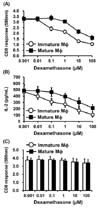
In order to examine the effects of Dex on the class I MHC-restricted processing of exogenous antigens, macrophages were allowed to phagocytose OVA-nanospheres in the presence or absence of Dex for 2 h and then fixed with paraformaldehyde. The efficacy of class Kb-restricted-OVA peptide presentation was evaluated using OVA-specific CD8 T cell hybridoma cells (B3Z), which express β-galactosidase when activated by OVA[257-264]-H-2Kb complexes. Dex dose-dependently inhibited class I MHC-restricted presentation of exogenous OVA (Fig. 1A). The inhibitory effect of Dex on class I MHC-restricted exogenous antigen presentation was much more potent in immature macrophages than in mature macrophages; the IC50 of Dex was approximately 1µM for immature macrophages and 10µM for mature macrophages.
In order to examine the effects of Dex on the class II MHC-restricted processing of exogenous antigens, macrophages were allowed to phagocytose OVA-nanospheres in the presence or absence of Dex for 2 h. The macrophages were then fixed with paraformaldehyde, and the amount of OVA peptide-class II MHC complexes was measured using OVA-specific CD4 T cell hybridoma DOBW cells. Dex dosedependently inhibited class II MHC-restricted presentation of exogenous OVA (Fig. 1B). This effect was also much more potent in immature macrophages than in mature macrophages; the IC50 of Dex was approximately 0.4µM for immature macrophages and 20µM for mature macrophages.
The exogenously added OVA peptide SIINFEKL can bind to cell surface Kb molecules, and thus does not require intracellular processing. In order to prove that Dex inhibits intracellular events of antigen processing pathways, macrophage cells were incubated with the peptide (1µM) for 2 h in the presence of different Dex concentrations. The cells were then washed, fixed, and examined for their stimulatory activities on T cell hybridoma B3Z cells. Dex did not inhibit presentation of the exogenously added synthetic peptide in immature or mature macrophages (Fig. 1C).
Short-term treatment of macrophages with Dex does not affect their phagocytic activity or the expression of cell surface molecules
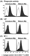
In order to test whether the antigen presentation-inhibitory activity of Dex (Fig. 1) was due to prevention of phagocytic activity, nanospheres containing both OVA and FITC were added to immature and mature macrophages pre-treated with Dex for 4 h. Cells were washed, cooled on ice, and then harvested by gentle pipetting. Flow cytometric analysis of the harvested cells demonstrated that Dex did not inhibit phagocytic activity of either immature or mature macrophages (Fig. 2A).
The effects of Dex on the expression of class I (K-2Kb) and II MHC molecules (I-Ab) and co-stimulatory molecules such as B7-1 and B7-2 were also examined with 4 h Dex-treated macrophages. Short-term treatment with Dex did not result in discernible effects on the expression levels of MHC molecules (Fig. 2B) or co-stimulatory molecules (Fig. 2C).
DISCUSSION
The major question addressed in the present study was whether Dex can directly inhibit MHC-restricted antigen processing pathways. Several studies have shown that Dex severely impairs the antigen-presenting function of macrophages and DCs (15,16,17). Nonetheless, these studies used macrophages or DCs that were differentiated or matured in the presence of Dex. Thus, the direct effect of Dex on MHC-restricted antigen processing has not yet been clarified. In the present study, we show evidence that Dex directly inhibits intracellular processing events of phagocytosed antigens in macrophages. Short-term exposure (4 h) of macrophages to Dex sufficed to inhibit both class I and class II MHC-restricted antigen processing pathways. We also discovered that immature macrophages are more sensitive to the Dex-mediated suppression of MHC-restricted antigen processing.
Exposure of macrophages or DCs to Dex during differentiation or maturation suppresses the phagocytic activity and the expression of co-stimulatory molecules such as CD80, CD86, and CD40 receptors (22,23,24,25). In our experimental conditions, where Dex inhibits MHC-restricted antigen processing, Dex did not exert inhibitory effects on the phagocytic activity or the total MHC expression levels or co-stimulatory molecules. It is noteworthy that we pre-treated macrophages with Dex for 2 h, and then allowed 2 h for phagocytosis and processing of exogenous OVA in the presence of Dex. Furthermore, Dex did not inhibit the presentation of the exogenously added OVA peptide SIINFEKL under our experimental conditions. These results demonstrate that Dex directly inhibits intracellular processing events of phagocytosed antigens in macrophages.
 XML Download