Journal List > Immune Netw > v.14(5) > 1033437

Lee, Kim, Jung, Lee, and Kim: Immunomodulatory Effects of ZYM-201 on LPS-stimulated B Cells

Abstract

ZYM-201 is a methyl ester of triterpenoid glycoside from Sanguisorba officinalis which has been used for treatment of inflammatory and metabolic diseases. In this study, immunomodulatory effects of ZYM-201 on B cells were examined in vitro and in vivo. When splenocytes were activated with lipopolysaccharide (LPS), the major population which had shown an increase in cell numbers was B cells. However, when the B cells were treated with ZYM-201 after LPS activation, their cell numbers and the expression of major costimulatory molecules, CD80 and CD86, were decreased. Furthermore, the effect of LPS, which induces activation of NF-κB, was abolished by ZYM-201: LPS-stimulated B cells showed decrease of phosphorylation after treatment of ZYM-201. The same results were shown in vivo experiments. These results suggest that ZYM-201 may play a role in the modulation of inflammatory responses through inhibiting NF-κB activation and downregulating the expression of costimulatory molecules on B cells.

Abbreviations

LPS

lipopolysaccharide

NF-κB

nuclear factor κB

MFI

mean fluorescence intensity

INTRODUCTION

Sanguisorba officinalis L. (Rosaceae), a well known traditional medicinal plant in East Asia, is used for treatment of inflammatory and metabolic disease (1). Recent studies have been reported that various extracts of S. officinalis have medicinal effects such as anti-allergic, anti-cancer, anti-thrombin, anti-wrinkle, and neuroprotective properties (1,2,3,4,5,6,7). The root of S. officinalis includes various saponin components including triterpenoids and their glycosides, gallic acid, and disaccharide (8,9). ZYM-201 is a modified compound of ziyuglycoside, one of triterpene glycoside originated from the root of S. officinalis, through deglycosidation and esterification (10). Previous studies have shown that ZYM-201 plays a role in inhibiting tissue factor activity, tumor necrosis factor (TNF)-α production and modulating hyperlipidemic conditions (10,11,12).
LPS is the major component of the outer membrane of Gram-negative bacteria and commonly used to study inflammation because it initiates acute inflammatory responses in animals (13,14). The binding of LPS to Toll-like receptor 4 triggers signal transduction pathways and finally induces phosphorylation and translocation of nuclear factor (NF)-κB (15) leading to regulating the expression of a large number of genes which have important functions in immune responses (16). The stimulation of B cells by LPS induces proliferation and upregulates the expression of CD80 and CD86, which results in enhancing their antigen-presenting capacity (15,17,18,19). Increased expression of these costimulatory molecules is associated with activation of NF-κB (20,21).
Since ZYM-201 has been reported to inhibit the production of TNF-α, we investigated the effects of ZYM-201 on the activation of lymphocytes in LPS-induced inflammation. We found that the activation of LPS-stimulated B cells was abolished by ZYM-201 through inhibiting NF-κB activation and downregulating costimulatory molecules.

MATERIALS AND METHODS

Preparation of cells

All experiments were performed in accordance with the approval of Soongsil University Institutional Animal Care and Use Committee. 6-week-old C57BL/6 mice were purchased from Young Bio (Seongnam, Korea). To make cell suspensions, extracted spleens from mice were cut into small fragments and crushed between gauze. After depletion of red blood cells with Gey's solution, the cell suspensions were cultured in RPMI1640 (GIBCO, Grand Island, NY, USA) supplemented with 10% fetal bovine serum, 100 U/ml of penicillin and 100 µg/ml of streptomycin (GIBCO).

Chemicals

ZYM-201 is a methyl ester of triterpenoid glycoside and modified compound through deglycosidation and esterification from ziyu-glycoside, which is an originated component from the root of S. officinalis (10). Extraction, modification and purification of ZYM-201 were performed as previously described (10).

Flow cytometry

Splenocytes were cultured with 1 µg/ml LPS for 24 hours and/or ZYM-201 (50 or 100 µM) was added 6 hours after LPS treatment. mAbs for CD80 (clone 16-10A1), CD86 (clone GL1), and B220 (clone RA3-6B2) were purchased from eBioscience (San Diego, CA).

Western blot

Splenocytes were positively enriched by using FITC-conjugated B220 mAbs and anti-FITC microbeads according to the manufacturer's instructions (Miltenyi Biotec Ltd., Bergisch Gladbach, Germany). B220+ cells were cultured with 1 µg/ml LPS and/or 100 or 200 µM ZYM-201 for 30 minutes, and lysed in RIPA buffer (Sigma-Aldrich, St.Louis, MO, USA) supplemented with protease inhibitor cocktail tablets and PhosSTOP (Roche, Mannheim, Germany). Cell lysates were separated using SDS-PAGE and transferred on to polyvinylidene difluoride membrane (Millipore, Darmstadt, Germany). mAbs for HSP90 (clone C45G5), NF-κB p65 (clone L8F6), and phospho-NF-κB p65 (Ser536) (clone 93H1) were purchased from Cell Signaling Technology (Danvers, MA, USA). As the second step reagents, HRP-linked anti-rabbit IgG and anti-mouse IgG Abs were used (Cell Signaling Technology). Proteins were visualized by enhanced chemiluminescence (Bio-Rad, Hercules, CA, USA) and x-ray films (AGFA, Dusseldorf, Germany) were used for detection.

in vivo experiment

7-week-old C57BL/6 mice, weighing 22±2 g were used. Three groups were analyzed and there were four mice in each group. Mice in the control group were intraperitoneally injected with sterile PBS and mice in the other groups were intraperitoneally injected with 1 mg/kg LPS (Sigma-Aldrich) and then orally administrated with 20 mg/kg ZYM-201 dissolved in 0.5% carboxymethylcellulose (CMC) solution (Sigma-Aldrich) or with 0.5% CMC solution twice (1 and 7 hours after LPS injection). 24 hours after LPS injection, spleens were taken for analysis.

RESULTS AND DISCUSSION

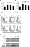
To investigate the effects of ZYM-201 on the cell number and activation of LPS-stimulated lymphocytes, cells from mouse spleens were cultured with 50, or 100 µM ZYM-201 after LPS treatment (Fig. 1A and 1B). The number of lymphocytes was increased from 100% (control) to 157% by LPS treatment, but decreased in the presence of ZYM-201 in a dose dependent manner: 142% with 50 µM ZYM-201 and 127% with 100 µM ZYM-201 (Fig. 1A). In particular, the number of B cells compared to other cell populations showed a big increase from 100% (control) to 185% by LPS treatment. However, this effect was abolished in the presence of ZYM-201; B cell numbers were decreased to 152% with 50 µM ZYM-201 and 132% with 100 µM ZYM-201 (Fig. 1B). In addition, the expression of CD80 and CD86 on LPS-stimulated B cells was down-regulated in the presence of ZYM-201; mean fluorescent intensity (MFI) of CD80 expression on LPS-stimulated B cells was downregulated from 330 to 222 followed by incubation with ZYM-201, and MFI of CD86 expression was also downregulated from 2410 to 1929 with ZYM-201 (Fig. 1C). These data indicate that ZYM-201 downregulates the activation of LPS-stimulated B cells. Because the upregulation of CD80 and CD86 costimulatory molecules is related with activation of NF-κB, we examined levels of NF-κB p65 phosphorylation on serine 536 (22) in B cells by western blot (Fig. 1D). Levels of NF-κB p65 phosphorylation in B cells were markedly increased by LPS stimulation, but dramatically decreased in the presence of ZYM-201 indicating that ZYM-201 has an inhibitory effect on LPS-stimulated B cell activation through inhibiting NF-κB phosphorylation and downregulating costimulatory molecules on B cells.
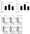
To confirm whether ZYM-201 has an inhibitory effect on LPS-stimulated B cell activation in vivo, cells from mice that had been injected with LPS and subsequently fed ZYM-201 twice were compared with those from mice that had not (Fig. 2). The numbers of lymphocytes and B cells were increased from 100% (control) to 138% and 143%, respectively, by LPS injection. Consistent with in vitro experiments, this effect was abolished after feeding ZYM-201; the numbers of lymphocytes and B cells were decreased to 92% and 90%, respectively, by ZYM-201 (Fig. 2A and 2B). Furthermore, the expression of constimulatory molecules on B cells in vivo showed the same pattern to that in vitro; the expression of CD80 and CD86 on LPS-stimulated B cells was downregulated after feeding ZYM-201 (Fig. 2C).
Taken together, these data indicate that ZYM-201, which has been known to have inhibiting tissue factor activity, TNF-α production and modulating hyperlipidemic conditions (10,11,12), has another function of anti-inflammatory activities via inhibition of NF-κB activation and downregulation of costimulatory molecules in B cells. In agreement with this, previous studies showed that inhibition of NF-κB activation suppress inflammatory responses (23) and have anti-inflammation and anticancer effects (24), and suggested that it could be used to treat immune diseases (25). Since ZYM-201 showed anti-inflammatory activities after LPS infection, we suggest that oral administration of ZYM-201 may be helpful to inflammatory diseases.

Figures and Tables

Figure 1
Effect of ZYM-201 on the activation of LPS-stimulated B cells in vitro. (A) and (B) Relative percentages of lymphocytes and B cells. Splenocytes were assessed after treatment with/without LPS (1 µg/ml) for 24 hours. In the case of ZYM-201 treatment, 50 and 100 µM ZYM-201 was added 6 hours after LPS treatment. Lymphocyte numbers were calculated by using SPHERO™ AccuCount Blank Particles (Spherotech, Lake Forest, IL) according to manufacturer's recommendations. Percentage of lymphocyte numbers was determined by comparison to control (100%). Percentage of B cells was decided using B220 mAbs and compared to control (100%). Error bars show standard deviation of six independent experiments. The statistical evaluation of the data was performed with Student's t-test and one-way analysis of variance. **p<0.01. (C) Flow cytometric analysis of CD80 and CD86 expression on B cells cultured with LPS and/or ZYM-201 (50 µM). Filled histograms show isotype-matched control Abs and numbers above bracketed line shows the percentage of positive cells for each Ab staining. Histograms are representative and mean fluorescence intensity (MFI) shown below each histogram is the average of six independent experiments. (D) Western blot analysis of levels of NF-κB p65 phosphorylation in B cells. Enriched B cells were cultured with LPS (1 µg/ml) and ZYM-201 (0 to 200 µM) for 30 minutes. Data shows total and phosporylated NF-κB p65 at serine536. HSP90 was used as loading control. Results are representative of six independent experiments.
in-14-260-g001
Figure 2
in vivo analysis of ZYM-201 effects on B cells of LPS-injected mice. Mice were intraperitoneally injected with LPS (1 mg/kg) and/or orally administrated with ZYM-201 (20 mg/kg). Twenty four hours after LPS injection, spleens were taken. (A) and (B) Relative percentages of lymphocytes and B cells. The numbers of lymphocytes and B220+ B cells were calculated by using SPHERO™ AccuCount Blank Particles and the percentage of the cells was determined by comparison to control (100%). Error bars show standard deviation of four independent experiments. **p<0.01; ***p<0.005. (C) CD80 and CD86 expression on B cells. Filled histograms show isotype-matched control Abs and numbers above bracketed line shows the percentage of positive cells for each Ab staining. Histograms are representative and mean fluorescence intensity (MFI) shown below each histogram is the average of four independent experiments.
in-14-260-g002

Abbreviations

LPS

lipopolysaccharide

NF-κB

nuclear factor κB

MFI

mean fluorescence intensity

ACKNOWLEDGEMENTS

This research was supported by Basic Science Research Program through the National Research Foundation of Korea (NRF) funded by the Ministry of Education, Science and Technology (NRF-2013R1A1A2007282).

Notes

The authors have no financial conflict of interest.

References

1. East J. The effect of certain plant preparations on the fertility of laboratory mammals. 4. Sanguisorba officinalis L. J Endocrinol. 1955; 12:273–276.
crossref
2. Park KH, Koh D, Kim K, Park J, Lim Y. Antiallergic activity of a disaccharide isolated from Sanguisorba officinalis. Phytother Res. 2004; 18:658–662.
crossref
3. Goun EA, Petrichenko VM, Solodnikov SU, Suhinina TV, Kline MA, Cunningham G, Nguyen C, Miles H. Anticancer and antithrombin activity of Russian plants. J Ethnopharmacol. 2002; 81:337–342.
crossref
4. Kim YH, Chung CB, Kim JG, Ko KI, Park SH, Kim JH, Eom SY, Kim YS, Hwang YI, Kim KH. Anti-wrinkle activity of ziyuglycoside I isolated from a Sanguisorba officinalis root extract and its application as a cosmeceutical ingredient. Biosci Biotechnol Biochem. 2008; 72:303–311.
crossref
5. Ban JY, Nguyen HT, Lee HJ, Cho SO, Ju HS, Kim JY, Bae K, Song KS, Seong YH. Neuroprotective properties of gallic acid from Sanguisorbae radix on amyloid beta protein (25--35)-induced toxicity in cultured rat cortical neurons. Biol Pharm Bull. 2008; 31:149–153.
crossref
6. Wang Z, Loo WT, Wang N, Chow LW, Wang D, Han F, Zheng X, Chen JP. Effect of Sanguisorba officinalis L on breast cancer growth and angiogenesis. Expert Opin Ther Targets. 2012; 16:Suppl 1. S79–S89.
7. Tsukahara K, Moriwaki S, Fujimura T, Takema Y. Inhibitory effect of an extract of Sanguisorba officinalis L. on ultraviolet-B-induced photodamage of rat skin. Biol Pharm Bull. 2001; 24:998–1003.
crossref
8. Liu X, Cui Y, Yu Q, Yu B. Triterpenoids from Sanguisorba officinalis. Phytochemistry. 2005; 66:1671–1679.
9. Mimaki Y, Fukushima M, Yokosuka A, Sashida Y, Furuya S, Sakagami H. Triterpene glycosides from the roots of Sanguisorba officinalis. Phytochemistry. 2001; 57:773–779.
crossref
10. Cho JY, Yoo ES, Cha BC, Park HJ, Rhee MH, Han YN. The inhibitory effect of triterpenoid glycosides originating from Sanguisorba officinalis on tissue factor activity and the production of TNF-alpha. Planta Med. 2006; 72:1279–1284.
crossref
11. Choi J, Kim MY, Cha BC, Yoo ES, Yoon K, Lee J, Rho HS, Kim SY, Cho JY. ZYM-201 sodium succinate ameliorates streptozotocin-induced hyperlipidemic conditions. Planta Med. 2012; 78:12–17.
crossref
12. Choi J, Yu T, Cha BC, Rhee MH, Yoo ES, Kim MY, Lee J, Cho JY. Modulatory effects of ZYM-201 sodium succinate on dietary-induced hyperlipidemic conditions. Pharmazie. 2011; 66:791–797.
13. Lu YC, Yeh WC, Ohashi PS. LPS/TLR4 signal transduction pathway. Cytokine. 2008; 42:145–151.
crossref
14. Alexander C, Rietschel ET. Bacterial lipopolysaccharides and innate immunity. J Endotoxin Res. 2001; 7:167–202.
crossref
15. Akira S, Sato S. Toll-like receptors and their signaling mechanisms. Scand J Infect Dis. 2003; 35:555–562.
crossref
16. Buss H, Dorrie A, Schmitz ML, Hoffmann E, Resch K, Kracht M. Constitutive and interleukin-1-inducible phosphorylation of p65 NF-{kappa}B at serine 536 is mediated by multiple protein kinases including I{kappa}B kinase (IKK)-{alpha}, IKK{beta}, IKK{epsilon}, TRAF family member-associated (TANK)-binding kinase 1 (TBK1), and an unknown kinase and couples p65 to TATA-binding protein-associated factor II31-mediated interleukin-8 transcription. J Biol Chem. 2004; 279:55633–55643.
crossref
17. Suvas S, Singh V, Sahdev S, Vohra H, Agrewala JN. Distinct role of CD80 and CD86 in the regulation of the activation of B cell and B cell lymphoma. J Biol Chem. 2002; 277:7766–7775.
crossref
18. Ogata H, Su I, Miyake K, Nagai Y, Akashi S, Mecklenbrauker I, Rajewsky K, Kimoto M, Tarakhovsky A. The toll-like receptor protein RP105 regulates lipopolysaccharide signaling in B cells. J Exp Med. 2000; 192:23–29.
crossref
19. Collins AV, Brodie DW, Gilbert RJ, Iaboni A, Manso-Sancho R, Walse B, Stuart DI, van der Merwe PA, Davis SJ. The interaction properties of costimulatory molecules revisited. Immunity. 2002; 17:201–210.
crossref
20. Giannoukakis NC, Bonham A, Qian S, Chen Z, Peng L, Harnaha J, Li W, Thomson AW, Fung JJ, Robbins PD, Lu L. Prolongation of cardiac allograft survival using dendritic cells treated with NF-kB decoy oligodeoxyribonucleotides. Mol Ther. 2000; 1:430–437.
crossref
21. Zhang G, Ghosh S. Toll-like receptor-mediated NF-kappaB activation: a phylogenetically conserved paradigm in innate immunity. J Clin Invest. 2001; 107:13–19.
crossref
22. Viatour P, Merville MP, Bours V, Chariot A. Phosphorylation of NF-kappaB and IkappaB proteins: implications in cancer and inflammation. Trends Biochem Sci. 2005; 30:43–52.
crossref
23. Pierce JW, Read MA, Ding H, Luscinskas FW, Collins T. Collins. Salicylates inhibit I kappa B-alpha phosphorylation, endothelial-leukocyte adhesion molecule expression, and neutrophil transmigration. J Immunol. 1996; 156:3961–3969.
24. Yin MJ, Yamamoto Y, Gaynor RB. The anti-inflammatory agents aspirin and salicylate inhibit the activity of I(kappa)B kinase-beta. Nature. 1998; 396:77–80.
crossref
25. Yamamoto Y, Gaynor RB. Therapeutic potential of inhibition of the NF-kappaB pathway in the treatment of inflammation and cancer. J Clin Invest. 2001; 107:135–142.
crossref
TOOLS
Similar articles